. 2022 Jul 20;14(654):eabn1413.
doi: 10.1126/scitranslmed.abn1413. Epub 2022 Jul 20.

Intranasal vaccination with lipid-conjugated immunogens promotes antigen transmucosal uptake to drive mucosal and systemic immunity

Affiliations

Intranasal vaccination with lipid-conjugated immunogens promotes antigen transmucosal uptake to drive mucosal and systemic immunity

Brittany L Hartwell et al. Sci Transl Med. .

Abstract

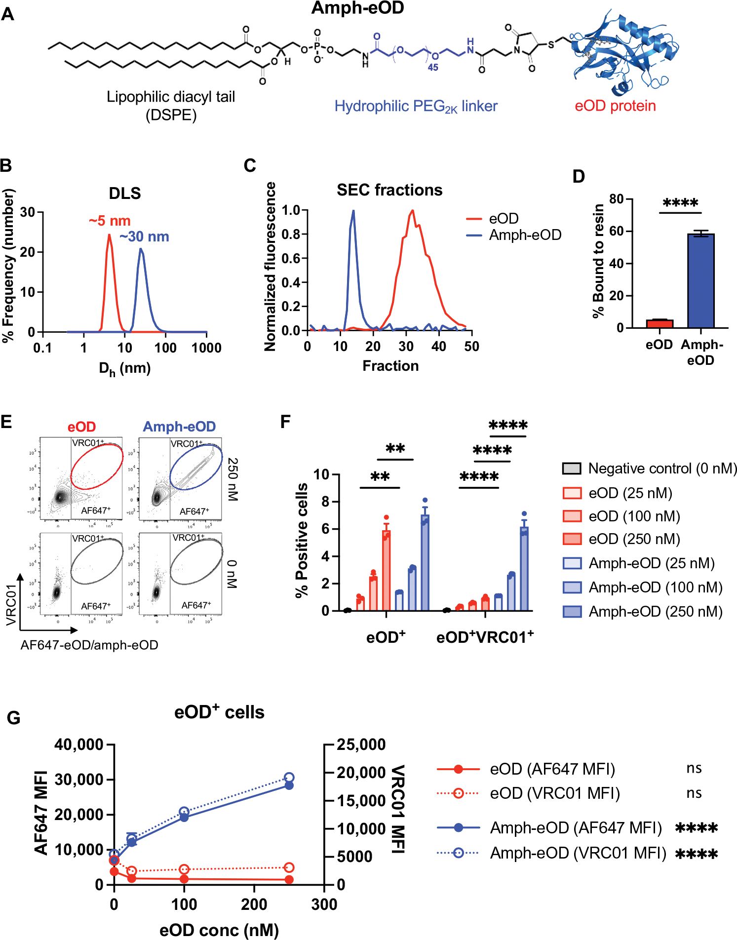
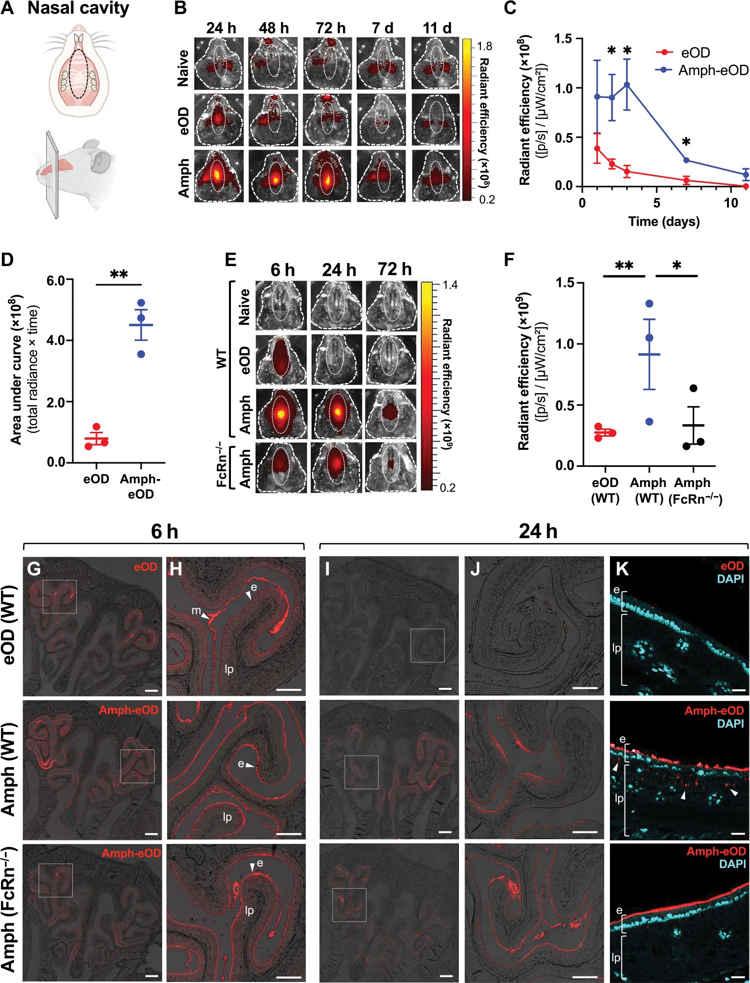
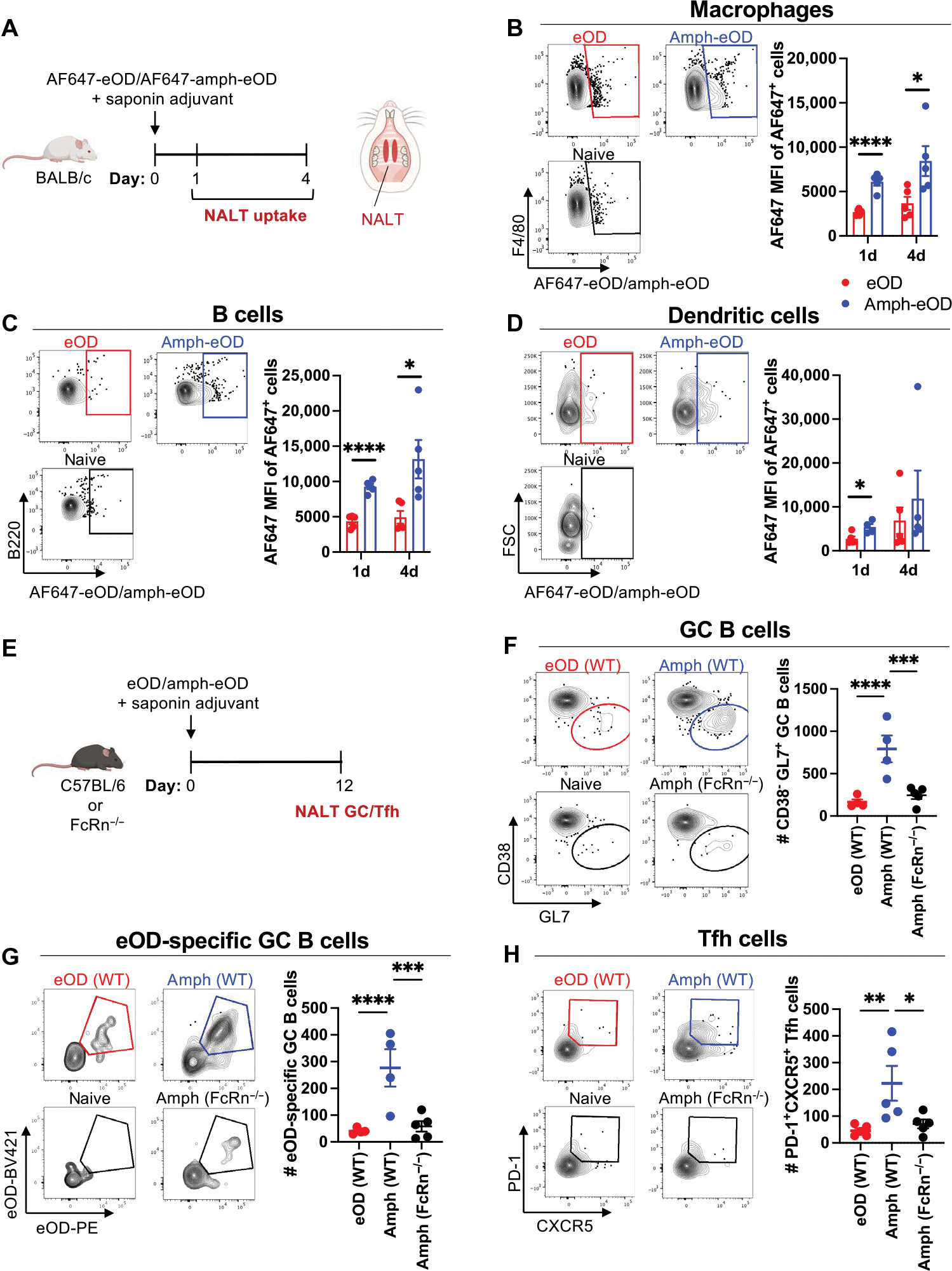
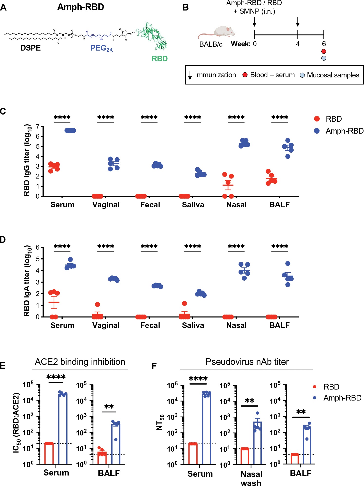
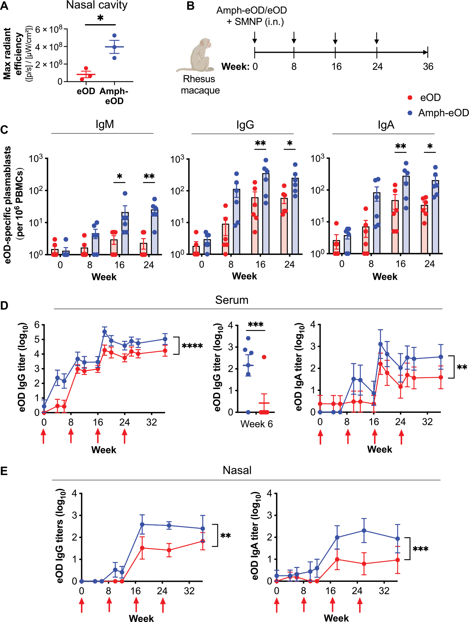
To combat the HIV epidemic and emerging threats such as SARS-CoV-2, immunization strategies are needed that elicit protection at mucosal portals of pathogen entry. Immunization directly through airway surfaces is effective in driving mucosal immunity, but poor vaccine uptake across the mucus and epithelial lining is a limitation. The major blood protein albumin is constitutively transcytosed bidirectionally across the airway epithelium through interactions with neonatal Fc receptors (FcRn). Exploiting this biology, here, we demonstrate a strategy of "albumin hitchhiking" to promote mucosal immunity using an intranasal vaccine consisting of protein immunogens modified with an amphiphilic albumin-binding polymer-lipid tail, forming amph-proteins. Amph-proteins persisted in the nasal mucosa of mice and nonhuman primates and exhibited increased uptake into the tissue in an FcRn-dependent manner, leading to enhanced germinal center responses in nasal-associated lymphoid tissue. Intranasal immunization with amph-conjugated HIV Env gp120 or SARS-CoV-2 receptor binding domain (RBD) proteins elicited 100- to 1000-fold higher antigen-specific IgG and IgA titers in the serum, upper and lower respiratory mucosa, and distal genitourinary mucosae of mice compared to unmodified protein. Amph-RBD immunization induced high titers of SARS-CoV-2-neutralizing antibodies in serum, nasal washes, and bronchoalveolar lavage. Furthermore, intranasal amph-protein immunization in rhesus macaques elicited 10-fold higher antigen-specific IgG and IgA responses in the serum and nasal mucosa compared to unmodified protein, supporting the translational potential of this approach. These results suggest that using amph-protein vaccines to deliver antigen across mucosal epithelia is a promising strategy to promote mucosal immunity against HIV, SARS-CoV-2, and other infectious diseases.


Conflict of interest statement

Competing interests: A U.S. utility patent has been issued (US9,107,904 B2) on which D.J.I. is an inventor related to the vaccine technology described here. This patent has been licensed to Elicio Therapeutics. D.J.I. is a consultant and holds equity in Elicio Therapeutics. D.J.I. also consults for Repertoire Immune Medicines, Ankyra Therapeutics, Strand Therapeutics, Window Therapeutics, Venn Therapeutics, Alloy Therapeutics, Livzon Pharmaceuticals, SQZ Biotechnologies, Jupiter Therapeutics, Surge Therapeutics, Third Rock Ventures, and Senda Biosciences. N.C.D., S.R.A., and J.C.L. have filed a patent related to the RBD-L452K-F490W sequence (U.S. Patent Application No. 17/512,902). B.L.H. and D.J.I. have filed a patent related to intranasal delivery of amph-protein conjugates (U.S. Patent Application No. 63/316,919).

Figures

Fig. 1.
Fig. 1.. Synthesis of albumin-binding amphiphile-protein immunogen conjugates.
(A) A schematic of amph-eOD structure is shown. (B) Dynamic light scattering (DLS) analysis of eOD and amph-eOD is shown as number-weighted % frequency. Dh, hydrodynamic diameter. (C) Size exclusion chromatography (SEC) profiles of eOD and amph-eOD are shown. (D) AF647-eOD or AF647-amph-eOD protein were incubated with albumin-functionalized agarose resin at 37°C, and the quantity of each protein bound to the resin after 2 hours was quantified. Statistical significance was determined by unpaired t test. (E to G) Fluorescent eOD or amph-eOD were incubated with murine C57BL/6 splenocytes for 1 hour at 37°C at a range of concentrations and then washed and stained with fluorescent VRC01 antibody. (E) Representative flow cytometry plots are shown of eOD/amph-eOD and VRC01 binding to the cells. (F) The percentage of cells positive for eOD alone or double positive for eOD and VRC01 was quantified; statistical significance was determined by two-way ANOVA followed by Sidak’s post hoc test. (G) Mean fluorescence intensity (MFI) of eOD and VRC01 is shown as a function of eOD concentration; statistically significant nonzero slope was determined by simple linear regression. All data are presented as means ± SEM. **P < 0.01 and ****P < 0.0001; ns, not significant.
Fig. 2.
Fig. 2.. Amph-protein conjugates exhibit enhanced persistence in the nasal mucosa and transport across the mucosal surface.
(A) Schematics illustrating (top) the ventral view of mouse upper palate and underside of top jaw, showing regions of interest (ROIs) used to quantify IVIS signals in (B) and (E), and (bottom) the sagittal view of mouse skull and nasal cavity showing approximate location of corresponding coronal cross sections used for histology in (G) and (H). (B) Representative IVIS images are shown of fluorescent signal in the nasal cavity of BALB/c mice (n = 3 animals per group) over time after intranasal administration of 5 μg of AF647-eOD or AF647-amph-eOD mixed with 5 μg of SMNP adjuvant. ROIs used to quantify IVIS signal are marked with dotted white oval. h, hour; d, day. (C) Quantified IVIS signals from (B) in nasal cavity over time are shown as average radiant efficiency. p, photon. Statistical significance was determined by unpaired t test at each time point. Data shown are from one representative of two independent experiments. (D) Quantified IVIS signal area under the curve (AUC; total radiance × time) from (C) was calculated. Statistical significance was determined by an unpaired t test. (E) Representative IVIS images show vaccine uptake and retention in the nasal cavity over time after intranasal administration of 5 μg of AF647-eOD or AF647-amph-eOD mixed with 5 μg of SMNP adjuvant in WT C57BL/6 versus FcRn−/− mice (n = 3 animals per group). (F) Quantified IVIS signal from (E) is shown for the nasal cavities of WT versus FcRn−/− mice at 6 hours after vaccination. Statistical significance was determined by two-way ANOVA followed by Tukey’s post hoc test. (G and H) Representative histology images of vaccine in nasal cavity in WT versus FcRn−/− mice at 6 hours are shown. Images in (H) are higher magnification views of dashed areas marked in (G). Scale bars, 1 mm (G) and 500 μm (H). (I to K) Representative histology images of vaccine in nasal cavity in WT versus FcRn−/− mice at 24 hours and immunization. Images in (J) are higher magnification views of dashed areas noted in (I). (K) High-magnification views stained with DAPI to identify the epithelial cell barrier; white arrows denote vaccine uptake. e marks epithelium, lp marks lamina propria, and m marks mucus. Scale bars, 1 mm (I), 500 μm (J), and 100 μm (K). All data are presented as means ± SEM. *P < 0.05 and **P < 0.01.
Fig. 3.
Fig. 3.. Amph-protein conjugates prime enhanced GC B cell and Tfh responses in the NALT in an FcRn-dependent manner.
(A to D) Groups of BALB/c mice (n = 5 animals per group) were immunized intranasally with 10 μg of AF647-amph-eOD or AF647-eOD mixed with 5 μg of saponin adjuvant, and NALT tissue was isolated 1 or 4 days later for flow cytometry analysis of antigen uptake. (A) Schematics illustrating (left) experimental timeline and (right) NALT tissue location are shown. (B to D) Representative flow cytometry plots of eOD signal gating and mean fluorescence intensities are shown for F4/80+ macrophages (B), B cells (C), and CD11c+ dendritic cells (D). FSC, forward scatter. Statistical significance was determined by unpaired t tests. (E to H) Groups of C57BL/6 (WT) or FcRn−/− mice (n = 5 animals per group) were immunized with 5 μg of eOD or amph-eOD mixed with 5 μg of saponin adjuvant; GC and Tfh responses were analyzed by flow cytometry on day 12. (E) The schematic shows experimental timeline. (F to H) Representative flow cytometry gating and enumeration of total GC B cells (F), antigen-specific GC B cells (G), and Tfh cells (H) are shown. Data shown are from one representative of two independent experiments. Statistical significance was determined by ordinary one-way ANOVA followed by Tukey’s post hoc test. All data are presented as means ± SEM. *P < 0.05, **P < 0.01, ***P < 0.001, and ****P < 0.0001.
Fig. 4.
Fig. 4.. Amph-protein conjugates elicit enhanced systemic and mucosal immune responses after intranasal vaccination.
(A to E) BALB/c mice (n = 5 animals per group) were immunized intranasally with 5 μg of eOD or amph-eOD mixed with 25 μg of cdGMP adjuvant and boosted 6 weeks later with the same formulations. (A) A schematic illustrating the experimental timeline is shown. (B to D) IgG and IgA titers were measured in the serum (B), vaginal wash (C), and feces (D). Red arrows indicate vaccination. (E) FRT and BM eOD-specific IgA ASCs were assessed by ELISPOT greater than 1 year after immunization. Data shown are from one representative of two independent experiments. (F to J) BALB/c mice (n = 5 animals per group) were immunized with 5 μg of eOD or amph-eOD mixed with 5 μg of SMNP adjuvant and boosted 6 weeks later with the same formulations. (F) A schematic illustrating the experimental timeline is shown. (G to I) IgG and IgA titers were measured in the serum (G), vaginal wash (H), and feces (I). (J) FRT and BM eOD-specific IgA ASCs were assessed by ELISPOT 35 weeks after immunization. Data shown are from one representative of two independent experiments (minus naïve background). Statistical significance in (E) and (J) was determined by unpaired t test, and that in (B) to (D) and (G) to (I) was determined by ordinary two-way ANOVA followed by Sidak’s post hoc test, comparing eOD to amph-eOD at each time point. *P < 0.05, **P < 0.01, ***P < 0.001, and ****P < 0.0001. All data show means ± SEM.
Fig. 5.
Fig. 5.. Intranasal vaccination with an amph-RBD conjugate elicits enhanced systemic and mucosal neutralizing antibody responses to SARS-CoV-2 immunogens.
(A) A schematic of amph-RBD structure is shown. (B) BALB/c mice (n = 5 animals per group) were immunized intranasally with 5 μg of RBD or amph-RBD mixed with 5 μg of SMNP adjuvant and boosted 4 weeks later with the same formulations. (C) IgG and (D) IgA titers in the serum, vaginal wash, fecal wash, saliva, nasal wash, and bronchoalveolar lavage fluid (BALF) were measured at 6 weeks after immunization. (E) ACE2:RBD binding inhibition (IC50) was measured for antibodies in serum and BALF at 6 weeks after immunization. (F) Pseudovirus neutralizing antibody (nAb) titers (NT50) were measured in the serum, nasal wash, and BALF at 6 weeks after immunization. Dotted lines in (E) and (F) represent the limit of quantitation. Data shown are from one representative of two independent experiments. Statistical significance in (C) and (D) was determined by two-way ANOVA followed by Sidak’s post hoc test, and that in (E) and (F) was determined by unpaired t test. **P < 0.01 and ****P < 0.0001. All data are presented as means ± SEM.
Fig. 6.
Fig. 6.. Intranasal immunization with amph-protein conjugates leads to improved humoral immune responses in nonhuman primates.
(A) Rhesus macaques (n = 3 animals per group) were immunized intranasally with 100 μg of AF647-eOD or AF647-amph-eOD mixed with 375 μg of SMNP adjuvant. Shown is quantified fluorescence signal of vaccine immunogens in the nasal cavity after 24 hours by IVIS imaging. Statistical significance was determined by unpaired t test. (B) Rhesus macaques (n = 6 animals per group) were immunized intranasally with 100 μg of eOD or amph-eOD mixed with 375 μg of SMNP adjuvant and boosted at 8, 16, and 24 weeks with the same formulations. (C) Frequencies of antigen-specific IgM, IgG, and IgA secreting plasmablasts of total PBMCs were measured by ELISPOT. (D) IgG and IgA titers in the serum were quantified over time; statistical significance shows an overall comparison across all time points. Individual IgG titers at 6 weeks are shown in the middle plot. (E) IgG and IgA titers in the nasal wash were quantified over time; statistical significance shows an overall comparison across all time points. Statistical significance in (C) to (E) was determined by two-way ANOVA. *P < 0.05, **P < 0.01, ***P < 0.001, and ****P < 0.0001. All data are presented as means ± SEM.

Comment in

References

    1. Neutra MR, Kozlowski PA, Mucosal vaccines: The promise and the challenge. Nat. Rev. Immunol. 6, 148–158 (2006). - PubMed
    1. Holmgren J, Czerkinsky C, Mucosal immunity and vaccines. Nat. Med. 11, S45–S53 (2005). - PubMed
    1. Lycke N, Recent progress in mucosal vaccine development: Potential and limitations. Nat. Rev. Immunol. 12, 592–605 (2012). - PubMed
    1. McGhee JR, Mestecky J, Dertzbaugh MT, Eldridge JH, Hirasawa M, Kiyono H, The mucosal immune system: From fundamental concepts to vaccine development. Vaccine 10, 75–88 (1992). - PubMed
    1. Mitragotri S, Immunization without needles. Nat. Rev. Immunol. 5, 905–916 (2005). - PubMed

Publication types