Review
. 2022 Dec;74(12):1893-1905.
doi: 10.1002/art.42296. Epub 2022 Oct 26.

Artificial Intelligence and Deep Learning for Rheumatologists

Affiliations
Review

Artificial Intelligence and Deep Learning for Rheumatologists

Christopher McMaster et al. Arthritis Rheumatol. 2022 Dec.

Abstract

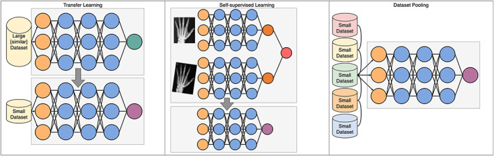
Deep learning has emerged as the leading method in machine learning, spawning a rapidly growing field of academic research and commercial applications across medicine. Deep learning could have particular relevance to rheumatology if correctly utilized. The greatest benefits of deep learning methods are seen with unstructured data frequently found in rheumatology, such as images and text, where traditional machine learning methods have struggled to unlock the trove of information held within these data formats. The basis for this success comes from the ability of deep learning to learn the structure of the underlying data. It is no surprise that the first areas of medicine that have started to experience impact from deep learning heavily rely on interpreting visual data, such as triaging radiology workflows and computer-assisted colonoscopy. Applications in rheumatology are beginning to emerge, with recent successes in areas as diverse as detecting joint erosions on plain radiography, predicting future rheumatoid arthritis disease activity, and identifying halo sign on temporal artery ultrasound. Given the important role deep learning methods are likely to play in the future of rheumatology, it is imperative that rheumatologists understand the methods and assumptions that underlie the deep learning algorithms in widespread use today, their limitations and the landscape of deep learning research that will inform algorithm development, and clinical decision support tools of the future. The best applications of deep learning in rheumatology must be informed by the clinical experience of rheumatologists, so that algorithms can be developed to tackle the most relevant clinical problems.


Figures

Figure 1
Figure 1
Neural network architectures. The first layer of a neural network consists of the data. These data are then passed to the first “hidden layer.” Each node, represented by a circle, is a weighted linear combination of all the nodes in the layer before. It is the weights that the model “learns.” Apart from a classic neural network where all nodes from 1 layer are connected to the next (otherwise known as a multilayer perceptron), other common architectures include recurrent networks with connections between nodes within a layer, usually used for sequence data (e.g., time‐series or text), and residual networks, where information from 1 layer can “skip” the next layer, giving the network a way to bypass inefficient layers.
Figure 2
Figure 2
Visualization of attention model (ref. 94). Two attention layers are shown with text input for NLP (top). The original input text reads, “There was swelling and redness of the joint. The joint was also stiff and tender, with reduced range of motion.” This text is converted into tokens, sometimes splitting words into more than one token (here “redness” is split into “red” and “##ness”—the “##” signifying that this token belongs with the preceding token). On the left, a lower layer of the attention‐based model relied on the words “range” and “motion” to interpret the word “reduced.” On the right, at a higher layer, the word “reduced” also depends strongly on the word “swelling” in the previous sentence. An attention model can be used for any sequence data (bottom). Here, these numbers could be laboratory values, with the task of predicting the next value in the sequence. The attention layer used the values “16” and “90” to predict the next value in the sequence. In this instance, attention is used to focus on a similar pattern to anticipate a future value.
Figure 3
Figure 3
A vertical edge detector convolution kernel. Edges that transition from dark to light (as shown in the input image) will be light in the output image. The pixel values (representing light intensity) are shown as pink numbers. No values in the output image are <0. This is because all values <0 are turned into 0 by a function known as a rectified linear unit (ref. 95)—this is known as an activation function and is a common technique in deep learning. The rim of zeroes around the input image—known as “padding”—allows the output image to retain the same dimensions as the input image. Color figure can be viewed in the online issue, which is available at http://onlinelibrary.wiley.com/doi/10.1002/art.42296/abstract.
Figure 4
Figure 4
Three unique deep learning methods used in rheumatology. A, A cascade of convolutional neural networks (CNNs) used to classify power Doppler images. At each step, the CNN classifies the image as either a certain EULAR Outcome Measures in Rheumatology synovitis scoring class or any higher class (e.g., the first step classifies to either a class of 0 or >0). If the CNN determines that it belongs to a higher class, it is passed along to the next CNN, which performs the same task for the next highest class. Eventually, the final CNN simply classifies images as either class 2 or class 3. B, A simplified diagram of the U‐Net architecture (49). An image begins as an “N × N × C” shape, where “N × N” is the image size (e.g., 224 × 224 pixels) and “C” is the number of channels (typically 3 channels of red/green/blue for a color image). The model gradually reduces the size, while increasing the number of channels, until the bottom of the architecture is reached, and then the reverse occurs. Connections across the architecture (dashed lines) act as a “memory.” The image recovered at the end is a segmented image, partitioning the original into the relevant parts. In this example, the bones of 2 metacarpal joints are segmented from the plain radiograph. C, A single coronal radiograph of the knee joint split into 2 images: the right half of the knee and the horizontally flipped left half. Both images are passed through the same CNN before joining up to produce a Kellgren/Lawrence (K/L) composite score as the model output. Color figure can be viewed in the online issue, which is available at http://onlinelibrary.wiley.com/doi/10.1002/art.42296/abstract.
Figure 5
Figure 5
Three methods to overcome the complications of limited data sets. Transfer learning takes a model trained on a large data set and repurposes it for a new task, replacing only the final layer. Self‐supervised learning is a type of transfer learning; however, the data set used in pre‐training does not need to have labels—here the task is simply to recognize that 2 versions of the same image are indeed the same image, and in doing so the model learns to recognize invariant features. Increasing data set size can be done in a number of ways; however, pooling data across institutions has technical, logistical, and privacy issues that must be overcome. Circles represent individual nodes. Color figure can be viewed in the online issue, which is available at http://onlinelibrary.wiley.com/doi/10.1002/art.42296/abstract.

References

    1. LeCun Y, Bengio Y, Hinton G. Deep learning. Nature 2015;521:436–44. - PubMed
    1. American College of Radiology . AI Central. URL: https://aicentral.acrdsi.org/.
    1. Silver D, Hubert T, Schrittwieser J, et al. Mastering chess and shogi by self‐play with a general reinforcement learning algorithm. arXiv [cs.AI] 2017. URL: http://arxiv.org/abs/1712.01815. - PubMed
    1. Zhang J, Zhao Y, Saleh M, et al. PEGASUS: Pre‐training with Extracted Gap‐sentences for Abstractive Summarization. arXiv [cs.CL] 2019. URL: http://arxiv.org/abs/1912.08777.
    1. Chen K, Oldja R, Smolyanskiy N, et al. MVLidarNet: Real‐time multi‐class scene understanding for autonomous driving using multiple views. arXiv [cs.CV] 2020. URL: http://arxiv.org/abs/2006.05518.