. 2022 Jul 26;119(30):e2207414119.
doi: 10.1073/pnas.2207414119. Epub 2022 Jul 20.

Phosphorylation of Arl4A/D promotes their binding by the HYPK chaperone for their stable recruitment to the plasma membrane

Affiliations

Phosphorylation of Arl4A/D promotes their binding by the HYPK chaperone for their stable recruitment to the plasma membrane

Ming-Chieh Lin et al. Proc Natl Acad Sci U S A. .

Abstract

The Arl4 small GTPases participate in a variety of cellular events, including cytoskeleton remodeling, vesicle trafficking, cell migration, and neuronal development. Whereas small GTPases are typically regulated by their GTPase cycle, Arl4 proteins have been found to act independent of this canonical regulatory mechanism. Here, we show that Arl4A and Arl4D (Arl4A/D) are unstable due to proteasomal degradation, but stimulation of cells by fibronectin (FN) inhibits this degradation to promote Arl4A/D stability. Proteomic analysis reveals that FN stimulation induces phosphorylation at S143 of Arl4A and at S144 of Arl4D. We identify Pak1 as the responsible kinase for these phosphorylations. Moreover, these phosphorylations promote the chaperone protein HYPK to bind Arl4A/D, which stabilizes their recruitment to the plasma membrane to promote cell migration. These findings not only advance a major mechanistic understanding of how Arl4 proteins act in cell migration but also achieve a fundamental understanding of how these small GTPases are modulated by revealing that protein stability, rather than the GTPase cycle, acts as a key regulatory mechanism.

Keywords: Arf-like GTPase; HYPK; Pak1; cell migration; protein stability.


Conflict of interest statement

The authors declare no competing interest.

Figures

Fig. 1.
Fig. 1.
Pak1 kinase is required to stabilize fast-degrading Arl4A/D under FN stimulation. (A) C33A cells were either kept in complete Dulbecco’s modified Eagle’s medium (mock) or were serum starved (SS) for 16 h before treatment with FN at 20 μg/mL for the indicated hours. (C) Cell lines with relatively abundant endogenous Arl4A (C33A), Arl4C (A549), and Arl4D (HeLa) were treated with CHX for the indicated times. The Arl4 protein level at each time point was normalized to the protein level at time 0 and is shown in the line graph, with error bars representing the mean ± SD; n = 3. (D) Mock and FN treatments were conducted in short interfering control (siCtrl) and short interfering Pak1 (siPak1) C33A cells. (F) Mock and FN treatments were further conducted in siCtrl-, siPak1-, and Pak1-rescued C33A cells. Res, siRNA-resistant form. (B, E, G) Quantification of Arl4 protein levels as determined by Western blotting in (A, D, F). The statistical results are shown in dot plots with error bars representing the means ± SD; n = 3. *P < 0.05; **P < 0.01; ***P < 0.001. One-way ANOVA with Dunnett’s post hoc multiple comparison test was applied.
Fig. 2.
Fig. 2.
Pak1 phosphorylates Arl4A/D on S143/S144 under FN signaling. (A) SILAC phosphoproteomic analysis of Arl4A upon FN stimulation. The [U-13C6]–labeled “heavy” and unlabeled “light” HeLa cells were transfected with Arl4A and reseeded on FN-uncoated (FN−) or -coated (FN+) coverslips for 1.5 h before SILAC liquid chromatography–tandem mass spectrometry. (Left) The immunofluorescence images indicate the same phenotype of Arl4A (green) in the two swapping groups upon reseeding on FN. Scale bar, 25 μm. (Right) The phosphorylation ratios of FN+/FN− of S141/S143 in Arl4A. The dots represent two biological replicates in which the heavy and light Arg/Lys-labeled chains were swapped. (B) HeLa cells equally overexpressing Arl4A/D WT, S143D/S144D, and S143A/S144A mutants were harvested for immunoblotting with anti–phospho-Arl4A/D (pArl4A/D) (S143/S144) antiserum. The anti-Arl4A/D antiserum was used to indicate the total Arl4A/D protein expression levels. (C and D) HeLa cells transfected with the indicated proteins were seeded on FN− or FN+ surfaces for 1.5 h and harvested to detect pArl4A/D (S143/S144) by Western blotting. pPak1 (T423) indicated the activation of Pak1 under FN treatment. (E and F) Experiments in (C and D) were conducted in short interfering control (siCtrl) and short interfering Pak1 (siPak1) cells. The pArl4A/D signals were quantified by normalizing to total Arl4A/D and are shown in the dot plots with error bars representing the means ± SD. **P < 0.01; ***P < 0.001; n = 3 to 5. The statistical results were analyzed by two-sample t test. (G) The in vitro kinase assay was performed by incubating HA-Pak1-CAAX purified from HeLa cells and GST and GST-Arl4A/D purified from Escherichia coli at 30 °C for 30 min. GST-DLC1 served as positive controls. (H) The kinase assay was performed as described for G, with equal amounts of GST, GST-Arl4A/D WT, and GST-Arl4A/D S143A/S144A phospho-defective mutants. CB, Coomassie blue; Exp, experiment.
Fig. 3.
Fig. 3.
Arl4A/D S143/S144 phosphorylation influences protein stability and ubiquitination. (A) Arl4A S143D/S143A and Arl4D S144D/S144A mutants were transfected into HeLa cells for the CHX chase assay for the indicated times. (B) One-phase exponential decay and T1/2 of each phosphomutant assayed in A are shown separately; n = 3. (C and D) HeLa cells were transfected with the indicated proteins and treated with MG132 for 4 h before Ub-IP. The Myc signals of phosphomimetic mutants (S143/144D) were compared with the signals of phosphodefective mutants (S143/144A) after immunoprecipitation (IP). A two-sample t test was applied for the results shown in the dot plots, with error bars representing the means ± SD; n = 3. *P < 0.05. HA, hemagglutinin; LC, light chain of HA antibody.
Fig. 4.
Fig. 4.
The chaperone-like protein HYPK specifically interacts with Arl4A/D. (A) Arl4s-HA was equally expressed with enhanced green fluorescent protein (EGFP) or EGFP-HYPK in HeLa cells and treated with dithiobis(succinimidyl propionate) at 1 mM for 2 h before coimmunoprecipitation (co-IP) and Western blotting analysis. (B) In vitro binding of His-tagged Arl4A, Arl4C, and Arl4D with GST or GST-HYPK. His-Arl4 proteins pulled down by GST-fusion proteins were analyzed by Western blotting. (C and D) HeLa cells expressing either Arl4A Q79L and T51N (C) or Arl4D Q80L and T52N (D) were coexpressed with EGFP or EGFP-HYPK for co-IP. QL/TN, GTP-bound/GTP-deficient mimics. (E) Mutated residues of Arl4A/D binding defective HYPKs A1 and A3. (F and G) HeLa cells expressing either Arl4A (F) or Arl4D (G) were coexpressed with EGFP, EGFP-HYPK, EGFP-A1, or EGFP-A3 for co-IP. Naa10 and Naa15, which are components of the NatA complex, were the positive controls for co-complexation with HYPK. The co-IP signals of Arl4 proteins and NatA components were quantified and are shown in the dot plots of A, C, D and H, with error bars indicating the mean ± SD; n = 3 to 5. *P < 0.05; **P < 0.01; ***P < 0.001. The statistical results were analyzed by two-sample t test or one-way ANOVA with Dunnett’s post hoc multiple comparison test for groups larger than two. CB, Coomassie blue; HA, hemagglutinin.
Fig. 5.
Fig. 5.
The HYPK–Arl4A/D interaction contributes to Arl4A/D protein stabilization. (A and B) CHX was applied to C33A and HeLa cells expressing empty vector, HYPK WT, and HYPK A3 for the indicated times, and endogenous Arl4A in C33A cells (A) and Arl4D in HeLa cells (B) were examined by Western blotting. (C and D) The protein levels of the Arl4s from A and B were normalized to α-tubulin and the protein level at time 0. The one-phase exponential decay of each group was plotted; n = 3. The half-lives (t1/2) of groups expressing the empty vector, HYPK WT, and the HYPK A3 mutant are shown. (E) Short hairpin control (shCtrl) RNA– and short hairpin HYPK (shHYPK) RNA–expressing HeLa cells were transfected with the indicated proteins and treated with MG132 before Ub-IP and Western blotting. Arl4A/D-Myc signals from immunoprecipitation (IP) were quantified by normalizing to immunoprecipitated hemagglutinin–ubiquitination (HA-Ub) signals and Arl4 inputs. The results are shown in dot plots, with error bars representing the mean ± SD; n = 4. **P < 0.01. The statistical results were analyzed by two-sample t test.
Fig. 6.
Fig. 6.
HYPK efficiently binds to Arl4A/D through the recognition of phospho-S143/S144 under FN stimulation. (A and B) HeLa cells were transfected with phospho-memetic or -defective Arl4A/D S143/S144 mutants for coimmunoprecipitation (co-IP) by enhanced green fluorescent protein (EGFP) or EGFP-HYPK. (C and D) Co-IP of Arl4A/D and HYPK in the cytosolic and membrane fractions upon FN treatment. HeLa cells were transfected with the indicated proteins before reseeding on FN− or FN+ dishes for 3 h. Dithiobis(succinimidyl propionate) cross-linking was performed before cell fractionation and immunoprecipitation (IP). α-Tubulin, cytosolic fraction protein; Na+/K+ ATPase, membrane fraction protein. (Right) The co-IP levels of HYPK and pArl4A/D (S143/S144) are quantified. The results, after normalization, are presented in dot plots with error bars showing the mean ± SD; n = 3 to 4. *P < 0.05; **P < 0.01; ***P < 0.001. The statistical results were analyzed by two-sample t test or one-way ANOVA with Dunnett’s post hoc multiple comparison test when more than two groups were compared.
Fig. 7.
Fig. 7.
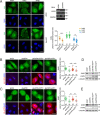
HYPK consolidates the membrane targeting signal of Arl4A and Arl4D. (A) Arl4 proteins expressed in short hairpin control (shCtrl) or short hairpin HYPK (shHYPK) HeLa cells were stained with Arl4A-, Arl4C-, and Arl4D-specific antibodies (green) and DAPI (blue; stains the nuclei). Scale bar, 25 μm. The efficiency of HYPK knockdown was determined by RT-PCR. GAPDH was used as the internal control. The plasma membrane to cytosol ratios of Arl4A, Arl4C, and Arl4D were quantified as described in SI Appendix, Materials and Methods. The ratios in the shCtrl- and shHYPK-expressing cells were compared by two-sample t test, with error bars showing the means ± SD. ***P < 0.001. The cell numbers analyzed are marked in the plot. (B and C) HYPK knockdown cells were rescued with the resistant form (Res) of HA-HYPK WT or the HA-HYPK A3 mutant (red) in HeLa cells. Scale bar, 25 μm. The plasma membrane to cytosol ratios of Arl4A/D in each group were calculated and are shown in dot plots. The ratio differences were analyzed by one-way ANOVA with Dunnett’s post hoc multiple comparison test. Error bars represent the means ± SD. ***P < 0.001. (D and E) Immunoblotting of the protein expression levels in B and C.
Fig. 8.
Fig. 8.
The model of FN-Pak1–triggered signaling for Arl4A/D protein stabilization. In Arl4A/D-expressing cells, Arl4A/D undergo rapid degradation. Under FN stimulation, Pak1-dependent phosphorylation at S143/S144 of Arl4A/D on the plasma membrane potentiates local and efficient HYPK binding for protein protection. HYPK-mediated protection not only stabilizes Arl4A/D from fast proteasome degradation but also strengthens their membrane targeting for cell migration. Ub, ubiquitination.

Similar articles

Cited by

References

    1. Patel M., Chiang T. C., Tran V., Lee F. J., Côté J. F., The Arf family GTPase Arl4A complexes with ELMO proteins to promote actin cytoskeleton remodeling and reveals a versatile Ras-binding domain in the ELMO proteins family. J. Biol. Chem. 286, 38969–38979 (2011). - PMC - PubMed
    1. Chiang T. S., et al. , ADP-ribosylation factor-like 4A interacts with Robo1 to promote cell migration by regulating Cdc42 activation. Mol. Biol. Cell 30, 69–81 (2019). - PMC - PubMed
    1. Chen K. J., Chiang T. C., Yu C. J., Lee F. S., Cooperative recruitment of Arl4A and Pak1 to the plasma membrane contributes to sustained Pak1 activation for cell migration. J. Cell Sci. 133, jcs233361 (2020). - PubMed
    1. Chiang T. S., Wu H. F., Lee F. S., ADP-ribosylation factor-like 4C binding to filamin-A modulates filopodium formation and cell migration. Mol. Biol. Cell 28, 3013–3028 (2017). - PMC - PubMed
    1. Chen Q., et al. , ARL4C stabilized by AKT/mTOR pathway promotes the invasion of PTEN-deficient primary human glioblastoma. J. Pathol. 247, 266–278 (2019). - PubMed

Publication types