. 2022 Jul 26;119(30):e2200512119.
doi: 10.1073/pnas.2200512119. Epub 2022 Jul 20.

EBNA2-EBF1 complexes promote MYC expression and metabolic processes driving S-phase progression of Epstein-Barr virus-infected B cells

Affiliations

EBNA2-EBF1 complexes promote MYC expression and metabolic processes driving S-phase progression of Epstein-Barr virus-infected B cells

Sophie Beer et al. Proc Natl Acad Sci U S A. .

Abstract

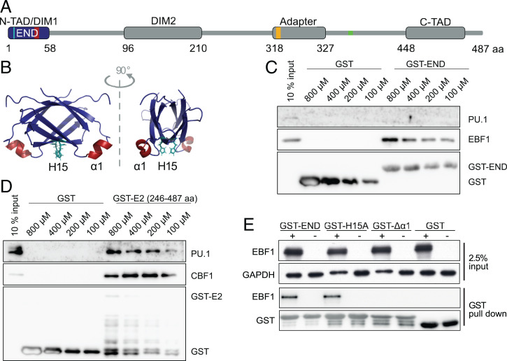
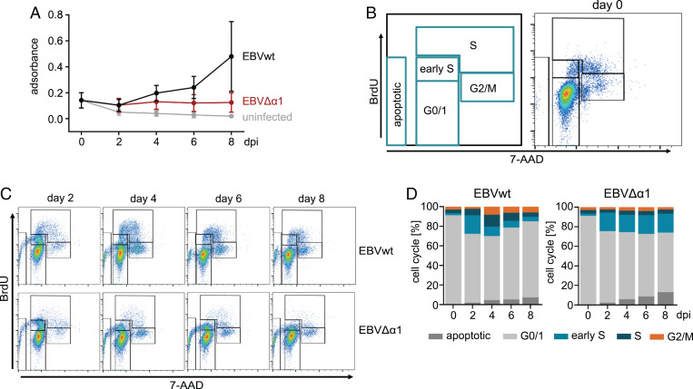
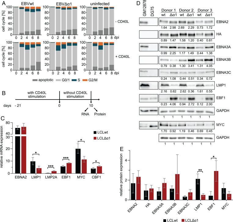
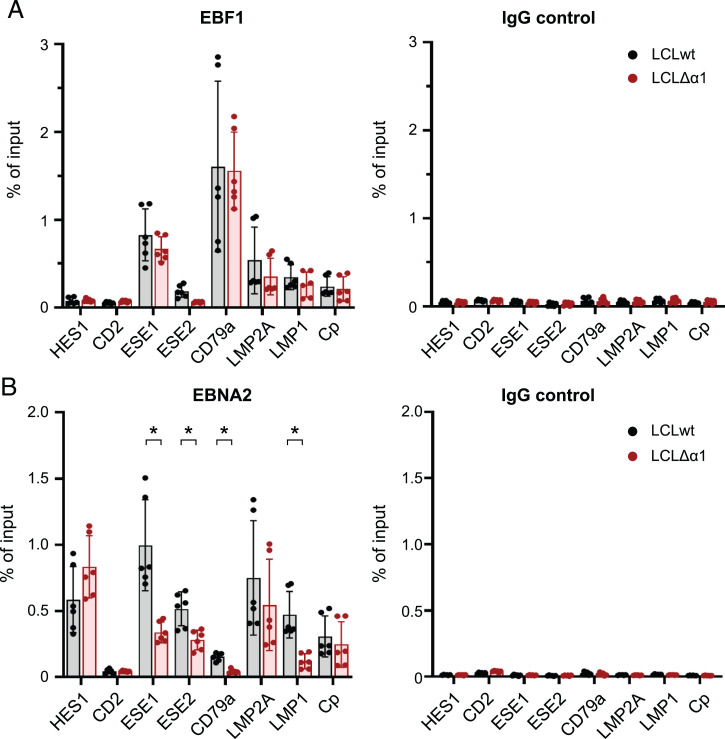
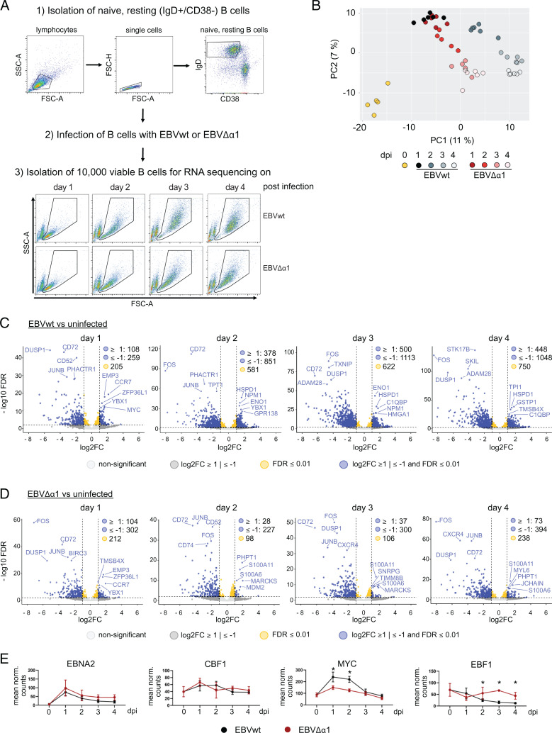
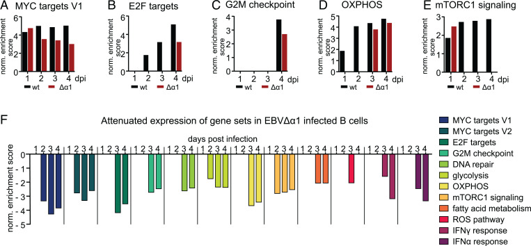
Epstein-Barr virus (EBV) is a human tumor virus which preferentially infects resting human B cells. Upon infection in vitro, EBV activates and immortalizes these cells. The viral latent protein EBV nuclear antigen 2 (EBNA2) is essential for B cell activation and immortalization; it targets and binds the cellular and ubiquitously expressed DNA-binding protein CBF1, thereby transactivating a plethora of viral and cellular genes. In addition, EBNA2 uses its N-terminal dimerization (END) domain to bind early B cell factor 1 (EBF1), a pioneer transcription factor specifying the B cell lineage. We found that EBNA2 exploits EBF1 to support key metabolic processes and to foster cell cycle progression of infected B cells in their first cell cycles upon activation. The α1-helix within the END domain was found to promote EBF1 binding. EBV mutants lacking the α1-helix in EBNA2 can infect and activate B cells efficiently, but activated cells fail to complete the early S phase of their initial cell cycle. Expression of MYC, target genes of MYC and E2F, as well as multiple metabolic processes linked to cell cycle progression are impaired in EBVΔα1-infected B cells. Our findings indicate that EBF1 controls B cell activation via EBNA2 and, thus, has a critical role in regulating the cell cycle of EBV-infected B cells. This is a function of EBF1 going beyond its well-known contribution to B cell lineage specification.

Keywords: B cell activation; Epstein-Barr virus; MYC expression; RNA sequencing; transcription factor.


Conflict of interest statement

The authors declare no competing interest.

Figures

Fig. 1.
Fig. 1.
The α1-helix on the surface of the END binds EBF1. (A) Schematic representation of functional EBNA2 modules: the END domain (blue) with positions of the α1-helix (amino acid residues 35 through 39; red) and histidine 15 (H15; turquoise), N- and C-terminal transactivation domains (N-TAD and C-TAD, respectively), two dimerization domains (DIM1 and DIM2), and an adaptor region that confers DNA binding via the CBF1 protein. The CBF1 binding region is indicated in orange. The PU.1 binding region is indicated in green. (B) NMR structure of the END domain homodimer, H15 (turquoise), and α1-helix (red). (C and D) GST pull-down experiments and (C) GST-END or (D) C-terminal half (amino acid residues 246 through 487) of EBNA2 (GST-E2). Whole-cell lysates of DG75 cells were incubated with GST–EBNA2 fusion proteins purified from Escherichia coli: the C-terminal half (amino acid residues 246 through 487) of EBNA2 (GST-E2), END domain (GST-END), or GST. (C) Western blot using PU.1-, CBF1-, or GST-specific antibodies. (D) Western blot using PU.1- or EBF1-specific antibodies. (E) GST pull-down experiments with the END domain and EBF1. Whole-cell lysates of DG75 cells transfected with an EBF1 expression plasmid (+) or an empty vector control (−) were incubated with GST-END fusion proteins purified from E. coli: wild-type END (GST-END), END-H15A missense mutant (GST-H15A), END α1-helix deletion mutant proteins (GST-Δα1), or GST. GAPDH served as a loading control for input lysates.
Fig. 2.
Fig. 2.
EBVΔα1-infected B cells arrest in the early S phase. (A) MTT assay of primary B cells infected with EBVwt or EBVΔα1; the assay was performed on days 0, 2, 4, 6, 8 postinfection. The mean of three biological replicates is plotted. Error bars indicate the SD. Day 0 refers to noninfected B cells. (B) Gating strategy for the cell cycle analysis with BrdU and 7-amino-actinomycin D (7-AAD) and one representative FACS plot for noninfected cells (day 0). All cells were gated on lymphocytes and single cells. (C) Results of the BrdU assays of one representative experiment. The assay was performed on days 2, 4, 6, and 8 postinfection with B cells infected with EBVwt or EBVΔα1. (D) Summary of the cell cycle analysis showing the mean results of three biological replicates.
Fig. 3.
Fig. 3.
Long-term cultures of LCLΔα1 established by cocultivation of primary B cells infected with EBVΔα1 on CD40L feeder cells. (A) Cell cycle analyses of primary B cells infected with EBVwt or EBVΔα1. Noninfected or infected primary B cells were either cultured without (−) CD40L feeder cells or with (+) CD40L feeder cells. The cell cycle distribution was analyzed by propidium iodide staining and flow cytometry on days 0, 2, 4, 6, and 8 postinfection. The mean of three biological replicates is plotted. See SI Appendix, Fig. S2 for one representative experiment. (B) Experimental setup for RNA and protein preparation. LCLwt or LCLΔα1 were cultured without CD40L feeder cells for 10 d prior to RNA and protein isolation. (C) Real-time qPCR of viral and cellular genes in LCLwt or LCLΔα1 generated from three donors cultivated for 10 d without CD40L stimulus, as in B. Transcript levels of viral and cellular genes were analyzed and normalized to RNA pol II transcript levels. The mean of three biological replicates is plotted; error bars indicate the SD. See SI Appendix, Fig. S3E for the messenger RNA (mRNA) expression level of RNA pol II. (D) Protein expression of latent viral and cellular proteins in LCLwt or LCLΔα1 generated from three donors after 10 d without CD40L activation. Cell lysates of EBV-infected GM12878 or EBV-negative DG75 cells served as positive or negative controls, respectively. The signals were normalized to the corresponding GAPDH signal and the values are indicated below each Western blot signal. (E) Relative expression of latent viral and cellular proteins. Bar charts were generated with values in D. The mean of three biological replicates is plotted; error bars indicate the SD. *P < 0.05; **P < 0.01; ***P < 0.001. dpi, days postinfection; wt, wild type.
Fig. 4.
Fig. 4.
EBNA2’s α1-helix assists in EBF1-dependent EBNA2-chromatin binding. (A and B) ChIP-qPCR for (A) EBF1 or immunoglobulin G (IgG) control and (B) EBNA2 or IgG control in established LCLwt and LCLΔα1 at cellular and viral genomic regions. LCLwt and LCLΔα1 were cultured without CD40L-expressing feeder cells for 1 d prior to chromatin preparation. CD2 was used as a negative control. Mean of three biological replicates is plotted; error bars indicate the SD. *P < 0.05.
Fig. 5.
Fig. 5.
Dynamic changes of gene expression patterns in B cells infected with EBVwt or EBVΔα1. (A) Workflow for cell preparation and infection. Naïve resting B cells (IgD+/CD38−) were isolated from adenoids and infected with EBVwt or EBVΔα1. A total of 10,000 noninfected, naïve, resting B cells were collected to serve as the day 0 sample. On days 1, 2, 3, and 4 after infection, 10,000 viable cells were isolated by flow cytometry sorting and collected for RNA sequencing. Flow cytometry plots of one representative experiment are shown. (B) Principal component analysis of all protein-coding genes. The percentage of variance explained by the first and second principal components (PCs; PC1 and PC2, respectively) are shown in parentheses. (C and D) Volcano plots of DE protein-coding genes comparing (C) EBVwt- or (D) EBVΔα1-infected B cells on days 1, 2, 3, and 4 postinfection with noninfected samples (day 0). Dotted lines indicate log2 fold change = 1 and FDR = 0.01. Gene names of the top five up- and down-regulated genes according to the FDR are indicated. € Mean normalized expression of EBNA2, CBF1, MYC, and EBF1 in EBVwt- and EBVΔα1-infected B cells on days 0, 1, 2, 3, and 4 postinfection. Error bars indicate SD, and asterisks indicate FDR < 0.001 calculated by DESeq2. dpi, days postinfection.
Fig. 6.
Fig. 6.
EBVΔα1 cannot efficiently induce critical cellular and metabolic processes. GSEA was performed with DE protein-coding genes with a P value of ≤0.01, as calculated by DESeq2. Gene sets with an FDR < 0.05 were considered significant. (A–E) Gene sets that are present in both EBVwt- and EBVΔα1-infected B cells on at least 1 d postinfection. Bar graphs show the normalized enrichment score (NES) at each day postinfection. DE genes of the analysis of EBVwt vs. noninfected or EBVΔα1 vs. noninfected were included for these GSEA. (F) GSEA with DE genes defined by the analysis EBVΔα1 vs. EBVwt. The bar graph shows the NES for each gene set at each day postinfection. A negative NES correlates with an enrichment in EBVwt-infected B cells over EBVΔα1-infected B cells. dpi, days postinfection; norm., normalized; ROS, reactive oxygen species.

Similar articles

Cited by

References

    1. Shannon-Lowe C., Rickinson A. B., Bell A. I., Epstein-Barr virus-associated lymphomas. Philos. Trans. R. Soc. Lond. B Biol. Sci. 372, 20160271 (2017). - PMC - PubMed
    1. Läderach F., Münz C., Epstein Barr virus exploits genetic susceptibility to increase multiple sclerosis risk. Microorganisms 9, 2191 (2021). - PMC - PubMed
    1. Bjornevik K., et al. , Longitudinal analysis reveals high prevalence of Epstein-Barr virus associated with multiple sclerosis. Science 375, 296–301 (2022). - PubMed
    1. Münz C., Latency and lytic replication in Epstein-Barr virus-associated oncogenesis. Nat. Rev. Microbiol. 17, 691–700 (2019). - PubMed
    1. Kaiser C., et al. , The proto-oncogene c-myc is a direct target gene of Epstein-Barr virus nuclear antigen 2. J. Virol. 73, 4481–4484 (1999). - PMC - PubMed

Publication types

MeSH terms

LinkOut - more resources