. 2022 Jul;12(7):e891.
doi: 10.1002/ctm2.891.

FBXW24 controls female meiotic prophase progression by regulating SYCP3 ubiquitination

Affiliations

FBXW24 controls female meiotic prophase progression by regulating SYCP3 ubiquitination

Yang Wang et al. Clin Transl Med. 2022 Jul.

Abstract

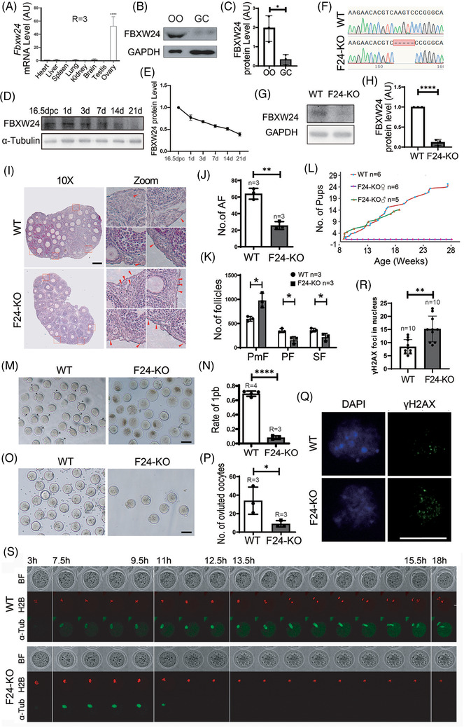
Background: An impeccable female meiotic prophase is critical for producing a high-quality oocyte and, ultimately, a healthy newborn. SYCP3 is a key component of the synaptonemal complex regulating meiotic homologous recombination. However, what regulates SYCP3 stability is unknown.

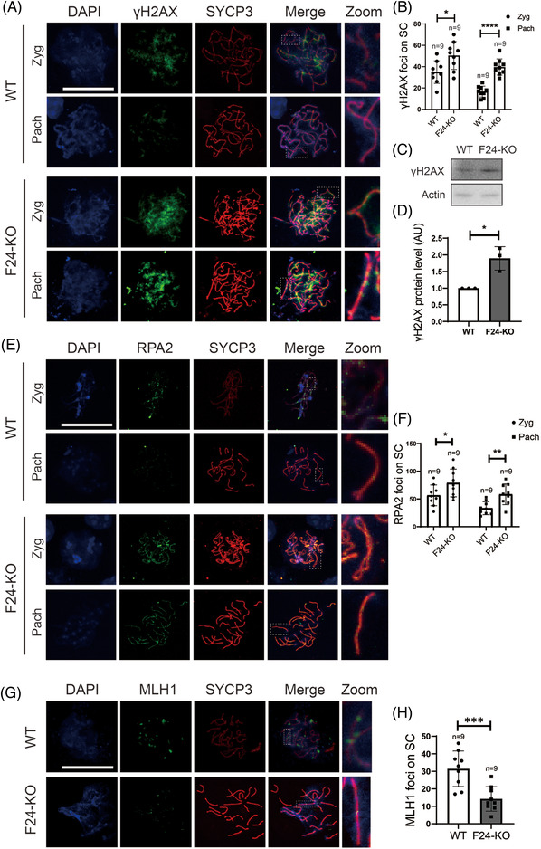
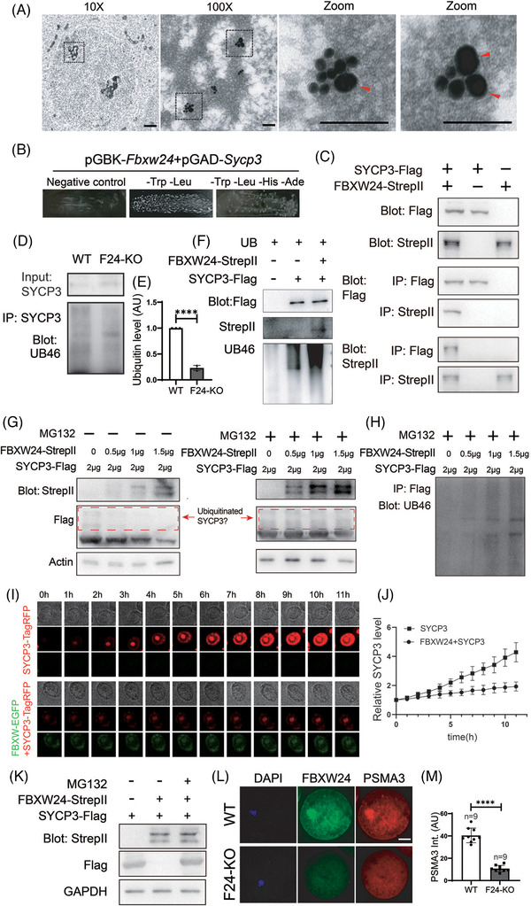
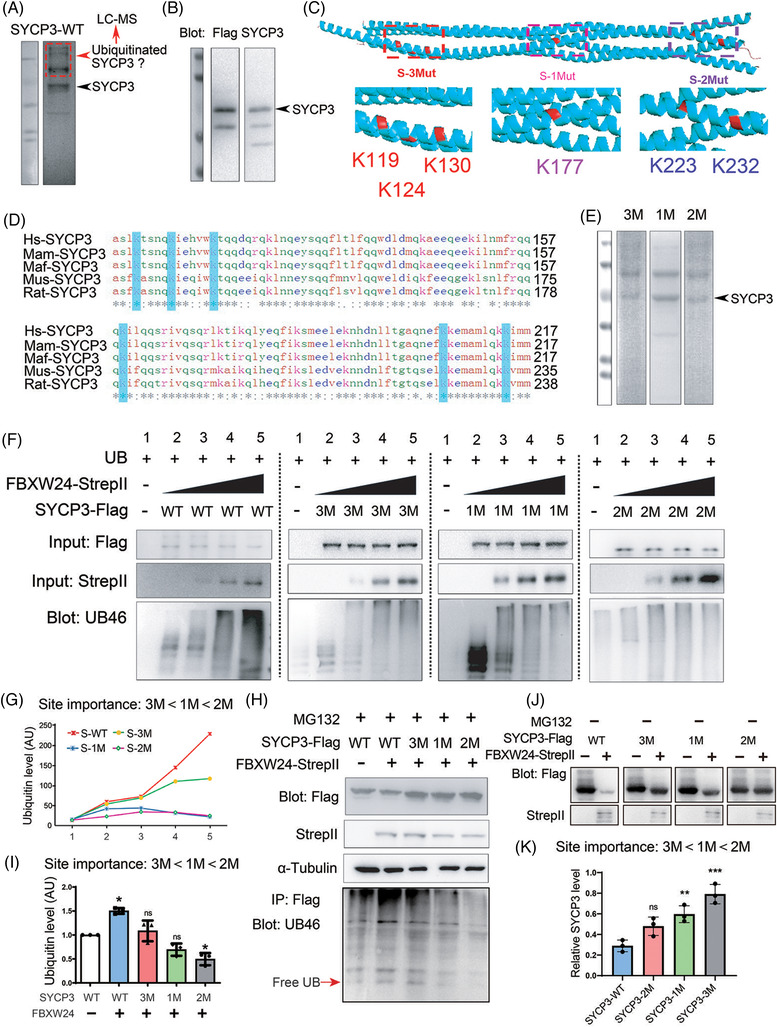
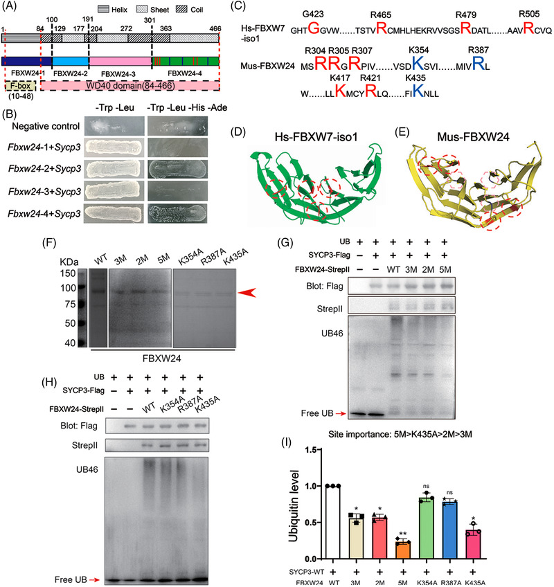
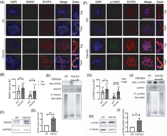
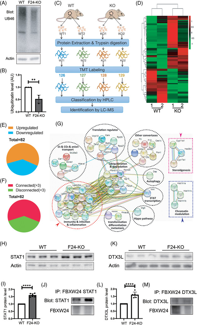
Methods: Fertility assays, follicle counting, meiotic prophase stage (leptotene, zygotene, pachytene and diplotene) analysis and live imaging were employed to examine how FBXW24 knockout (KO) affect female fertility, follicle reserve, oocyte quality, meiotic prophase progression of female germ cells, and meiosis of oocytes. Western blot and immunostaining were used to examined the levels & signals (intensity, foci) of SYCP3 and multiple key DSB indicators & repair proteins (γH2AX, RPA2, p-CHK2, RAD51, MLH1, HORMAD1, TRIP13) after FBXW24 KO. Co-IP and immuno-EM were used to examined the interaction between FBXW24 and SYCP3; Mass spec was used to characterize the ubiquitination sites in SYCP3; In vivo & in vitro ubiquitination assays were utilized to determine the key sites in SYCP3 & FBXW24 for ubiquitination.

Results: Fbxw24-knockout (KO) female mice were infertile due to massive oocyte death upon meiosis entry. Fbxw24-KO oocytes were defective due to elevated DNA double-strand breaks (DSBs) and inseparable homologous chromosomes. Fbxw24-KO germ cells showed increased SYCP3 levels, delayed prophase progression, increased DSBs, and decreased crossover foci. Next, we found that FBXW24 directly binds and ubiquitinates SYCP3 to regulate its stability. In addition, several key residues important for SYCP3 ubiquitination and FBXW24 ubiquitinating activity were characterized.

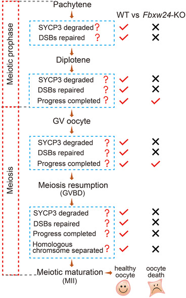
Conclusions: We proposed that FBXW24 regulates the timely degradation of SYCP3 to ensure normal crossover and DSB repair during pachytene. FBXW24-KO delayed SYCP3 degradation and DSB repair from pachytene until metaphase II (MII), ultimately causing failure in oocyte maturation, oocyte death, and infertility.

Keywords: FBXW24; SYCP3; meiotic prophase; oocyte; ubiquitination.


Conflict of interest statement

The authors declare no conflict of interest.

Figures

FIGURE 1
FIGURE 1
Oocyte‐predominant FBXW24 is essential for female fertility. (A) Q‐PCR shows that Fbxw24 mRNA was predominant in ovaries. (B and C) Western blots and quantification demonstrate that FBXW24 is more dominant in oocytes than in granular cells (GCs). (D and E) Western blots and quantification show that FBXW24 protein peaked at 16.5 DPC and then gradually decreased from PND 1 to 21. (F–H) Five bases from the third exon of the Fbxw24 gene were deleted through Cas9 and caused a frame‐shift of the Fbxw24 gene; western blots show that in Fbxw24‐KO ovaries, FBXW24 protein is completely eliminated. (I–K) At PND 21, Fbxw24 knockout significantly increased primordial follicles (PMF) while it decreased primary follicles (PF), secondary follicles (SF), and antral follicles (AF). Four selected regions (red dot‐line square) from WT and KO ovaries were zoomed and placed on the right, and primordial follicles were arrow‐pointed. (L) Curves for cumulative pups from 2‐month‐old to 7‐month‐old showed that Fbxw24‐KO male mice had normal fertility, while Fbxw24‐KO female mice were completely infertile. (M and N) Fbxw24 knockout significantly decreased oocyte maturation rate (first polar body, 1 pb). (O and P) Fbxw24 knockout significantly decreased numbers of ovulated oocytes. (Q and R) Immunofluorescence and quantification showed that Fbxw24 knockout significantly increased DSBs (by γH2AX foci) in the GV oocytes. DNA in blue, γH2AX in green. (S) Live imaging shows that oocytes were not able to go through anaphase because homologous chromosomes did not separate at all. Red fluorescence signals are H2B, and green fluorescence signals are α‐Tubulin. Scale bar in Q, 20 μm; Scale bar in other panels, 100 μm. GAPDH was used as a loading control. *, p < 0.05; **, p < 0.01; ****, p < 0.0001. AU, arbitrary unit
FIGURE 2
FIGURE 2
Fbxw24 knockout delayed meiotic prophase progression due to increased SYCP3 level. (A and B) Immunofluorescence showed that in WT female mice, SYCP3 intensity on spread MII chromosomes was only 20% of the SYCP3 intensity on pachytene chromosomes; while in Fbxw24‐KO female mice, SYCP3 intensity on spread MII chromosomes was about 75% of the SYCP3 intensity on pachytene chromosomes. SYCP3 intensity on Fbxw24‐KO spread MII chromosomes was about 4‐fold higher than on WT MII chromosomes. DNA in blue, SYCP3 in red. (C and D) Immunofluorescence and quantification showed that Fbxw24 knockout significantly increased SYCP3 level in meiotic prophase cells at leptotene, zygotene, and pachytene stages within the 16.5 DPC female genital ridge. DNA in blue, FBXW24 in green, SYCP3 in red. "Int." is an abbreviation of "intensity". (E and F) Western blots and quantification showed that the SYCP3 level in Fbxw24‐KO 16.5 DPC genital ridge was over a fold higher than in WT. (G) In the WT 19.5 DPC genital ridge, most of the meiotic germ cells are at pachytene or diplotene stages, while in the Fbxw24‐KO 19.5 DPC genital ridge, significantly more meiotic germ cells were still at leptotene or zygotene stages. (H and I) Injection of exogenous Sycp3 mRNA into WT GV oocytes significantly decreased oocyte maturation (first polar body extrusion), while injection of exogenous Fbxw24 mRNA into Fbxw24‐KO oocytes significantly recovered oocyte maturation. MII oocytes were labelled by black arrowheads. b. Quantification of A. 1pb, first polar body. Scale bar in H, 100 μm; Scale bar in other panels, 20 μm. **, p < 0.01; ***, p < 0.001; ****, p < 0.0001. Different lower‐case letters in I indicate significant differences. AU, arbitrary unit
FIGURE 3
FIGURE 3
Fbxw24 knockout increased DSBs and decreased crossover (A and B). Immunofluorescence and quantification showed that the number of γH2AX foci significantly increased in the Fbxw24‐KO zygotene or pachytene meiotic germ cells. DNA in blue, γH2AX in green, SYCP3 in red. (C and D) Western blots and quantification showed that the γH2AX level significantly increased in the 16.5 DPC Fbxw24‐KO genital ridge. (E and F) Immunofluorescence and quantification showed that the number of RPA2 foci significantly increased in the Fbxw24‐KO zygotene or pachytene meiotic germ cells. DNA in blue, RPA2 in green, SYCP3 in red. (G and H) Immunofluorescence and quantification showed that the number of MLH1 foci significantly increased in the Fbxw24‐KO pachytene meiotic germ cells. DNA in blue, MLH1 in green, SYCP3 in red. FBXW24 is shortened as "F24" when needed. Actin was used as a loading control. Scale bar, 20 μm. *, p < 0.05; ***, p < 0.01; ***, p < 0.001; ****, p < 0.0001. AU, arbitrary unit
FIGURE 4
FIGURE 4
FBXW24 directly binds and ubiquitinates SYCP3. (A) Immuno‐EM shows that in the chromatin regions of meiotic cells in the 16.5 DPC genital ridge, some SYCP3 signals localized closely to FBXW24 (<20 nm). One region (dot‐line square) from a 10x image was photographed at 100x to show the overall signal within the chromatin region; two regions (dot‐line square) from the 100x image were further magnified to show the close adjacency between SYCP3 and FBXW24. SYCP3 and FBXW24 were detected by secondary antibodies conjugated with 15 nm gold (small dots) and 35 nm gold (big dots, arrow‐pointed), respectively. (B) Yeast‐two‐hybridization (Y2H) assay shows that FBXW24 can directly bind SYCP3. (C) Co‐IP between in‐vitro purified FBXW24 and SYCP3 shows that FBXW24 directly binds SYCP3. 1 μg SYCP3‐Flag protein and/or FBXW24‐StrepII protein were/was used in each reaction. (D and E) SYCP3 IP, western blot and quantification demonstrate that Fbxw24 knockout significantly decreased SYCP3 ubiquitination level. (F) In‐vitro ubiquitination assay showed that in the presence of ubiquitin and other components (Table S5), FBXW24 can significantly increase the SYCP3 ubiquitination level. 1 μg SYCP3‐Flag protein and/or FBXW24‐StrepII protein were/was in each reaction. (G and H) 293T cells were transfected with Fbxw24‐StrepII and Sycp3‐Flag plasmid (Amounts of plasmid used were in the image), and western blot was done under two conditions. Left of G, in the absence of MG132, as the FBXW24 level increased, the SYCP3 level gradually decreased. Right of G and H, in the presence of MG132, as the FBXW24 level increased, the SYCP3 level remained unchanged, while ubiquitinated SYCP3 gradually increased. Red dot‐line square labelled probable ubiquitinated SYCP3. (I and J) SYCP3 titer were first added into the Sf9 medium to be expressed to a medium level (at about Day 1.5), then FBXW24 titer was added, and SYCP3 intensity almost remained unchanged; In contrast, in the sf9 medium without FBXW24 supplement, SYCP3 intensity keep raising, and big SYCP3 aggregates kept increasing. (K) Western blot shows that FBXW24 expression substantially reduced SYCP3 level; once proteasome activity was inhibited with MG132, SYCP3 level is recovered. (L and M) In WT GV oocytes, PSMA3, a sub‐unit of the proteasome, was enriched within nuclear chromatin and highly overlapped with FBXW24; in Fbxw24‐KO GV oocytes, PSMA3 did not show any abundance within chromatin. For all plasmids except B, Sycp3 is fused to TagRFP and Flag, and Fbxw24 is fused to EGFP and Strep II. To save space, SYCP3‐TagRFP‐Flag was shortened as "SYCP3‐TagRFP" or "SYCP3‐Flag", and FBXW24‐EGFP‐stepII was shortened as "FBXW24‐EGFP" or "FBXW24‐Strep II" as needed. Scale bar for 10X image in A, 1 μm; for 100X and zoom images in A, 100 nm. Scale bar for other panels, 20 μm. Actin or GAPDH was used as a loading control. ****, p < 0.0001. AU, arbitrary unit
FIGURE 5
FIGURE 5
Characterization of key sites in SYCP3 for ubiquitination. (A) Purified WT SYCP3‐TagRFP‐Flag protein was subjected to SDS‐PAGE and Coomassie staining. The stained gel shows that the purified WT SYCP3 demonstrated multiple bands; the lower band (arrow‐pointed) corresponded to the expected size band, and the other upper bands were cut altogether for the characterization of ubiquitination sites within SYCP3 through mass spectrometry (Figure S11A–I and Table S4). (B) The expected right‐size band in A was verified through Flag and SYCP3 western blots (arrow‐pointed). (C) Based on the known SYCP3 structure model, K119, K124, K130, K177, K223, and K232, which are on the surface of the SYCP3 tetramer, were subdivided into three groups according to their positional proximity, and three non‐ubiquitinated mutants were made: K119A, K124A, and K130A – simplified as S‐3 M, K177A – simplified as S‐1 M, and K223A and K232A – simplified as S‐2 M. (D) Protein sequence alignment among SYCP3 from Homo sapiens (Hs), Macaca mulatta (Mam), Macaca fascicularis (Maf), Mus musculus (Mus), and Rattus norvegicus (Rat) shows that all six selected ubiquitination sites are conserved. (E) SDS‐PAGE and Coomassie staining demonstrate that the purified SYCP3 mutant proteins also showed multiple bands; the right‐size band (arrow‐pointed) is at similar positions to SYCP3 WT protein. (F) In vitro dose‐dependent ubiquitination assay shows that each mutant showed decreased ubiquitination, but the extent of this reduction differed for these three mutants. SYCP3 WT and each mutant reaction were separated by a vertical dot‐line. 2 μg SYCP3 WT or each mutant protein was used in each reaction, 0 μg, 0.5 μg, 1 μg, 1.5 μg, or 2 μg FBXW24 protein was used in each transfection from left to right. (G) Quantification of F shows that the site importance ranking for ubiquitination appeared to be 3 M < 1 M < 2 M. (H and I) In vivo side‐by‐side comparative ubiquitination assay in the presence of MG132 also illustrates that the site importance ranking for ubiquitination appeared to be 3 M < 1 M < 2 M. 2 μg Sycp3 and/or Fbxw24 plasmid were/was used in each transfection. (J) In vivo side‐by‐side comparison of FBXW24‐dependent SYCP3 degradation among WT, 3 M, 1 M and 2 M. The degradation extent is also 3 M < 1 M < 2 M. 2 μg Sycp3 and/or Fbxw24 plasmid were/was used in each transfection. α‐tubulin was used as a loading control. *, p < 0.05; **, p < 0.01; ***, p < 0.001. AU, arbitrary unit
FIGURE 6
FIGURE 6
Characterization of key sites in FBXW24 required for SYCP3 ubiquitination. (A) I‐Tasser was used to predict the FBXW24 structure and FBXW24 was sub‐divided into four parts based on its secondary structure. FBXW24‐1 (1–99 AA, dark blue) is primarily α‐helix; FBXW24‐2 (100–191 AA, light blue), FBXW24‐3 (192–300 AA, pink), and FBXW24‐4 (301–466 AA, green) are mostly β‐sheet; and residues between them are primarily coils. Predicted F‐box‐like, from 10–48 AA, and WD40 domain, from 84–486 AA, was also mapped. (B) Y2H showed that FBXW24‐2 or FBXW24‐4 could directly bind SYCP3 through Y2H. (C–E) I‐Tasser showed that the R‐rich C‐terminal region of FBXW24 is structurally similar to the R & K‐rich C‐terminal region of FBXW7. We focused on two groups of R & K‐rich sites (red type in C) within FBXW24‐4 (red dot‐line circles in E) and made three FBXW24 mutants: R304A, R305A, and R307A – simplified as 3 M; K417A and R421A – simplified as 2 M; and R304A, R305A, R307A, K417A, and R421A – simplified as 5 M. We also selected three R & K sites (violet type in C) within FBXW24‐4 (pink dot‐line circles in E) that are spatially similar to the enzymatic sites (red type in C) in the C‐terminal WD domain of human FBXW7‐iso1 (isoform 1, red dot‐line circles in D), and made another three mutants: K354A, R387A, and K435A. (F) SDS‐PAGE and coomassie staining demonstrate the purity of WT, 3 M, 2 M, 5 M, K354A, R387A, and K435A (arrow‐pointed). (G) In vitro side‐by‐side comparative ubiquitination assay shows that all three mutants (3 M, 2 M, and 5 M) have decreased ubiquitinating capacity. 1 μg SYCP3‐Flag protein and/or FBXW24‐StrepII protein were/was used in each reaction. (H) In vitro side‐by‐side comparative ubiquitination assay shows that among K354A, R387A, and K435A, only K435A has significantly decreased ubiquitinating capacity. 1 μg SYCP3‐Flag protein and/or FBXW24‐StrepII protein were/was used in each reaction. (I) Quantification of G and H showed that the site importance is 5 M > K435A > 2 M > 3 M. Different lower‐case letters above the graph column indicate significant differences. Free ubiquitin was arrow‐pointed. AU, arbitrary unit
FIGURE 7
FIGURE 7
Fbxw24 knockout increased RAD51 and p‐CHK2 foci in female germ cells. (A and B) Immunofluorescence and quantification showed that RAD51 foci significantly increased in the Fbxw24‐KO zygotene or pachytene germ cells. DNA in blue, RAD51 in green, and SYCP3 in red. (C and D) Western blots and quantification show that the RAD51 level significantly increased in the Fbxw24‐KO 16.5 DPC genital ridge. (E) RAD51 antibody immunoprecipitation and UB46 western blots demonstrate that RAD51 can be ubiquitinated, while Fbxw24 knockout substantially reduced RAD51 ubiquitination level. Further, exogenous FBXW24 protein (1 μg) supplementation increased RAD51 ubiquitination level and reduced RAD51 level close to WT. (F and G) Immunofluorescence and quantification showed that the p‐CHK2 foci significantly increased in the Fbxw24‐KO zygotene or pachytene germ cells. DNA in blue, p‐CHK2 in green, SYCP3 in red. (H and I) Western blots and quantification show that the p‐CHK2 level significantly increased in the Fbxw24‐KO 16.5 DPC genital ridge. (J) p‐CHK2 antibody immunoprecipitation and UB46 western blots demonstrate that RAD51 can be ubiquitinated, while Fbxw24 knockout substantially reduced p‐CHK2 ubiquitination level. Further, exogenous FBXW24 protein (1μg) supplementation increased p‐CHK2 ubiquitination level and reduced RAD51 level close to WT. α‐tubulin or GAPDH was used as a loading control. *, p < 0.05; **, p < 0.01. AU, arbitrary unit
FIGURE 8
FIGURE 8
Quantitative proteomics revealed that Fbxw24 knockout altered the level of various proteins. (A and B) Fbxw24 knockout substantially reduced pan‐ubiquitination levels in PND 21 ovaries. (C) TMT‐labeled quantitative proteomics was performed to comprehensively investigate the effects of Fbxw24 knockout on mouse ovaries at the protein level. Peptides from two repeats of WT or Fbxw24‐KO ovaries were isobaric‐mass tagged by TMT6‐126, 127, 128, and 129, respectively, and analyzed by LC‐MS. (D) Heat map of 82 differentially expressed proteins (DEPs) at a 1.2‐fold threshold (Fbxw24‐KO / WT > 1.2 or < 0.833). (E) Among the 82 DEPs, 60.98% (50 out of 82) were up‐regulated, and 39.02% (32 out of 82) were down‐regulated. (F) By string 2.0, among the 82 DEPs, 60.98% (50 out of 82) connect with each other. (G) KEGG and protein interaction analysis show that six ubiquitination and degradation‐related proteins are in the centre of the interaction network (green dot‐line circle), and around them, there are eight groups of proteins; 14 proteins are involved in immunity, infection, and inflammation processes and consist of the largest group (red dot‐line circle). In particular, there are two separate groups: one group containing Cyp11a1, Cyp17a1, and Hsd3b1, all of which are involved in steroidogenesis (pink dot‐line rectangle); and the other contains Hist1h1b, Hist1h1c, and Hist1h1e, which are all involved in chromatin modulation (blue dot‐line rectangle). (H and I) Western blot verified that Fbxw24 knockout increased STAT1 protein level. (J) Co‐IP and western blot showed that FBXW24 interacted with STAT1. (K and L) Western blot verified that Fbxw24 knockout increased DTX3L protein level. (M) Co‐IP and western blot showed that FBXW24 interacted with DTX3L. Actin was used as a loading control. *, p < 0.05. AU, arbitrary unit
FIGURE 9
FIGURE 9
FBXW24 working model. This model showed the correlation between FBXW24, SYCP3 degraded, DSB repaired, and meiotic progression from pachytene to MII. In WT female germ cells, an average level of FBXW24 promotes SYCP3 degradation during pachytene, which subsequently promotes DSB repair. Therefore, germ cells can keep progressing till MII. In Fbxw24‐KO female germ cells, Fbxw24 knockout delayed SYCP3 degradation, subsequently, DSB cannot be repaired, pachytene progression is delayed; delayed Sycp3 degradation and unrepaired DSB remained until MII, which conduced to non‐separated homologous chromosomes and unrepaired DSB, ultimately resulted in MII oocyte death

Similar articles

Cited by

References

    1. Gao J, Colaiácovo MP. Zipping and unzipping: protein modifications regulating synaptonemal complex dynamics. Trends Genet. 2018;34(3):232‐245. - PMC - PubMed
    1. Zickler D, Kleckner N. Recombination, pairing, and synapsis of homologs during meiosis. Cold Spring Harb Perspect Biol. 2015;7(6):a016626. - PMC - PubMed
    1. Qiao H, Chen JK, Reynolds A, Höög C, Paddy M, Hunter N. Interplay between synaptonemal complex, homologous recombination, and centromeres during mammalian meiosis. PLoS Genet. 2012;8(6):e1002790. - PMC - PubMed
    1. Miyamoto T, Hasuike S, Yogev L, et al. Azoospermia in patients heterozygous for a mutation in SYCP3. Lancet. 2003;362(9397):1714‐1719. - PubMed
    1. Bolor H, Mori T, Nishiyama S, et al. Mutations of the SYCP3 gene in women with recurrent pregnancy loss. Am J Hum Genet. 2009;84(1):14‐20. - PMC - PubMed

Publication types