

Review
doi: 10.5435/JAAOSGlobal-D-21-00255.
eCollection 2022 Jul 1.
A Review of the Lateral Patellofemoral Joint: Anatomy, Biomechanics, and Surgical Procedures
Affiliations
- PMID: 35858252
- PMCID: PMC9302287
- DOI: 10.5435/JAAOSGlobal-D-21-00255
Item in Clipboard
Review
A Review of the Lateral Patellofemoral Joint: Anatomy, Biomechanics, and Surgical Procedures
J Am Acad Orthop Surg Glob Res Rev.
.
Abstract
The lateral patellofemoral joint soft tissues contain key structures that surround and balance the joint. These structures can affect joint tracking, stability, and force distribution. It is important to understand the lateral patellofemoral anatomy and biomechanics, and their relationship with patellofemoral instability, anterior knee pain, and osteoarthritis. Lateral-sided surgical procedures such as lateral release, lateral retinacular lengthening, and partial lateral patellar facetectomy can be useful in the treatment of such patellofemoral problems.
Copyright © 2022 The Authors. Published by Wolters Kluwer Health, Inc. on behalf of the American Academy of Orthopaedic Surgeons.
Figures

Illustration showing the arciform fibers and the insertions of the iliotibial band to the patella and Gerdy tubercle: (a) vastus lateralis muscle, (b) superficial oblique retinaculum (arciform fibers), (c) superficial layer of the iliotibial band, and (d) biceps femoris muscle.

Lateral view of the knee with the superficial layer reflected showing (a) the patella, (b) the quadriceps aponeurosis, (c) the anchor of (d) the deep fascia to que quadriceps aponeurosis, and (e) the vastus lateralis.

Illustration showing the lateral and medial patellofemoral ligaments (LPFL and MPFL), the lateral and medial patellomeniscal ligaments (LPML and MPML), and the lateral and medial patellotibial ligaments (LPTL and MPTL) [This drawing represents the authors' and an artist's rendering of the anatomy, and the capsule has been removed to simplify the visualization of these structures, but they are not intra-articular. In dissections, there is confluence of these ligaments with the capsule, they blend with the capsule but are not intracapsular].

Illustration showing the lateral joint capsule in the back, the deep retinacular layer reflected up, and the superficial retinacular layer reflected down.
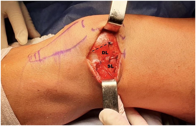
Photograph showing the surgical approach, lateral to the patella. The incision shows the superficial layer (SL) and the deep layer (DL) of the lateral retinaculum, sutured, and lengthened 15 mm. The dotted line shows the original attachment of the superficial layer.
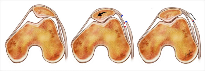
Illustrations showing the axial views of the patellofemoral joint and the effect of lateral retinacular lengthening. On the left, the original patellofemoral joint shows lateral tilt of the patella. On the center, the superficial (blue) and deep (red) layers of the lateral retinaculum have been incised with the correction of the lateral tilt. On the right, the superficial and deep layers are sutured lengthening the lateral retinaculum.

Photograph showing how the lateral facetectomy is done with an oscillating saw, while the lateral facet is held with a clamp.

Photograph showing the final result after the lateral facetectomy has been done and the fragment is separated from the rest of the patella.
References
-
- Hinckel BB, Arendt EA: Lateral retinaculum lengthening or release. Oper Tech Sports Med 2015;23:100-106.
-
- Sherman SL, Rund JM, Farr J: Partial lateral patella facetectomy and management of the lateral soft tissues. Patellofemoral Pain, Instability, and Arthritis 2020:479-495.
-
- Vieira EL, Vieira EA, Da Silva RT, Berlfein PA, Abdalla RJ, Cohen M: An anatomic study of the iliotibial tract. Arthroscopy 2007;23:269-274. - PubMed
Publication types
MeSH terms
LinkOut - more resources
Full Text Sources
Medical
