. 2022 Jul 19;119(29):e2115129119.
doi: 10.1073/pnas.2115129119. Epub 2022 Jul 11.

Single-molecule analysis of actin filament debranching by cofilin and GMF

Affiliations

Single-molecule analysis of actin filament debranching by cofilin and GMF

Johnson Chung et al. Proc Natl Acad Sci U S A. .

Abstract

Eukaryotic cells contain branched actin networks that are essential for endocytosis, motility, and other key cellular processes. These networks, which are formed by filamentous actin and the Arp2/3 complex, must subsequently be debranched to allow network remodeling and to recycle the Arp2/3 complex. Debranching appears to be catalyzed by two different members of the actin depolymerizing factor homology protein family: cofilin and glial maturation factor (GMF). However, their mechanisms of debranching are only partially understood. Here, we used single-molecule fluorescence imaging of Arp2/3 complex and actin filaments under physiological ionic conditions to observe debranching by GMF and cofilin. We demonstrate that cofilin, like GMF, is an authentic debrancher independent of its filament-severing activity and that the debranching activities of the two proteins are additive. While GMF binds directly to the Arp2/3 complex, cofilin selectively accumulates on branch-junction daughter filaments in tropomyosin-decorated networks just prior to debranching events. Quantitative comparison of debranching rates with the known kinetics of cofilin-actin binding suggests that cofilin occupancy of a particular single actin site at the branch junction is sufficient to trigger debranching. In rare cases in which the order of departure could be resolved during GMF- or cofilin-induced debranching, the Arp2/3 complex left the branch junction bound to the pointed end of the daughter filament, suggesting that both GMF and cofilin can work by destabilizing the mother filament-Arp2/3 complex interface. Taken together, these observations suggest that GMF and cofilin promote debranching by distinct yet complementary mechanisms.

Keywords: Arp2/3 complex; actin depolymerizing factor homology; branched actin networks; glial maturation factor; tropomyosin.


Conflict of interest statement

The authors declare no competing interest.

Figures
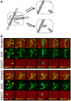
Fig. 1.
Fig. 1.
Debranching of Arp2/3-mediated actin branches by GMF. (A) Cartoon illustrating the two-color debranching assay with dye (red star/asterisk)-labeled Arp2/3 complex (white) and actin (gray). (B) Examples of debranching induced by 50 nM GMF in physiological buffer in the two-color assay. Each triplet of rows of images depicts a sequence of consecutive video frames showing a branch and its subsequent debranching in merged view (Top), actin channel (Middle), and Arp2/3 channel (Bottom). The time stamp (seconds) for each image is indicated (Scale bars:, 2.4 µm). Yellow arrows indicate the branches of interest; cyan squares mark the former branch site on the mother (m) filament immediately after the dissociation of the daughter (d) filament and the disappearance of the Arp2/3 complex.
Fig. 2.
Fig. 2.
Cofilin-induced debranching. (A) Examples of debranching mediated by 200 nM cofilin in physiological buffer. Each triplet of image rows depicts a sequence of consecutive frames showing a branch and its subsequent debranching in merged view (Top), actin channel (Middle), and Arp2/3 channel (Bottom). The time stamp (seconds) for each image is indicated (Scale bars, 2.4 µm). Yellow arrows indicate the branches of interest; cyan squares mark the former branch site on the mother (m) filament immediately after the dissociation of the daughter (d) filament and the simultaneous disappearance of the Arp2/3 complex. (B) Debranching at varying concentrations of cofilin in physiological buffer in the presence or absence of 2 µM Tpm1.7. Points indicate lifetimes of individual branches seen to debranch (Top) or total time of observation for branches that lasted until the end of the experiment or diffused away without being seen to debranch (Bottom). Whisker plots indicate median (red), 25th and 75th percentiles (blue), and maximum and minimum (black). Lifetime distributions and fits are presented in SI Appendix, Fig. S2, and censoring-corrected debranching rates are given in SI Appendix, Table S2. (C) Debranching rates (± SE) calculated from the data in B for experiments without (black circles) Tpm1.7. Also shown are debranching rates predicted by a model in which cofilin binding to only a single site at the end of an actin filament (for example, to the last actin subunit at the daughter filament pointed end) is enough to trigger debranching (black points). Also shown are results for analogous models in which debranching requires simultaneous binding of cofilin molecules to at least two (blue points) or three (red points) consecutive binding sites at the filament end. Analogous results are obtained (SI Appendix, Fig. S3) for models in which the cofilin binding site(s) that triggers debranching is in the middle of a filament (e.g., on the mother filament adjacent to Arp2/3) rather than at the end. Note that the simulations are not fits to the data; they are predictions with no free parameters calculated from known cofilin binding/dissociation rate constants (see Materials and Methods). Shading shows the SE of the model predictions. (D) The same as for C, but for debranching rates measured in the presence of 2 µM Tpm1.7. In this case, the simulated model includes the additional assumption that the single actin site is obstructed by Tpm 75% of the time, a value that was chosen for good agreement with the data.
Fig. 3.
Fig. 3.
GMF and cofilin independently promote debranching. Debranching rates (± SE) mediated by 50 nM GMF and 100 nM cofilin individually and together were measured and analyzed as in SI Appendix, Figs. S1 and S2 in physiological buffer. Experiments were performed in the absence of Tpm. Lifetime distributions and fits are presented in SI Appendix, Fig. S2C, and censoring-corrected debranching rates are given in SI Appendix, Table S2.
Fig. 4.
Fig. 4.
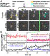
Example of a debranching event with fluorescently labeled cofilin and Arp2/3 complex in the presence of 2 µM Tpm1.7, which occurs at the designated time (green line). The same event is shown in Movie S1; other examples are shown in SI Appendix, Fig. S4 and corresponding Movies S2–S4. The 3-color experiment used fluorescently labeled actin (blue-excited AF488-actin), cofilin (green-excited Cy3-Cof1), and Arp2/3 complex (red-excited JF646-Arp2/3) in physiological buffer. In this experiment, green and red lasers were on continuously, whereas the blue laser was only on intermittently (only at 32.9 s, in the images shown here). (A) Image pairs at selected time points showing cofilin (Top) and the Arp2/3 complex (Bottom) before debranching (at 39.6 s and 44.6 s) and after debranching (at 53.2 s). The top panel of the first image pair (32.9 s) shows superimposed actin and cofilin fluorescence. The position of the pointed end of the daughter filament is inferred (orange square) from the position of the JF646-Arp2/3 fluorescence spot (before debranching), and from tracking the daughter pointed end Cy3-Cof1 decoration (after debranching). After debranching, the position on the mother filament at which the branch had been present (cyan square) is inferred by interpolating between the Cy3-Cof1 decorated mother filament ends. A 9.7 µm × 9.7 µm area centered at the mother branch site of each image is displayed. The images displayed here were spatially filtered by a 2 × 2 matrix of ones for clarity. (B) Fluorescence recorded over a time period spanning the debranching event in A. Plots show Cy3-Cof1 and JF646-Arp2/3 intensities integrated over 3 × 3 pixel areas tracking with the daughter pointed end and mother branch site. For each protein, only a single record is shown to the left of the green line since the locations of the daughter pointed end and mother branch site are identical before debranching.
Fig. 5.
Fig. 5.
GMF and cofilin can both debranch by breaking the Arp2/3–mother filament interaction. (A) Cartoon illustrating alternative possible debranching pathways. (B) Two examples of frame sequences showing that Arp2/3 is transiently retained on the daughter filament after GMF-mediated debranching recorded in physiological buffer (Top) or imidazole low-salt buffer (Bottom). The yellow arrows mark branches of interest with indicated mother (m) and daughter (d) filaments; squares mark the position of the daughter pointed end in the first frame after debranching showing the retention of Arp2/3. (C) Two examples of Arp2/3 retention on the daughter filament after cofilin-mediated debranching in imidazole low-salt buffer. The yellow arrows mark branches of interest with indicated mother (m) and daughter (d) filaments; squares mark the position of the daughter pointed end in the first frame after debranching showing the retention of Arp2/3 (Scale bars, 2.4 µm).
Fig. 6.
Fig. 6.
Proposed independent pathways of GMF- and cofilin-stimulated debranching. GMF (green) triggers branch dissociation by binding to Arp2/3 complex (white), while cofilin (orange) acts by binding to a site at the pointed end of the daughter filament.

Similar articles

Cited by

References

    1. Senju Y., Lappalainen P., Regulation of actin dynamics by PI(4,5)P2 in cell migration and endocytosis. Curr. Opin. Cell Biol. 56, 7–13 (2019). - PubMed
    1. Bieling P., et al. , Force feedback controls motor activity and mechanical properties of self-assembling branched actin networks. Cell 164, 115–127 (2016). - PMC - PubMed
    1. Kage F., et al. , FMNL formins boost lamellipodial force generation. Nat. Commun. 8, 14832 (2017). - PMC - PubMed
    1. Papalazarou V., Machesky L. M., The cell pushes back: The Arp2/3 complex is a key orchestrator of cellular responses to environmental forces. Curr. Opin. Cell Biol. 68, 37–44 (2021). - PMC - PubMed
    1. Jaumouillé V., Waterman C. M., Physical constraints and forces involved in phagocytosis. Front. Immunol. 11, 1097 (2020). - PMC - PubMed

Publication types

MeSH terms

LinkOut - more resources