. 2022 Jul 26;119(30):e2122236119.
doi: 10.1073/pnas.2122236119. Epub 2022 Jul 12.

Tropism of SARS-CoV-2 for human cortical astrocytes

Affiliations

Tropism of SARS-CoV-2 for human cortical astrocytes

Madeline G Andrews et al. Proc Natl Acad Sci U S A. .

Abstract

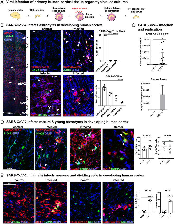
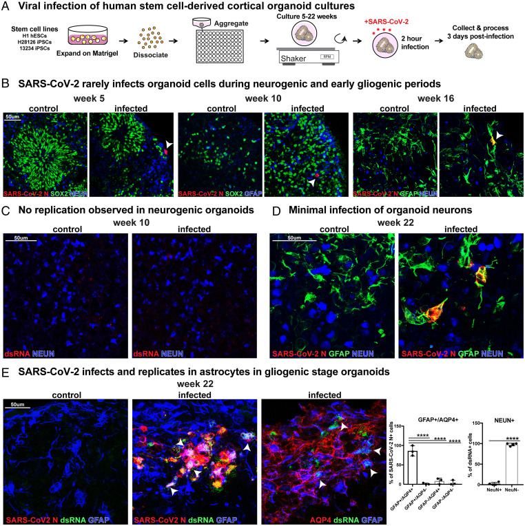
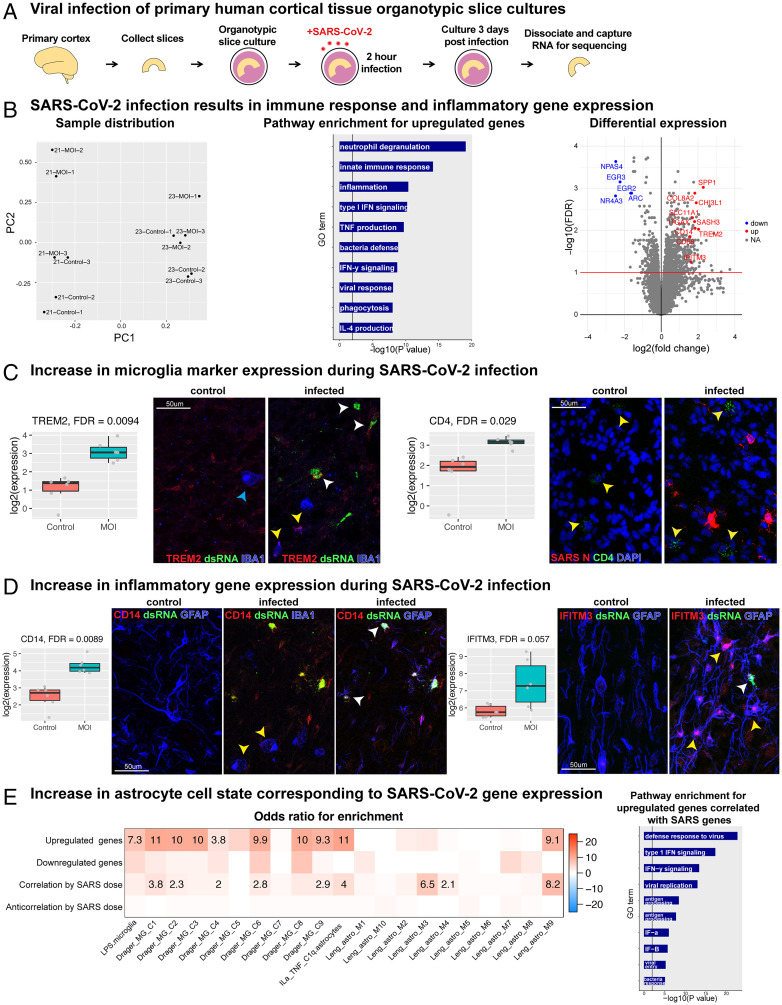
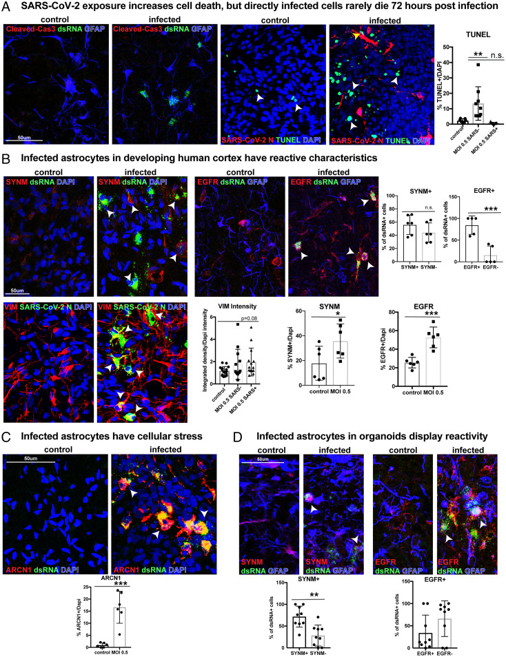
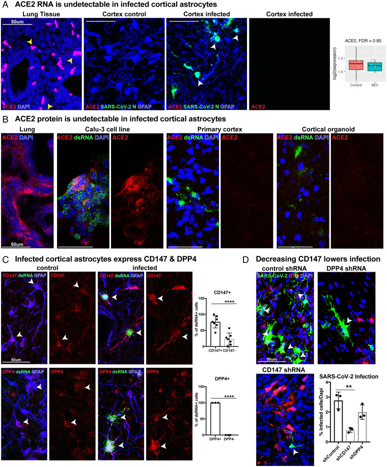
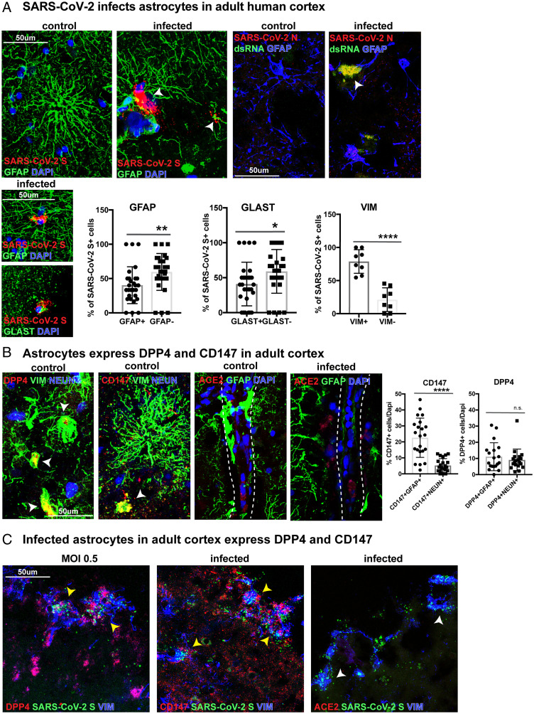
The severe acute respiratory syndrome coronavirus 2 (SARS-CoV-2) readily infects a variety of cell types impacting the function of vital organ systems, with particularly severe impact on respiratory function. Neurological symptoms, which range in severity, accompany as many as one-third of COVID-19 cases, indicating a potential vulnerability of neural cell types. To assess whether human cortical cells can be directly infected by SARS-CoV-2, we utilized stem-cell-derived cortical organoids as well as primary human cortical tissue, both from developmental and adult stages. We find significant and predominant infection in cortical astrocytes in both primary tissue and organoid cultures, with minimal infection of other cortical populations. Infected and bystander astrocytes have a corresponding increase in inflammatory gene expression, reactivity characteristics, increased cytokine and growth factor signaling, and cellular stress. Although human cortical cells, particularly astrocytes, have no observable ACE2 expression, we find high levels of coronavirus coreceptors in infected astrocytes, including CD147 and DPP4. Decreasing coreceptor abundance and activity reduces overall infection rate, and increasing expression is sufficient to promote infection. Thus, we find tropism of SARS-CoV-2 for human astrocytes resulting in inflammatory gliosis-type injury that is dependent on coronavirus coreceptors.

Keywords: SARS-CoV-2 tropism; astrocyte reactivity; organoid models.


Conflict of interest statement

Competing interest statement: A.R.K. is a co-founder, consultant, and member of the Board of Neurona Therapeutics. The other authors declare no competing interest.

Figures

Fig. 1.
Fig. 1.
SARS-CoV-2 infects astrocytes in developing human cortex. (A) Experimental paradigm for viral infection of human cortical tissue from GW 19 to 23. Cortical tissue was sliced, exposed to SARS-CoV-2 for 2 h, and cultured for 72 h. Samples were processed for immunostaining and qPCR. (B) SARS-CoV-2 infects GFAP+AQP4+ astrocytes in developing human cortex. All (100%) infected cells coexpress SARS-CoV-2+ nucleocapsid (N) and dsRNA, indicating viral infection and replication (one-way ANOVA, SARS-CoV-2+dsRNA+ vs. SARS-CoV-2-dsRNA+ ****P < 0.0001, SARS-CoV-2+dsRNA+ vs. SARS-CoV-2+dsRNA− ****P < 0.0001, error bars represent SD, n = 3 technical replicates from two biological samples). More than 90% of infected cells coexpress markers of astrocytes and SARS-CoV-2 infection (white arrowheads; one-way ANOVA: GFAP+AQP4+ vs. GFAP-AQP4− ****P < 0.0001, error bars represent SD, n = 4 technical replicates from three biological samples from stages GW 19, 22, 23). (C) Plaque assay was used to identify SARS-CoV-2 propagation in astrocytes. Astrocytes were exposed to the virus for 2 h and washed, and the media were changed. Supernatant from infected astrocytes was then cultured on Vero cells. Vero cells were fixed and plaque forming units quantified, where none were observed in control but >40,000 observed in cells treated with MOI 0.5 supernatant (n = 2, error bars represent SD). SARS-CoV-2 viral RNA can also be detected in slice cultures exposed to MOI 0.5 by qRT-PCR (unpaired Student’s t test, *P < 0.0149, n = 3 slices/condition from two biological replicates across three independent qRT-PCRs). (D) Mature S100B+ or GLAST+ and immature HOPX+ astrocytes demonstrate viral tropism, as approximately half of infected cells express these markers as indicated by white arrowheads (unpaired Student’s t test, S100B: P = 0.088 HOPX: P = 0.186, error bars represent SD, n = 3 biological samples from stages GW 19, 22, 23 from five technical replicates). (E) SARS-CoV-2 minimally infects neurons and dividing cells. Few infected dsRNA+ SARS-CoV-2+ cells are NEUN+ cortical neurons or KI67+ dividing cells, as fewer than 8% are NEUN+, and fewer than 11% are KI67+ (unpaired Student’s t test: NEUN ****P < 0.0001, KI67 ***P < 0.0001, error bars represent SD; n = 3 biological samples from six technical replicates).
Fig. 2.
Fig. 2.
SARS-CoV-2 infects astrocytes in cortical organoids. (A) Experimental paradigm for viral infection of induced pluriptotent stem cell (iPSC)-derived cortical organoids. Organoids from differentiation weeks 5, 10, 16, and 22 were infected with SARS-CoV-2. (B) Cells in organoids are only rarely infected at 5, 10, or 16 wk, as indicated by SARS-CoV-2 N expression (white arrowheads). These differentiation timepoints correspond to neurogenic and early gliogenic periods. (C) Although rare cells are infected and coexpress SARS-CoV-2 at neurogenic stages, there is no observed dsRNA+ viral replication at these timepoints. (D) In week 22 organoids, which correspond to the gliogenic period of differentiation, infection is readily observed. GFAP+ astrocytes coexpress SARS-CoV-2 N, but NEUN+ neurons are very rarely infected. (E) SARS-CoV-2 preferentially infects and replicates in astrocytes in gliogenic stage organoids. In total, 96% of infected cells stain positive for astrocyte markers (GFAP or AQP4), while only 2% express a neuronal marker (NEUN+). White arrowheads indicate SARS-CoV-2+ dsRNA+ GFAP+ AQP4+ astrocytes (one-way ANOVA: GFAP+AQP4+ vs. GFAP+AQP4−/GFAP−AQP4+/GFAP−AQP4− ****P < 0.0001, GFAP+AQP4+; NEUN: unpaired Student's t test: ****P < 0.00001, error bars represent SD, n = 4 organoids from two stem cell lines).
Fig. 3.
Fig. 3.
RNA-seq reveals an inflammatory response in human cortex after SARS-CoV-2 infection. (A) Primary organotypic cortical cultures from GW21 and GW23 were exposed to SARS-CoV-2 for 2 h and processed for RNA-seq 72 h later (three replicates for infection and control for each individual). (B) (Left) PC analysis revealed the first axis of variation as primarily due to age, while the second was related to SARS-CoV-2 infection. PC1 explained 15% of the variance and PC2 10% of the total variance. (Middle) Pathway enrichment analysis using Gene Ontology. A set of 22 genes are upregulated based on FDR < 0.1 and at least a 2-fold increase (100% increase or doubling) of normal expression. Upregulated genes in infected samples correspond to immune response, inflammation, neutrophil degranulation, and cytokine signaling. (Right) Volcano plot of significance versus effect size, with line drawn at FDR < 10%. The top downregulated genes are associated with transcription and cell growth (blue), and selected upregulated genes associated with inflammation, immune response, and microglia activation (red) are labeled. (C) We observe an increase in microglia markers TREM2 and CD4 expression after infection. Box plots contain expression values for the gene of interest (TREM2: FDR < 0.0094, CD4: FDR < 0.029, blue arrow: microglia without TREM2, yellow arrows: microglia with TREM2 or CD4, white arrow: dsRNA+-infected cell with TREM2). (D) There is an increase in inflammatory gene expression, including CD14 (FDR < 0.0089) and IFITM3 (FDR < 0.057), after infection. Protein staining indicates CD14 is increased in IBA1+ microglia (yellow arrow) and infected dsRNA+GFAP+ astrocytes (white arrow). IFITM3 is increased in infected (yellow arrow) and uninfected (white arrow) astrocytes. (E) There is an increase in astrocyte reactivity state corresponding to infection-related genes. Cell state enrichment analysis of differentially expressed genes from B and SARS-CoV-2 gene expression dose-responsive genes were compared with publicly available gene sets of activated microglia (24) and reactive astrocyte (25) cell states. Gene Ontology plots show an upregulated set of 918 genes correlated with SARS-CoV-2 dose based on FDR < 0.1 and at least a 2-fold increase of normal expression (GeneOntology up). When evaluating cell state by SARS-CoV-2 dose, our dataset most highly corresponds to an activated astrocyte population with high NF-κB signaling. Pathway enrichment of our viral dose-related dataset indicates genes that regulate viral defense, cytokine signaling, and immune response.
Fig. 4.
Fig. 4.
SARS-CoV-2 infection increases reactivity in cortical astrocytes. (A) In slice cultures exposed to SARS-CoV-2, we do not observe an increase in cleaved caspase-3 after infection. We observe an overall increase in the number of TUNEL+ cells within the infected culture (white arrow). However, the number of TUNEL+ SARS-CoV-2 N+-infected cells (yellow arrow) is not significantly different from control (ordinary one-way ANOVA with multiple comparisons: **P < 0.008, error bars represent SD, n = 3 biological samples from stages GW 19, 22, 23 from six technical replicates). (B) Infected astrocytes in the developing human cortex have reactive characteristics. Reactive markers SYNM and EGFR increase after infection compared to control, indicating an increase in reactivity postinfection (unpaired Student’s t test: SYNM: *P < 0.05, EGFR: ***P < 0.0003; error bars represent SD). More than 55% of infected primary astrocytes express SYNM, and 85% express EGFR (unpaired Student’s t test: SYNM P = 0.19, EGFR ***P < 0.0009, error bars represent SD, n = 3 biological samples from stages GW 19, 22, 23 from five technical replicates). The reactive astrocyte marker, VIM, also trends toward increased intensity in infected cells (ordinary one-way ANOVA with multiple comparisons: P = 0.08, error bars represent SD, n = 3 biological samples from 12 technical replicates). (C) Infected samples have a 15% increase in the ER stress marker, ARCN1. All ARCN1+ cells coexpress SARS-CoV-2 dsRNA (unpaired Student’s t test: control vs. MOI 0.5, ***P < 0.0002, error bars represent SD, n = 3 biological samples from stages GW 19, 22, 23 from six technical replicates). (D) Infected astrocytes in organoids display reactivity. In total, 72% of infected organoid cells express SYNM, and about one-third express EGFR (unpaired Student’s t test: SYNM **P < 0.0014, EGFR P = 0.109, error bars represent SD, n = 2 cell lines from four technical replicates).
Fig. 5.
Fig. 5.
Coronavirus receptors CD147 and DPP4, but not ACE2, are expressed in human cortex. (A) In situ hybridization for ACE2 RNA was performed in developing lung tissue (positive control) and developing cortical tissue. While ACE2 is highly expressed in the lung (yellow arrowheads), ACE2 was not detected in human cortical tissue or in infected cortical astrocytes (white arrowheads). ACE2 RNA also does not increase after infection (P = 0.85). (B) ACE2 protein is undetectable in cortical astrocytes. Immunohistochemistry for ACE2 protein was performed on lung tissue and a lung cell line, Calu-3, confirming efficient protein detection. We find no observable ACE2 protein in primary cortical tissue, cortical organoids, or infected cells in either sample type. (C) Cortical astrocytes express coronavirus receptors CD147 and DPP4. In total, 76% of infected dsRNA+ GFAP+ astrocytes express the coronavirus receptor CD147, and 100% express DPP4 (unpaired Student’s t test: CD147 ***P < 0.0002, DPP4 ****P < 0.0001, error bars represent SD, n = 3 biological samples from stages GW 19, 22, 23 from four technical replicates). (D) Organotypic cultures were treated with lentivirus containing shRNAs against CD147 or DPP4 to knock down CD147 or DPP4, respectively. After decreasing the abundance of CD147, there was a significant reduction in infection rate (ordinary one-way ANOVA: control vs. CD147 **P < 0.034, control vs. DPP4 P = 0.15, error bars represent SD, n = 3 technical replicates from two biological samples).
Fig. 6.
Fig. 6.
Astrocytes in the adult human cortex are vulnerable to SARS-CoV-2 infection. (A) Surgically resected adult human cortical tissue from 19 and 34 y of age was acutely sectioned and exposed to SARS-CoV-2 for 2 h and cultured for 48 to 72 h before collection. GFAP+, GLAST+, or VIM+ astrocytes in the adult cortex were infected and labeled by SARS-CoV-2 spike (S) riboprobe or SARS-CoV-2 nucleocapsid (N) antibody. Of the infected cells, 40% were GFAP+, 41% were GLAST+, and 79% were VIM+, indicating different populations of astrocytes are vulnerable to infection (unpaired Student’s t test, GFAP **P < 0.01, GLAST *P < 0.036, VIM ****P < 0.0001, n = 2 biological samples from four technical replicates). (B) In the adult cortex, coronavirus receptors DPP4 and CD147 are expressed on VIM+ astrocytes (white arrows), while ACE2 is localized to blood vessels. There is a greater abundance of CD147 in GFAP+ astrocytes than NEUN+ neurons, while DPP4 is expressed in both cell types (unpaired Student’s t test, CD147+GFAP+ vs. CD147+NEUN+ ****P < 0.0001, DPP4+GFAP+ vs. DPP4+NEUN+ P = 0.424). (C) Infected SARS-CoV-2 S+ VIM+ astrocytes in the adult cortex express DPP4 and CD147 (yellow arrows), but ACE2 is not observed in infected cells (white arrows).

Update of

Comment in

  • In SARS-CoV-2, astrocytes are in it for the long haul.
    Huang S, Fishell G. Huang S, et al. Proc Natl Acad Sci U S A. 2022 Jul 26;119(30):e2209130119. doi: 10.1073/pnas.2209130119. Epub 2022 Jul 18. Proc Natl Acad Sci U S A. 2022. PMID: 35858460 Free PMC article. No abstract available.

Similar articles

Cited by

References

    1. Varatharaj A., et al. ; CoroNerve Study Group, Neurological and neuropsychiatric complications of COVID-19 in 153 patients: A UK-wide surveillance study. Lancet Psychiatry 7, 875–882 (2020). - PMC - PubMed
    1. Solomon I. H., et al. , Neuropathological features of covid-19. N. Engl. J. Med. 383, 989–992 (2020). - PMC - PubMed
    1. Bartley C. M., et al. , Anti-SARS-CoV-2 and autoantibody profiles in the cerebrospinal fluid of 3 teenaged patients with COVID-19 and subacute neuropsychiatric symptoms. JAMA Neurol. 78, 1503–1509 (2021). - PMC - PubMed
    1. Helms J., et al. , Neurologic features in severe SARS-CoV-2 infection. N. Engl. J. Med. 382, 2268–2270 (2020). - PMC - PubMed
    1. Mao L., et al. , Neurologic manifestations of hospitalized patients with coronavirus disease 2019 in Wuhan, China. JAMA Neurol. 77, 683–690 (2020). - PMC - PubMed

Publication types

Substances