. 2022 Jul 19;40(3):111107.
doi: 10.1016/j.celrep.2022.111107.

Alphaherpesvirus US3 protein-mediated inhibition of the m6A mRNA methyltransferase complex

Affiliations

Alphaherpesvirus US3 protein-mediated inhibition of the m6A mRNA methyltransferase complex

Robert J J Jansens et al. Cell Rep. .

Abstract

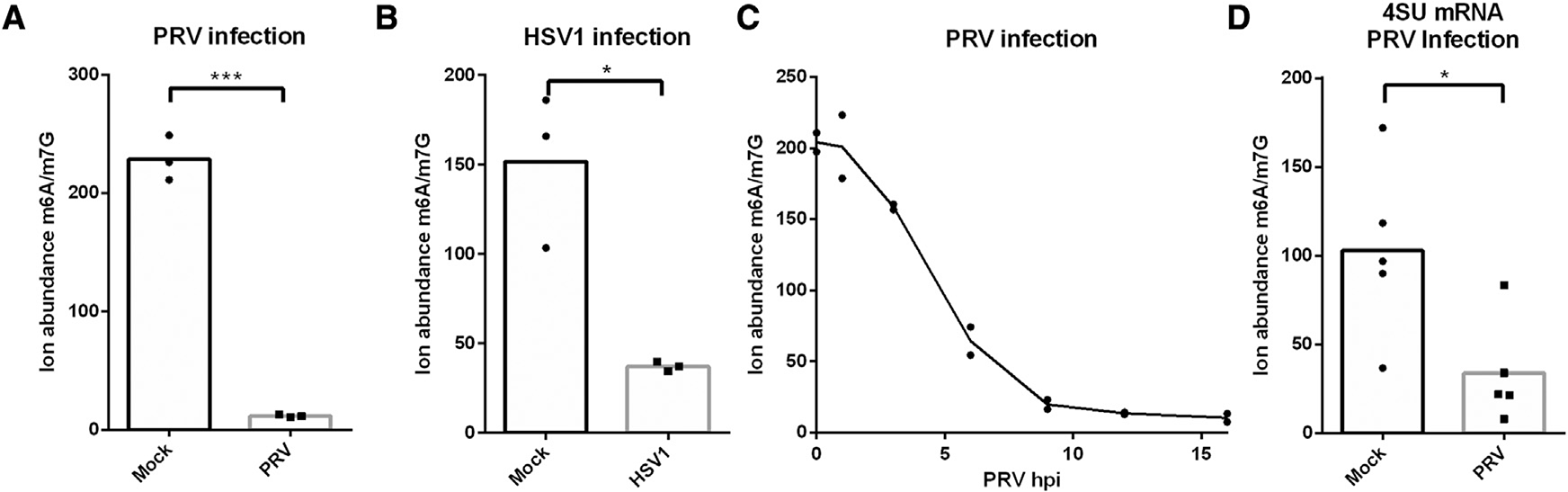
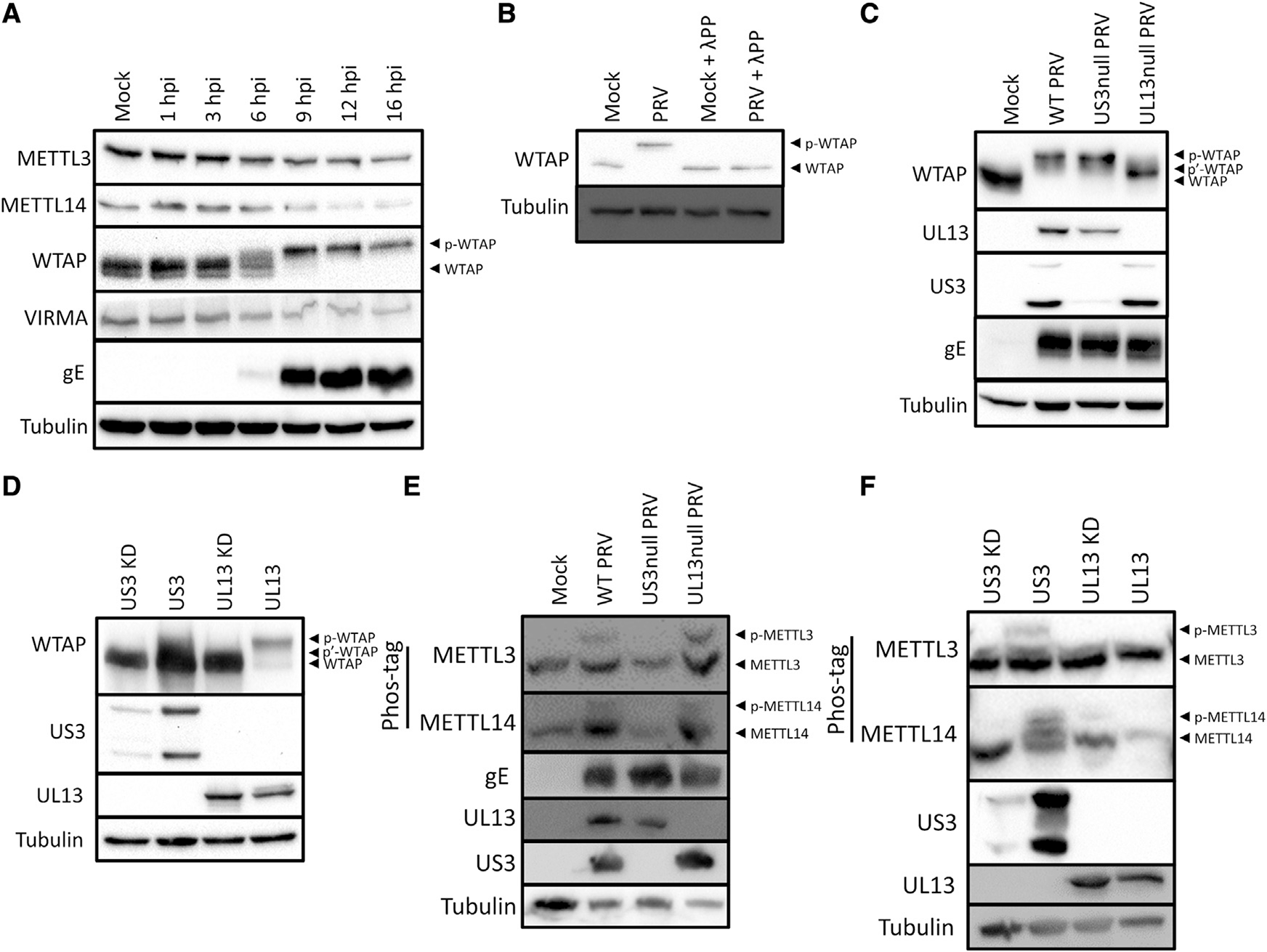
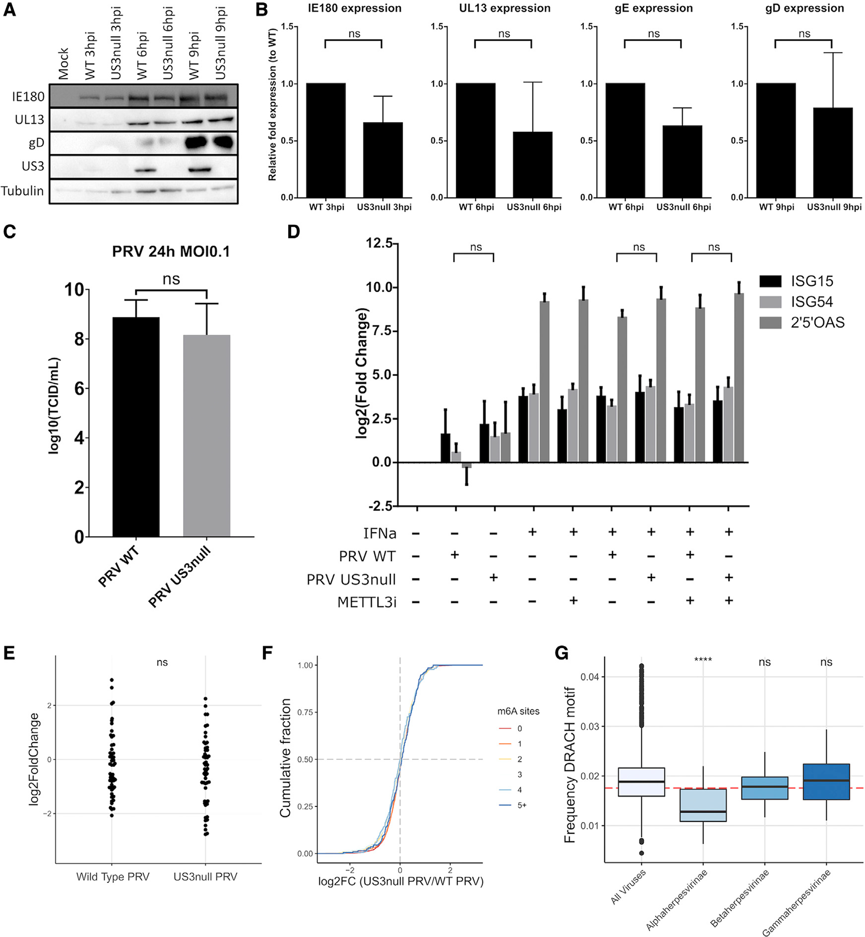
Chemical modifications of mRNA, the so-called epitranscriptome, represent an additional layer of post-transcriptional regulation of gene expression. The most common epitranscriptomic modification, N6-methyladenosine (m6A), is generated by a multi-subunit methyltransferase complex. We show that alphaherpesvirus kinases trigger phosphorylation of several components of the m6A methyltransferase complex, including METTL3, METTL14, and WTAP, which correlates with inhibition of the complex and a near complete loss of m6A levels in mRNA of virus-infected cells. Expression of the viral US3 protein is necessary and sufficient for phosphorylation and inhibition of the m6A methyltransferase complex. Although m6A methyltransferase complex inactivation is not essential for virus replication in cell culture, the consensus m6A methylation motif is under-represented in alphaherpesvirus genomes, suggesting evolutionary pressure against methylation of viral transcripts. Together, these findings reveal that phosphorylation can be associated with inactivation of the m6A methyltransferase complex, in this case mediated by the viral US3 protein.

Keywords: CP: Microbiology; CP: Molecular biology; PRV; US3; herpes; m6A; pseudorabies; writer complex.


Conflict of interest statement

Declaration of interests S.R.J. is an advisor to and owns equity in 858 Therapeutics and Lucerna Technologies.

Figures

Figure 1.
Figure 1.. Alphaherpesvirus infection results in a decrease in m6A-methylated mRNA
Quantification of m6A levels using mass spectrometry in (A) mRNA from mock-infected or PRV-infected ST cells at 16 hpi (n = 3 biological replicates), (B) mRNA from mock-infected or HSV-1-infected HEK293-T cells at 16 hpi (n = 3 biological replicates), (C) mRNA from PRV-infected ST cells at different time points post-inoculation (n = 2 biological replicates), and (D) 4SU-labeled mRNA produced between 7 and 9 hpi of mock-infected and PRV-infected ST cells (n = 5 biological replicates). Statistical significance was calculated by unpaired Student’s t test. *p < 0.05; ***p < 0.001.
Figure 2.
Figure 2.. PRV infection triggers phosphorylation of the m6A writer complex
(A) Western blotting of a time-course experiment of different components of the m6A writer complex upon infection of ST cells with PRV at an MOI of 10 (n = 2 biological replicates). (B) Western blotting of WTAP in mock- or PRV-infected ST cells at 16 hpi that were either or not treated with λ-phosphatase (n = 3 biological replicates). (C) Western blotting of ST cells infected with wild-type (WT) PRV or isogenic PRV strains lacking expression of either of the viral protein kinases at 16 hpi (n = 3 biological replicates). (D) Western blotting of HEK293-T cells transfected with either viral protein kinase at 48 h post-transfection (hpt) (n = 3 biological replicates). Kinase-dead (KD) US3 and UL13 contain a point mutation in the ATP binding site or the catalytic site, respectively. (E) Phos-tag assays of ST cells mock-infected or infected with WT PRV or isogenic PRV strains lacking expression of US3 or UL13 at 16 hpi. (F) Phos-tag assays of HEK293-T cells transfected with either of the viral protein kinases at 48 hpt. See also Figure S1.
Figure 3.
Figure 3.. PRV infection leads to US3-dependent inactivation of the m6A writer complex
(A) Mass spectrometry-based quantification of m6A levels in total mRNA from ST cells infected with wild-type PRV strain NIA3 or isogenic PRV lacking either of the viral protein kinases at 9 hpi (n = 5 biological replicates). (B) 4SU-labeled nascent mRNA produced between 7 and 9 hpi from mock-infected ST cells or ST cells infected with wild-type PRV strain NIA3 or isogenic US3null or UL13null PRV (n = 5 biological replicates). (C) Total mRNA from HEK293-T cells transfected with an empty vector or either of the active or kinase-dead viral kinases (n = 3 biological replicates). (D) Total mRNA from ST cells infected with wild-type PRV strain Becker or isogenic PRV lacking the US3 protein or PRV expressing a kinase-inactive US3 protein (KD) at 9 hpi (n = 3 biological replicates). (E) Phos-tag assays of mock-infected ST cells or ST cells infected with wild-type PRV strain Becker or isogenic PRV lacking the US3 protein or expressing a kinase-inactive US3 protein at 6 hpi. See also Figure S2. Statistical significance was calculated by one-way ANOVA. *p < 0.05; **p < 0.01; ***p < 0.001.
Figure 4.
Figure 4.. Chromatin dissociation of the m6A writer complex upon PRV infection
(A) Immunofluorescence imaging of METTL3 and WTAP in ST cells infected with wild-type PRV- or mock-infected for 8 h (n = 3 biological replicates). Scale bar, 5 μm. (B and C) ST cells were mock infected or infected with wild-type or isogenic US3null PRV for 16 h, after which WTAP (B) or METTL3 (C) was immunoprecipitated. The different components of the m6A writer complex were then detected using western blotting and quantified using densitometry (n = 2 biological replicates). (D) ST cells were mock infected or infected with wild-type PRV or isogenic US3null or UL13null PRV and at 16 hpi fractioned in soluble and chromatin-bound lysates, followed using Western blotting for several components of the writer complex. See also Figure S3.
Figure 5.
Figure 5.. Inactivation of the m6A writer complex is not essential for viral replication, but m6A consensus sites are significantly underrepresented in alphaherpesvirus genomes
(A) ST cells were infected with wild-type or isogenic US3null PRV for different times, and viral protein levels were determined using western blotting. (B) ST cells were infected with wild-type or isogenic US3null PRV for different times, and viral transcript levels were determined using qPCR. Data are represented as mean ± SEM (n = 3 biological replicates). (C) ST cells were infected with wild-type or isogenic US3null PRV at an MOI of 0.1 for 24 h, and infectious viral particle counts were determined by titrations. Data are represented as mean ± SEM (n = 3 biological replicates). (D) ST cells were infected with wild-type or isogenic US3null PRV for 8 h and treated with MG132 from 2 hpi. At 4 hpi, cells were either or not treated with 300 ng/mL IFNa. ISG transcript levels were determined using qPCR. Data are represented as mean ± SEM (n = 3 biological replicates). (E) Fold changes of all porcine ISGs were determined using RNA-seq in wild-type or US3null-infected ST cells compared with mock-infected ST cells (n = 2 biological replicates). (F) Cumulative fold change of m6A-methylated or non-methylated host transcripts as measured using RNA-seq comparing wild-type PRV-infected and isogenic US3null PRV-infected ST cells. ST cells were harvest at 16 h after infection. Host transcripts were binned on the basis of the number of m6A sites, with the red line representing unmethylated transcripts (n = 2 biological replicates). (G) All available eukaryotic virus genomes were downloaded (n = 5,516 genomes), and the frequency of the DRACH motif (in which D can be A, G, or U; R can be A or G; and H can be A, C, or U) was analyzed and compared with the subfamilies alphaherpesvirinae (n = 43 genomes), betaherpesvirinae (n = 23 genomes), and gammaherpesvirinae (n = 39 genomes). The red dotted line represents the theoretical random frequency of the DRACH motif. See also Figures S4 and S5. Statistical significance was calculated by unpaired Student’s t test or one-way ANOVA. ****p < 0.0001.

References

    1. Baquero-Perez B, Antanaviciute A, Yonchev ID, Carr IM, Wilson SA, and Whitehouse A (2019). The Tudor SND1 protein is an m6A RNA reader essential for replication of Kaposi’s sarcoma-associated herpesvirus. eLife 8, e47261. 10.7554/eLife.47261. - DOI - PMC - PubMed
    1. Barbieri I, and Kouzarides T (2020). Role of RNA modifications in cancer. Nat. Rev. Cancer 20, 303–322. 10.1038/s41568-020-0253-2. - DOI - PubMed
    1. Bartkoski M, and Roizman B (1976). RNA synthesis in cells infected with herpes simple virus. XIII. Differences in the methylation patterns of viral RNA during the reproductive cycle. J. Virol. 20, 583–588. 10.1128/jvi.20.3.583-588.1976. http://www.ncbi.nlm.nih.gov/pubmed/186635. - DOI - PMC - PubMed
    1. Bartkoski MJ, and Roizman B (1978). Regulation of herpesvirus macromolecular synthesis VII. Inhibition of internal methylation of mRNA late in infection. Virology 85, 146–156. 10.1016/0042-6822(78)90419-1. - DOI - PubMed
    1. Bokar JA, Rath-Shambaugh M, Ludwiczak R, Narayan P, and Rottman F (1994). Characterization and partial purification of mRNA N6-adenosine methyltransferase from HeLa cell nuclei. Internal mRNA methylation requires a multisubunit complex. J. Biol. Chem. 269, 17697–17704. 10.1016/s0021-9258(17)32497-3. - DOI - PubMed

Publication types