. 2022 Oct;59(10):6076-6090.
doi: 10.1007/s12035-022-02947-8. Epub 2022 Jul 20.

A Functional Interaction Between Y674-R685 Region of the SARS-CoV-2 Spike Protein and the Human α7 Nicotinic Receptor

Affiliations

A Functional Interaction Between Y674-R685 Region of the SARS-CoV-2 Spike Protein and the Human α7 Nicotinic Receptor

Juan Facundo Chrestia et al. Mol Neurobiol. 2022 Oct.

Abstract

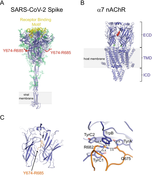
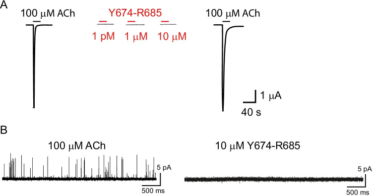
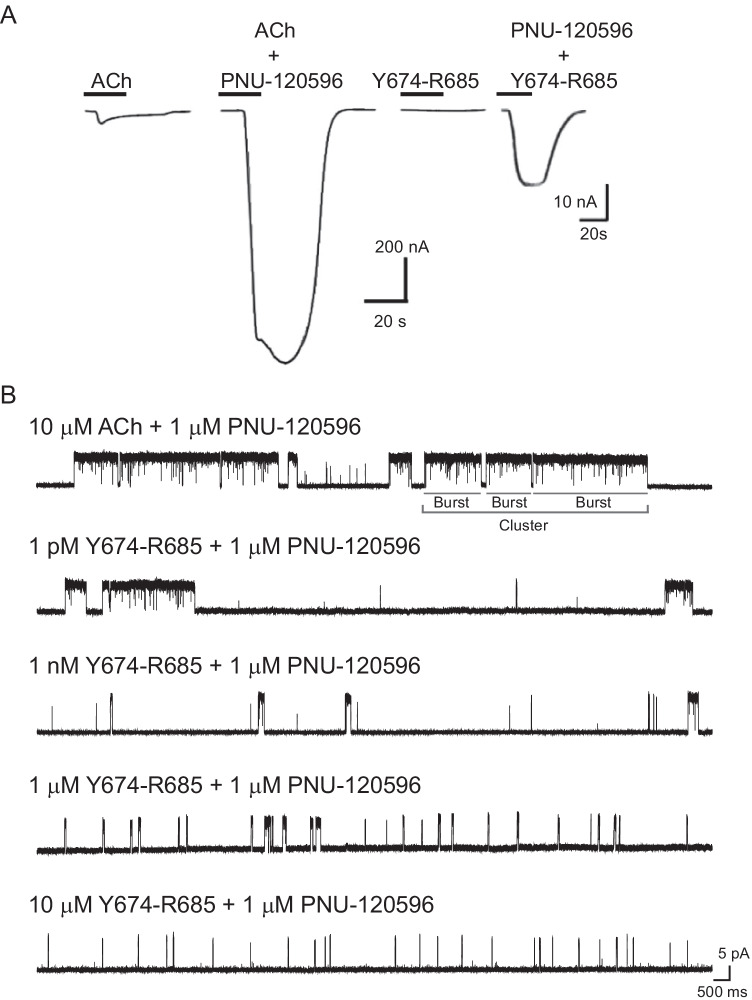
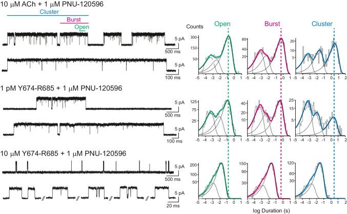
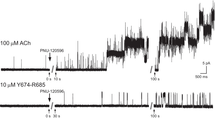
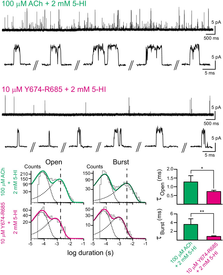
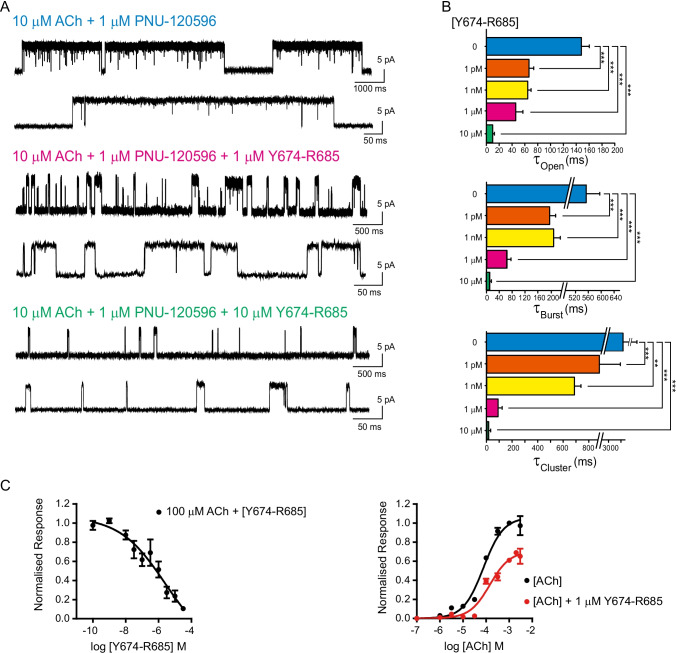
The α7 nicotinic acetylcholine receptor (nAChR) is present in neuronal and non-neuronal cells and has anti-inflammatory actions. Molecular dynamics simulations suggested that α7 nAChR interacts with a region of the SARS-CoV-2 spike protein (S), and a potential contribution of nAChRs to COVID-19 pathophysiology has been proposed. We applied whole-cell and single-channel recordings to determine whether a peptide corresponding to the Y674-R685 region of the S protein can directly affect α7 nAChR function. The S fragment exerts a dual effect on α7. It activates α7 nAChRs in the presence of positive allosteric modulators, in line with our previous molecular dynamics simulations showing favourable binding of this accessible region of the S protein to the nAChR agonist binding site. The S fragment also exerts a negative modulation of α7, which is evidenced by a profound concentration-dependent decrease in the durations of openings and activation episodes of potentiated channels and in the amplitude of macroscopic responses elicited by ACh. Our study identifies a potential functional interaction between α7 nAChR and a region of the S protein, thus providing molecular foundations for further exploring the involvement of nAChRs in COVID-19 pathophysiology.

Keywords: Neurotransmitter receptors; Nicotinic receptor; Patch-clamp; SARS-CoV-2 spike protein; Single-channel recordings.


Conflict of interest statement

The authors declare no competing interests.

Figures

Fig. 1
Fig. 1
Three-dimensional structures of the SARS-CoV-2 S protein and the human α7 nAChR. A The model represents the complete, fully glycosylated S protein in the closed state after furin cleavage [1]. The protein is rendered as a blue cartoon with the glycans depicted in green. The receptor binding motifs (residues S438-Q506) and the Y674-R685 region are highlighted in yellow and red, respectively. The Y674-R685 region was shown to be accessible for binding in previous MD simulations of the fully glycosylated S protein [8]. B Cryo-EM structure of the human α7 nAChR (PDB code: 7KOX) [10]. This receptor is a homopentamer formed of five α7 subunits. Each subunit is composed of an extracellular (ECD), transmembrane (TMD), and intracellular (ICD) domain. The agonist binding site is located in the ECD at the interface between two neighbouring subunits. In this structure, epibatidine (red spheres) is bound to the agonist binding site. The green spheres represent bound calcium ions. C MD simulations of Y674-R685 bound to the human α7 nAChR show favourable binding to the binding pocket [8]. Example of conformations from simulations in which the most important interactions with conserved key aromatic residues are present. Left: Overall view of the Y674-R685: α7(ECD) complex. Right: Close-up view of interactions formed by R682 and Q675 within agonist binding site. The α7 receptor and Y674-R685 are coloured in dark blue and orange, respectively. Interactions between side chains of R682 and Q675 and the aromatic rings of TrpB ((α7W171), TyrC1 (α7Y210), TyrC2 (α7Y217) and TyrA (α7Y115) are shown with dashed lines
Fig. 2
Fig. 2
The Y674-R685 fragment cannot elicit detectable α7 responses. A Macroscopic responses of the human α7 nAChR. Representative traces from single oocytes expressing human α7 nAChR to applications of ACh (100 μM) or 1 pM, 1 μM, or 10 μM of Y674-R685. Drug applications were for 20 s followed by a 300 s washout. B Single-channel currents of the human α7 nAChR recorded from cell-attached patches in the presence of 100 μM ACh or 10 μM Y674-R685. No channel activity was detected at a 1 pM to 100 μM Y674-R685 concentration range. Channel openings are shown as upward deflections. Pipette potential: 70 mV. Filter: 9 kHz
Fig. 3
Fig. 3
Activation of the human α7 nAChR by Y674-R685 in the presence of the PAM PNU-120596. A Macroscopic currents were recorded from oocytes expressing the human α7 nAChR after a pulse of 30 μM ACh (control) or 1 μM Y674-R685 in the absence or presence of 10 μM PNU-120596. Current traces shown are representative of n = 15 recordings from oocytes isolated from N = 3 donors. B Single-channel currents of the human α7 nAChR in the presence of the type II PAM PNU-120596 (1 μM) activated by 100 μM ACh (left), or Y674-R685 at different concentrations (1 pM, 1 nM, 1 μM, or 10 μM) (right). For each condition typical channel traces are shown. Channel openings are shown as upward deflections. Pipette potential: 70 mV. Filter: 3 kHz
Fig. 4
Fig. 4
Single-channel recordings of the human α7 nAChR in the presence of Y674-R685. Single-channel currents of human α7 nAChR in the presence of the type II PAM PNU-120596 (1 μM) activated by 100 μM ACh, or Y674-R685 at 1 pM or 10 μM. For each condition, channel traces at two different temporal scales are shown. Channel openings are shown as upward deflections. Representative open, burst, and cluster duration histograms are shown for each condition. The open, burst, and cluster durations correspond to the durations of the slowest components of each histogram. The dashed lines show how these mean durations change among different conditions. Pipette potential: 70 mV. Filter: 3 kHz
Fig. 5
Fig. 5
Channel activity elicited by Y674-R685 in the absence or presence of the PAM. Representative experiments in which channel activity from the same patch was recorded before and after addition of PNU-120596. ACh or 10 μM Y674-R685 were present in the pipette solution. The indicated time corresponds to the time of recording after addition of PNU-120596. Top: single-channel currents of human α7 activated by 100 μM ACh appeared mainly as brief isolated openings. Addition of 1 μM PNU-120596 to the extracellular solution surrounding a cell-attached patch resulted in a marked increase in current in the continued presence of 100 μM ACh in the patch pipette. This experiment is representative of 4 different patches. Bottom: channel activity was undetectable in the presence of Y674-R685. Addition of 1 μM PNU-120596 to the extracellular solution surrounding the silent patch revealed channel activity, indicating that only in the presence of the PAM 10 μM Y674-R685 can activate α7. This experiment is representative of 3 different patches. Channel openings are shown as upward deflections. Pipette potential: 70 mV. Filter 3 kHz
Fig. 6
Fig. 6
Activation of the human α7 nAChR by Y674-R685 in the presence of the type I PAM, 5-HI. Single-channel currents were recorded from cells expressing the human α7 nAChR in the presence of 2 mM 5-HI as the PAM and 100 μM ACh or 10 μM Y674-R685 as agonists. Traces at two different scales are shown for each condition. Pipette potential: 70 mV, Filter: 9 kHz. Representative open and burst duration histograms are shown for each agonist. The bar chart shows the mean durations ± SD of the slowest components of the open and burst duration histograms for each agonist (n = 37 for ACh and n = 3 for Y674-R685). *p < 0.05, **p < 0.01 (Student’s t-test)
Fig. 7
Fig. 7
Y674-R685 Inhibition of α7 nAChR channels activated by ACh. A Single-channel currents elicited by 10 μM ACh and potentiated by 1 μM PNU-120596 were recorded in the absence (control) or presence of 1 or 10 μM Y674-R685. Typical channel traces are shown at two different temporal scales. Channel openings are shown as upward deflections. Pipette potential: 70 mV. Filter: 3 kHz. B Bar chart showing the mean durations of openings (τopen), bursts (τburst), and clusters (τcluster) in the absence (blue) and in the presence of 1 pM (orange), 1 nM (yellow), 1 μM (pink), and 10 μM (green) Y674-R685. The data correspond to the mean duration (± SD) of the slowest components of open, burst, and cluster histograms. **p < 0.01, ***p < 0.001 (Student’s t-test) (see Supplementary Table 1). C Concentration response curve (CRC) for the inhibition of the α7 nAChR by Y674-R685. Increasing concentrations of Y674-R685 (0.1 nM to 30 μM) were co-applied with control ACh (100 μM). Responses were measured from the peak of ACh-elicited currents. Each data point represents the average normalized response of six cells (± SEM). Right panel: competition CRC data (red) for 1 μM Y674-R685 co-applied with different ACh concentrations (0.1–2000 μM). For comparison, ACh CRC data alone (black) are shown at the same concentrations. Data were fitted with the Hill equation, as described in the “Materials and methods” section. Data points represent the average normalized response of six oocytes (± SEM)

Similar articles

Cited by

References

    1. Casalino L, Gaieb Z, Goldsmith JA, et al. Beyond shielding: the roles of glycans in the SARS-CoV-2 spike protein. ACS Cent Sci. 2020;6:1722–1734. doi: 10.1021/acscentsci.0c01056. - DOI - PMC - PubMed
    1. Hoffmann M, Kleine-Weber H, Schroeder S, et al. SARS-CoV-2 cell entry depends on ACE2 and TMPRSS2 and is blocked by a clinically proven protease inhibitor. Cell. 2020;181(2):271–280.e8. doi: 10.1016/j.cell.2020.02.052. - DOI - PMC - PubMed
    1. Yan R, Zhang Y, Li Y, et al. Structural basis for the recognition of SARS-CoV-2 by full-length human ACE2. Science. 2020;367(6485):1444–1448. doi: 10.1126/science.abb2762. - DOI - PMC - PubMed
    1. Cantuti-Castelvetri L, Ojha R, Pedro LD, et al. Neuropilin-1 facilitates SARS-CoV-2 cell entry and infectivity. Science. 2020;370(6518):856–860. doi: 10.1126/science.abd2985. - DOI - PMC - PubMed
    1. Daly JL, Simonetti B, Klein K, et al. Neuropilin-1 is a host factor for SARS-CoV-2 infection. Science. 2020;370(6518):861–865. doi: 10.1126/science.abd3072. - DOI - PMC - PubMed

MeSH terms

Substances