. 2022 Jul 20;12(1):12448.
doi: 10.1038/s41598-022-14449-7.

Development of a rapid in vitro pre-screen for distinguishing effective liposome-adjuvant delivery systems

Affiliations

Development of a rapid in vitro pre-screen for distinguishing effective liposome-adjuvant delivery systems

Laura A J Feather et al. Sci Rep. .

Abstract

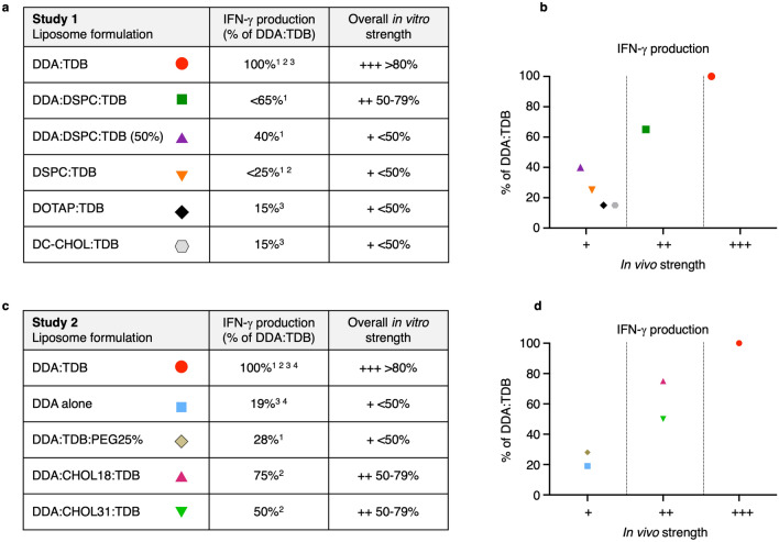
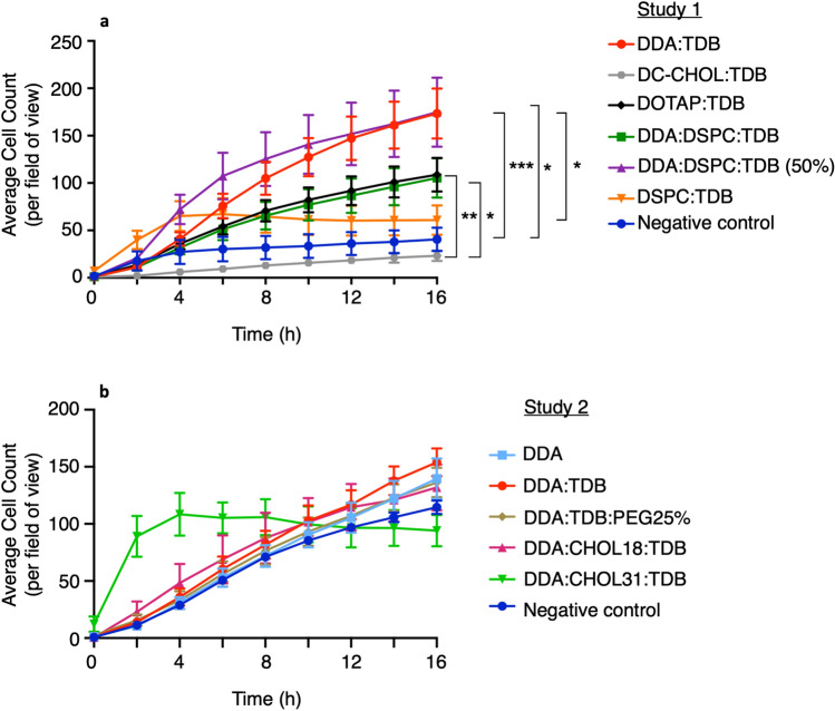
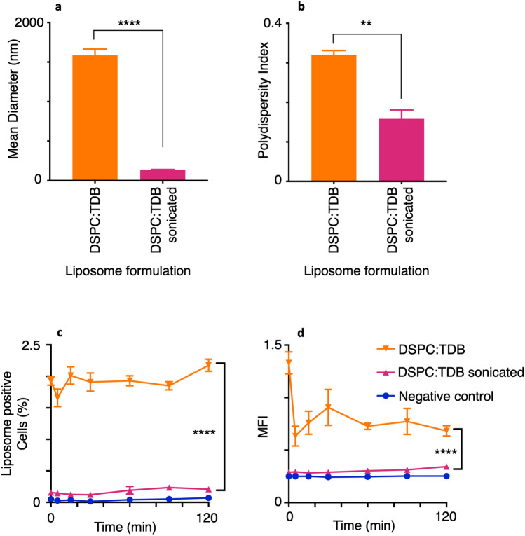
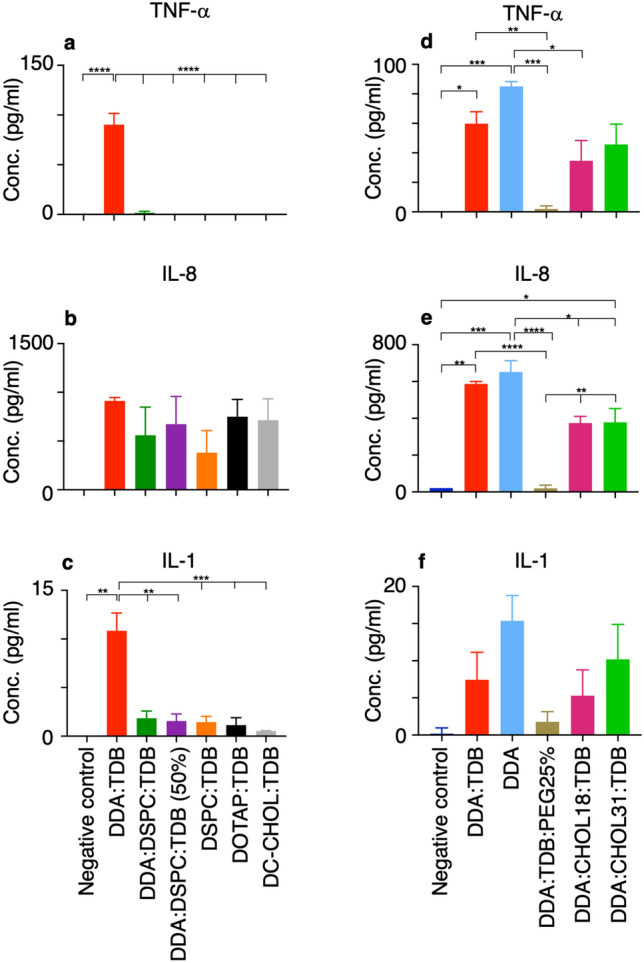
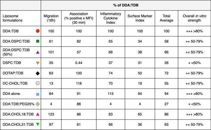
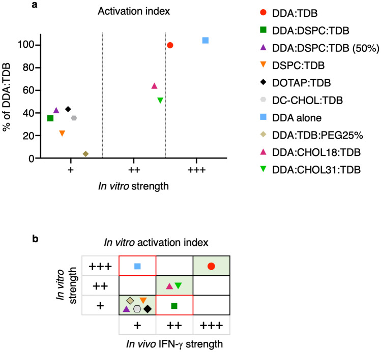
Liposomes are a strong supporting tool in vaccine technology, as they are a versatile system that not only act as antigen delivery systems but also adjuvants that can be highly effective at stimulating both innate and adaptive immune responses. Their ability to induce cell-mediated immunity makes their use in vaccines a useful tool in the development of novel, more effective vaccines against intracellular infections (e.g. HIV, malaria and tuberculosis). Currently, screening of novel liposome formulations uses murine in vivo models which generate data that often correlates poorly with human data. In addition, these models are both high cost and low throughput, making them prohibitive for large scale screening of formulation libraries. This study uses the cationic liposome formulation DDA:TDB (known as cationic adjuvant formulation 01 (CAF01)), as a lead formulation, along with other liposome formulations of known in vivo efficacy to develop an in vitro screening tool for liposome formulation development. THP-1-derived macrophages were the model antigen presenting cell used to assess the ability of the liposome formulations to attract, associate with and activate antigen presenting cells in vitro, crucial steps necessary for an effective immune response to antigen. By using a combination of in vitro functions, the study highlights the potential use of an in vitro screening tool, to predict the in vivo efficacy of novel liposome formulations. CAF01 was predicted as the most effective liposome formulation when assessing all in vitro functions and a measure of in vitro activation was able to predict 80% of the liposome correctly for their ability to induce an in vivo IFN-ү response.


Conflict of interest statement

The authors declare no competing interests.

Figures

Figure 1
Figure 1
In vivo IFN-ү production induced by exposure to liposome formulations, compared against DDA:TDB provides in vivo liposome efficacy. Data from in vivo papers referenced within the figure were taken for each liposome formulation. (a) Shows a summary table combining data, from all study 1 papers, of IFN-ү production shown as the percentage of DDA:TDB, and the corresponding in vivo strength category established as ‘+++’ > 80%, ‘++’ 50–79% and ‘+’ < 50%. (c) Shows a summary table combining data, from all study 2 papers, of IFN-ү production shown as the percentage of DDA:TDB, and the corresponding in vivo strength category established as ‘+++’ > 80%, ‘++’ 50–79% and ‘+’ < 50%. This data is represented in (b) and (d). Study 1 references: 1Hussain et al.. 2Henriksen-Lacey et al.. 3Henriksen-lacey et al.. Study 2 references: 1Kaur et al.; 2Kaur et al.; 3Henriksen et al.; 4Davidsen et al..
Figure 2
Figure 2
The extent of migration of THP-1-derived macrophages towards liposomes is dependent on the formulation. 700 µl of liposomes diluted to 20 µg/ml in serum-free RPMI were placed in the bottom well of a 24-well transwell culture plate and 300 μl of VD3-stimulated macrophages, at a cell density of 2.66 × 105 cells/ml placed in an 8 µm pourable insert above. The plate set up was incubated at 37 °C for over 16 h within the Cell IQ live cell imager. Images were taken every 2 h to enable the number of macrophages that had migrated towards the liposomes to be counted. (n ≥ 3 mean ± SEM). *P ≤ 0.05, **P ≤ 0.005, ***P ≤ 0.0005, ****P ≤ 0.0001 with repeated measures two-way ANOVA and Tukey’s multiple comparison test.
Figure 3
Figure 3
In vitro macrophage uptake of liposomes can reveal functional differences between formulations. Liposomes were diluted to 20 μg/ml in serum-free RPMI co-cultured 1:1 with VD3- stimulated macrophages at a density of 1 × 106/ml and incubated for 2 h. At indicated time points, 200 µl of co-culture was mixed with 200 µl ice cold sfRPMI medium before flow cytometric analysis of 10,000 events. (a) and (c) show percentage of macrophages positive for liposome association and (b) and (d) show the mean fluorescence intensity from cells positive for liposome association. Data shown for n = 3 (mean+/- SEM) with repeated measures ANOVA and Bonferroni post hoc test at 30 min, *P ≤ 0.05, **P ≤ 0.005, ***P ≤ 0.0005, ****P ≤ 0.0001. (Flow histograms shown in Supplementary Figs. S2 and S3).
Figure 4
Figure 4
Altering the size of DSPC:TDB does not influence in vitro cellular uptake. DSPC:TDB liposomes produced via lipid film hydration to make large (> 1000 nm) liposomes. Large liposomes then sonicated at 45 °C for 15 cycles using a Diaganode water bath sonicator to produce small (~ 200 nm) DSPC:TDB liposomes. Liposome characteristics; size (a) and polydispersity (b) of both sonicated and non-sonicated liposomes were measured using dynamic light scattering (Brookhaven ZetaPlus). THP-1-derived macrophages at 2 × 106/ml were co-cultured 1:1 with DilC-fluorescently labelled liposomes at 20 μg/ml. At indicated time points, 200 μl of co-culture was analysed using flow cytometry (10 000 events) for percentage of macrophages positive for association (c) and mean fluorescence intensity of macrophages (d). (n = 3, mean ± SEM) (a) and (b) show significance of ****P ≤ 0.0001 with a multiple T test and (c) and (d) show significance ****P ≤ 0.0001 with repeated measures ANOVA and Bonferroni post hoc at 30 min.
Figure 5
Figure 5
Study 1: Assessing changes in surface marker expression from VD3-stimulated macrophages allows differences to be seen between liposome formulations. Liposomes at 20 μg/ml were incubated with VD3-stimulated macrophages at a final cell density of 1 × 106/ml for 24 h. After incubation with PE-conjugated antibodies, analysis of the co-culture was conducted using flow cytometry to determine the percentage of the cell population to express surface markers and the mean fluorescence intensity (MFI) of the macrophages that had associated with liposomes. Macrophages not exposed to liposomes were used to set the negative and positive discriminator for each surface marker to allow for the effect different liposome formulations had on macrophage surface marker expression to be highlighted. Results shown for n = 4 ± SEM with significant results *P < 0.01, **P < 0.001, ***P < 0.0001 from One-way ANOVA and Tukey’s multiple comparison test.
Figure 6
Figure 6
Study 2: Assessing changes in surface marker expression from VD3-stimulated macrophages allows differences to be seen between liposome formulations. Liposomes at 20 μg/ml were incubated with VD3-stimulated macrophages at a final cell density of 1 × 106/ml for 24 h. After incubation with PE-conjugated antibodies, analysis of the co-culture was conducted using flow cytometry to determine the percentage of the cell population to express surface markers. Macrophages not exposed to liposomes were used to set the negative and positive discriminator for each surface marker to allow for the effect different liposome formulations had on macrophage surface marker expression to be highlighted. Results shown for n = 4 ± SEM with significant results *P < 0.01, **P < 0.001, ***P < 0.0001 from One-way ANOVA and Tukey’s multiple comparison test.
Figure 7
Figure 7
Different liposome formulations vary the level of pro-inflammatory cytokine release from macrophages. THP-1 monocytes were differentiated into macrophages with the addition of 100 nM VD3 and incubated at 37 °C for 48 h. 1 ml of liposomes at 20 μg/ml were added to 1 ml of macrophages at 2 × 106/ml and incubated for a further 24 h. Samples were centrifuged at 300×g for 5 min to pellet the cells. The supernatant was then used in ELISA assays to obtain levels of TNF-α (a,d), IL-8 (b,e), IL-1β (c,f). IL-12 and IL-10 results not shown as produced negative concentrations. Negative control = macrophages alone. Results shown for n = 3 (mean ± SEM) with One-Way ANOVA and Tukey’s multiple comparison test, *P ≤ 0.05, **P ≤ 0.005, ***P ≤ 0.0005, ****P ≤ 0.0001.
Figure 8
Figure 8
DDA:TDB and DOTAP:TDB significantly hinder macrophage viability within 2 h of exposure. 2 × 106/ml THP-1-derived macrophages were incubated at 37 °C with liposomes at 20 μg/ml, over 24 h. At various intervals, 50 μl of the co-culture was placed in 500 μl AxV binding buffer and stained with 5 μl of both annexin V (AxV-FITC) and propidium iodide (PI-PE). 10,000 events analysed via flow cytometry. Results shown as the percentage of cells positive for AxV-FITC (Black bars) and PI-PE (grey bars) for n = 2 (mean+/- SEM). *P ≤ 0.05, **P ≤ 0.005, ***P ≤ 0.0005 and ****P ≤ 0.0001 with two-way ANOVA with Tukey’s multiple comparison test. Negative control = macrophages alone.
Figure 9
Figure 9
Principal Component Analysis of all 10 liposome formulations and their in vitro functions differentiates DDA:TDB. All 10 liposome formulations and their corresponding in vitro responses were analysed together using principal component analysis and presented on a Biplot. The Biplot shows in vitro functions as loading factors on two principal components (PC1 and PC2) and liposomes are grouped according to functional similarity using PC scores. Ellipses indicate the profile for each liposomes and highlights functional overlap.
Figure 10
Figure 10
An in vitro combination index consisting of inflammatory cytokines, surface markers, association and migration predicts 5 out of 10 formulations correctly. (a) shows the % of DDA:TDB results obtained from the average number of macrophages migrated at 16 h, the in vitro association at 30 min (percentage positive cells X MFI), average co-stimulatory marker expression (in which expression for each individual marker was determined as the product of percentage positive and MFI) and the average cytokine production, categorised as a ‘+++’ ≥ 80%, ‘++’ 50–79%, ‘+’ < 50% in vitro strength. (b) Compares the in vitro strength obtained from all in vitro functions (a) against the in vivo efficacy of the formulations. Cells shaded in green highlight liposome formulations that were accurately predicted and those with red borders highlight those incorrectly predicted.
Figure 11
Figure 11
An activation index consisting of inflammatory cytokines and surface markers, CD40, CD80, CD86 and MHC II predicts 8/10 liposome formulations accurately for in vivo efficacy. Figure showing all 10 liposomes and their in vitro strength categorised as a percentage response of DDA:TDB, where ‘+++’ ≥ 80%, ‘++’ 50–79%, ‘+’ < 50%. (a) shows liposomes categorised using the in vitro activation index consisting of an average of CD40, CD80, CD86 and MHC II (in which expression for each individual marker was determined as the product of percentage positive and MFI) and TNF-α, IL-8 and IL-1β. The table in (b) shows the liposomes’ in vitro strength obtained from the activation index in (a) against the in vivo IFN-ү strength.

References

    1. Awate S, Babiuk LA, Mutwiri G. Mechanisms of action of adjuvants. Front. Immunol. 2013;4:114. doi: 10.3389/fimmu.2013.00114. - DOI - PMC - PubMed
    1. Di Pasquale A, et al. Vaccine Adjuvants: From 1920 to 2015 and beyond. Vaccines. 2015;3(2):320–343. doi: 10.3390/vaccines3020320. - DOI - PMC - PubMed
    1. Petrovsky N, Aguilar JC. Vaccine adjuvants: Current state and future trends. Immunol Cell Biol. 2004;82(5):488–496. doi: 10.1111/j.0818-9641.2004.01272.x. - DOI - PubMed
    1. O’Hagan DT, et al. The mechanism of action of MF59: An innately attractive adjuvant formulation. Vaccine. 2012;30(29):4341–4348. doi: 10.1016/j.vaccine.2011.09.061. - DOI - PubMed
    1. Glück R, Moser C, Metcalfe IC. Influenza virosomes as an efficient system for adjuvanted vaccine delivery. Expert Opin. Biol. Ther. 2004;4(7):1139–1145. doi: 10.1517/14712598.4.7.1139. - DOI - PubMed

Publication types