Case Reports
. 2022 Jul 1:27:101647.
doi: 10.1016/j.ajoc.2022.101647. eCollection 2022 Sep.

Longitudinal characterization and treatment response of retinal arterial macroaneurysms in adult-onset coats disease

Affiliations
Case Reports

Longitudinal characterization and treatment response of retinal arterial macroaneurysms in adult-onset coats disease

Amisha D Dave et al. Am J Ophthalmol Case Rep. .

Abstract

Purpose: To perform longitudinal analysis of retinal arterial macroaneurysms in 3 patients with adult-onset Coats disease.

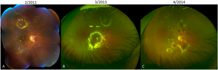
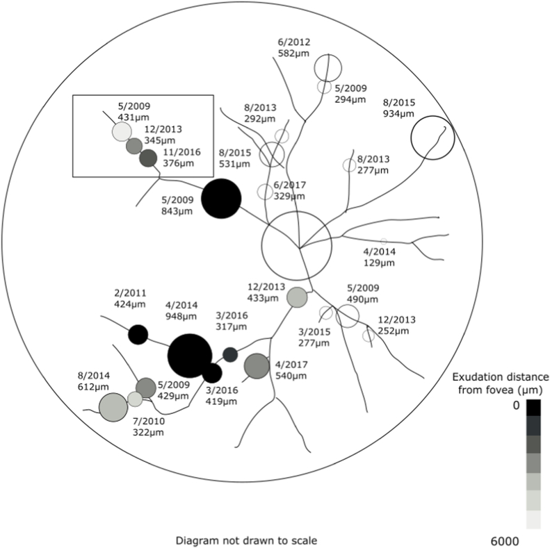
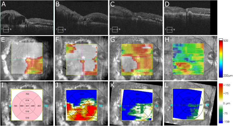
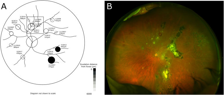
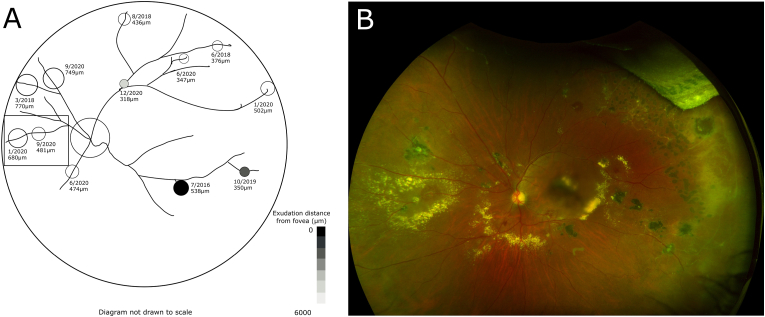
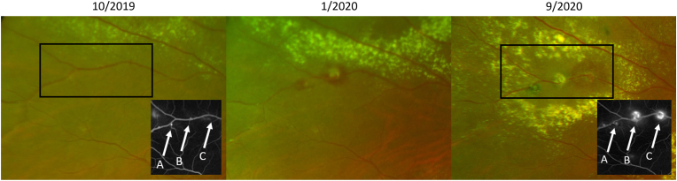
Observations: Three eyes of three patients with adult-onset Coats disease were followed longitudinally for 4-15 years. Ultra-widefield images and montage color fundus photographs of affected eyes were analyzed. Size, retinal location, and grading for predominant characteristic (hemorrhagic, exudative, or quiescent) of each individual macroaneurysm were followed longitudinally from the time of onset. Fifty-one individual retinal arterial macroaneurysms were identified. The distance of any lesion-associated hemorrhage or exudation present from the foveal center was measured. Macroaneurysms were located in all quadrants of the retina, with the majority (37/51) graded as hemorrhagic at lesion onset. Hemorrhagic and exudative macroaneurysms that entered the quiescent phase remained quiescent for an average of 26 months. Seven macroaneurysms were found to have hemorrhage or exudation that came within 125 μm of the fovea and all three eyes followed demonstrated a longitudinal decrease in visual acuity despite laser and intravitreal injection therapy. At the initial visit, visual acuities ranged from 20/40 to 20/200, but decreased to 20/80 to 20/320 by the last follow-up visit.

Conclusion and importance: There are many challenges in treating patients with adult-onset Coats disease. Long-term loss of visual acuity often results from sequelae of hemorrhage and exudation affecting the macula.

Keywords: Adult-onset; Coats disease; Intravitreal anti-vascular endothelial growth factor; Retinal arterial macroaneurysm.


Conflict of interest statement

None; The following authors have no financial disclosures: AD, AT, TD, HW, TK, WW, CC.

Figures

Fig. 1
Fig. 1
Montage color fundus photograph (A) and Optos ultra-widefield retinal images (B and C) showing widespread distribution of macroaneurysms with hemorrhage and/or exudates in all quadrants of the retina and longitudinal progression of activity. In 2011 (A), patient 1 presents with a macroaneurysm in the inferotemporal macula with a small amount of exudation, which is not responsive to focal laser or intravitreal bevacizumab and progresses to a large foveal-involving exudate in 2013 (B). After receiving intravitreal triamcinolone, the exudation is much improved in 2014 (C), but vision is limited by residual subfoveal scarring. (For interpretation of the references to color in this figure legend, the reader is referred to the Web version of this article.)
Fig. 2
Fig. 2
Each macroaneurysm is labeled with its date and diameter at appearance from case 1. Proximal progression of macroaneurysms is shown in the boxed area.
Fig. 3
Fig. 3
Optical coherence tomography scans (OCT; A-D), macular thickness analysis maps (E–H), baseline thickness analysis map from 12/2012 in microns (I), and relative change in macular thickness analysis maps from baseline (J–L) of the right eye of the patient in case 1. In 12/2012 (A and E), there is significant cystoid macular edema (CME). Following 3 injections of intravitreal bevacizumab, there is no improvement of CME in 3/2013 (B,F, and J). One month following a single intravitreal injection of preservative-free triamcinolone 2 mg, CME in 4/2013 is much improved (C, G, and K). With no additional intervention, there is continued response and resolution of CME in 8/2013 (D, H, and L), 5 months following intravitreal triamcinolone. Areas of white in panels E–H correspond to thicknesses greater than 500 μm.
Fig. 4
Fig. 4
(A) Each macroaneurysm is labeled with its date and diameter at appearance from case 2. Proximal progression of macroaneurysms is shown in the boxed area. Ultra-widefield retinal imaging from 3/2020 (B) demonstrates macroaneurysms with active hemorrhage superiorly, scarring within the macula and periphery, and temporal vascular sclerosis.
Fig. 5
Fig. 5
(A) Each macroaneurysm is labeled with its date and diameter at appearance from case 3. Proximal progression of macroaneurysms is shown in the boxed area. An ultra-widefield retinal image from 9/2020 (B) demonstrates multiple macroaneurysms with surrounding exudation and actively hemorrhaging macroaneurysms throughout the retina.
Fig. 6
Fig. 6
Proximal progression of macroaneurysms in case 3. Available fundus fluorescein angiography insets are shown for the regions highlighted by the black rectangles.

Similar articles

Cited by

References

    1. Dalvin L.A., Udyaver S., Lim L.S., et al. Coats disease: clinical features and outcomes by age category in 351 cases. J Pediatr Ophthalmol Strabismus. Sep 1 2019;56(5):288–296. doi: 10.3928/01913913-20190716-01. - DOI - PubMed
    1. Smithen L.M., Brown G.C., Brucker A.J., Yannuzzi L.A., Klais C.M., Spaide R.F. Coats' disease diagnosed in adulthood. Ophthalmology. Jun 2005;112(6):1072–1078. doi: 10.1016/j.ophtha.2004.12.038. - DOI - PubMed
    1. Zhang L., Ke Y., Wang W., Shi X., Hei K., Li X. The efficacy of conbercept or ranibizumab intravitreal injection combined with laser therapy for Coats' disease. Graefes Arch Clin Exp Ophthalmol. Jul 2018;256(7):1339–1346. doi: 10.1007/s00417-018-3949-1. - DOI - PMC - PubMed
    1. Rishi E., Rishi P., Appukuttan B., Uparkar M., Sharma T., Gopal L. Coats' disease of adult-onset in 48 eyes. Indian J Ophthalmol. Jul 2016;64(7):518–523. doi: 10.4103/0301-4738.190141. - DOI - PMC - PubMed
    1. Brockmann C., Lowen J., Schonfeld S., et al. Vascular findings in primarily affected and fellow eyes of middle-aged patients with Coats' disease using multimodal imaging. Br J Ophthalmol. Oct 2021;105(10):1444–1453. doi: 10.1136/bjophthalmol-2020-317101. - DOI - PubMed

Publication types

LinkOut - more resources