Review
. 2022 Jun 10;12(4):20210075.
doi: 10.1098/rsfs.2021.0075. eCollection 2022 Aug 6.

Time-keeping and decision-making in the cell cycle

Affiliations
Review

Time-keeping and decision-making in the cell cycle

John J Tyson et al. Interface Focus. .

Abstract

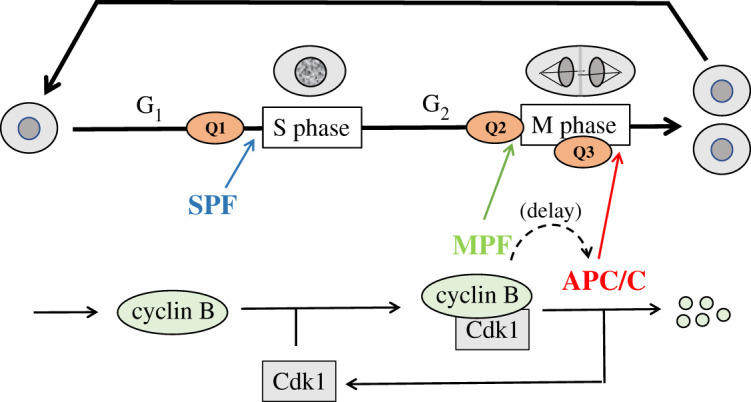
Cell growth, DNA replication, mitosis and division are the fundamental processes by which life is passed on from one generation of eukaryotic cells to the next. The eukaryotic cell cycle is intrinsically a periodic process but not so much a 'clock' as a 'copy machine', making new daughter cells as warranted. Cells growing under ideal conditions divide with clock-like regularity; however, if they are challenged with DNA-damaging agents or mitotic spindle disrupters, they will not progress to the next stage of the cycle until the damage is repaired. These 'decisions' (to exit and re-enter the cell cycle) are essential to maintain the integrity of the genome from generation to generation. A crucial challenge for molecular cell biologists in the 1990s was to unravel the genetic and biochemical mechanisms of cell cycle control in eukaryotes. Central to this effort were biochemical studies of the clock-like regulation of 'mitosis promoting factor' during synchronous mitotic cycles of fertilized frog eggs and genetic studies of the switch-like regulation of 'cyclin-dependent kinases' in yeast cells. In this review, we uncover some secrets of cell cycle regulation by mathematical modelling of increasingly more complex molecular regulatory networks of cell cycle 'clocks' and 'switches'.

Keywords: bistable switches; cell cycle checkpoints; cell cycle regulation; cyclin-dependent kinases; limit cycles; mathematical models.


Conflict of interest statement

We declare we have no competing interests.

Figures

Figure 1.
Figure 1.
The cell cycle. Upper half: in general, the eukaryotic cell cycle is divided into four phases: G1 (unreplicated DNA), S (DNA synthesis), G2 (replicated DNA) and M (mitosis). During S phase, every chromosome is replicated, and during M phase, the ‘sister chromatids’ are pulled to opposite poles of the mitotic spindle, followed by cell division. Progression through the cell cycle is monitored at three checkpoints: Q1 (Is DNA undamaged?), Q2 (Is DNA fully replicated?) and Q3 (Are chromosomes properly aligned on the mitotic spindle?). If the answer to Q1 is yes, then S phase is initiated by its ‘promoting factor’ SPF. If the answer to Q2 is yes, then M phase is initiated by its ‘promoting factor’ MPF. If the answer to Q3 is yes, then anaphase is initiated by its ‘promoting complex’ APC/C. The dynamics of these promoting factors are the subject of this review. Lower half: in the first few hours after fertilization, a frog egg proceeds through rapid mitotic cycles. As cyclin B is synthesized, MPF activity (CycB : Cdk1) rises and initiates mitosis. At the end of mitosis, APC/C is activated, and cyclin B is rapidly degraded after a significant time delay. In the next cycle, when MPF activity is low, SPF drives DNA replication. These early embryonic cycles alternate between S phase and M phase, lacking gap phases and checkpoints.
Figure 2.
Figure 2.
Time-delayed negative feedback loop. (a) Time courses of limit cycle oscillation. Parameter values: table 1; Jph,kin = Jdp,kin = 0.01, Jph,apc = Jdp,apc = 0.1, [PP] = [Kintot] = [APCtot] = 1. (b) Projection of limit cycle oscillation onto a pseudo-phase plane.
Figure 3.
Figure 3.
B55-ENSA-Gwl (BEG) pathway (see inset in (b)). APC/C is activated by a coherent feed-forward loop from MPF directly and indirectly through Gwl, ENSA and B55. In the figures, ‘APC/C’ refers to the active form of the ubiquitin ligase, namely APCP : Cdc20. (a) Time courses of limit cycle oscillation. Parameter values: table 1; Km = 0.0008, [dUb]= 0.75. (b) Projection of limit cycle oscillation (dotted curve) onto a pseudo-phase plane.
Figure 4.
Figure 4.
APC is regulated by a bistable switch. Inset to (b) shows the reaction network. As before, ‘APC/C’ is the variable [APCPC20]. (a) Time courses of limit cycle oscillation. MPF, B55 and APC/C are plotted with respect to the left axis and ENSA with respect to the right axis. Parameter values: table 1; Km = 0.0026, [dUb] = 0.5. (b) Projection of limit cycle oscillation onto a pseudo-phase plane. Notice that APC/C activity is now a bistable function of MPF.
Figure 5.
Figure 5.
APC and MPF are both regulated by bistable switches. (a) The reaction network. MPF + preMPF = CycBT. (b) Time courses of limit cycle oscillation. As before, ‘APC/C’ is the variable [APCPC20]. MPF, B55, CycBT and APC/C are plotted with respect to the left axis and ENSA with respect to the right axis. Parameter values: table 1; Km = 0.0026, [dUb] = 0.5, [CAP] = 0.3. (c) Pseudo-phase plane, [APCPC20] versus [MPF]. Dashed line shows projection of limit cycle oscillation in (b). Notice that MPF activity is now a bistable function of APC/C. (d) Pseudo-phase plane, [CycBT] versus [MPF]. Dashed line shows projection of limit cycle oscillation in (b). Grey region, [MPF] > [CycBT], is ‘unreachable’.
Figure 6.
Figure 6.
Checkpoint signalling. (a) The reaction network. uDNA, unreplicated DNA; uXs, unaligned chromosomes; Cdc20 and Cdh1, alternative targeting subunits of APC/C. (b) G2 checkpoint. Unreplicated DNA raises the peak of the N-shaped, MPF nullcline (green curve) so that it crosses the Z-shaped, CycBT nullcline (orange curve) at a stable steady state (filled circle) of high [CycBT] and low MPF activity (G2 arrest). Parameter values: table 1, Model no. 4; [Cdc25tot] = 0.4. (c) Spindle assembly checkpoint. Unaligned chromosomes inhibit Cdc20, thereby blocking APC/C-mediated degradation of polyubiquitinated CycB and creating a stable steady state (filled circle) of high [CycBT] and high MPF activity (mitotic arrest). Parameter values: table 1, Model no. 4; [Cdc20tot] = 0.1. (d) G1 checkpoint. Damaged DNA stabilizes Cdh1, the subunit that targets APC/C activity to cyclins in G1 phase. Hence, cyclins A and B cannot accumulate, and the cell is blocked at a stable steady state (filled circle) of low [CycBT] and low MPF and SPF activities (G1 arrest). Parameter values: table 1, Model no. 4; kde3,cycb = 0.2.
Figure 7.
Figure 7.
Size-controlled mitotic cycle in fission yeast. (a) Upper panel: time course of cell size, V(t), which increases exponentially (μ = 0.005 min−1, mass doubling time = 140 min) and drops by a factor of 2 at cell division (when [MPF] drops below 0.2). Lower panel: time courses of representative variables. All the variables are read off the left axis, except for ENSA, which is read off the right axis. (b) A one-parameter bifurcation diagram, for [MPF] as a function of V, assuming V is constant in the nonlinear ODEs. Solid red curve, stable steady states (nodes); intermediate dashed grey curve, unstable saddle points; upper dashed grey curve, unstable nodes; open green circles, unstable limit cycles emanating from a Hopf bifurcation at V ≈ 0.05; filled green circles, maxima and minima of stable limit cycle oscillations emanating from a SNIC bifurcation at V ≈ 0.85.

References

    1. Morgan DO. 2007. The cell cycle: principles of control. London, UK: New Science Press.
    1. Murray AW, Kirschner MW. 1989. Dominoes and clocks: the union of two views of the cell cycle. Science 246, 614-621. (10.1126/science.2683077) - DOI - PubMed
    1. Tyson JJ, Novak B. 2008. Temporal organization of the cell cycle. Curr. Biol. 18, R759-R768. (10.1016/j.cub.2008.07.001) - DOI - PMC - PubMed
    1. Nurse P. 1990. Universal control mechanism regulating onset of M-phase. Nature 344, 503-508. (10.1038/344503a0) - DOI - PubMed
    1. Coleman TR, Dunphy WG. 1994. Cdc2 regulatory factors. Curr. Opin. Cell Biol. 6, 877-882. (10.1016/0955-0674(94)90060-4) - DOI - PubMed

LinkOut - more resources