. 2022 Jun 22:26:189-206.
doi: 10.1016/j.omto.2022.06.007. eCollection 2022 Sep 15.

Development of STEAP1 targeting chimeric antigen receptor for adoptive cell therapy against cancer

Affiliations

Development of STEAP1 targeting chimeric antigen receptor for adoptive cell therapy against cancer

Yixin Jin et al. Mol Ther Oncolytics. .

Abstract

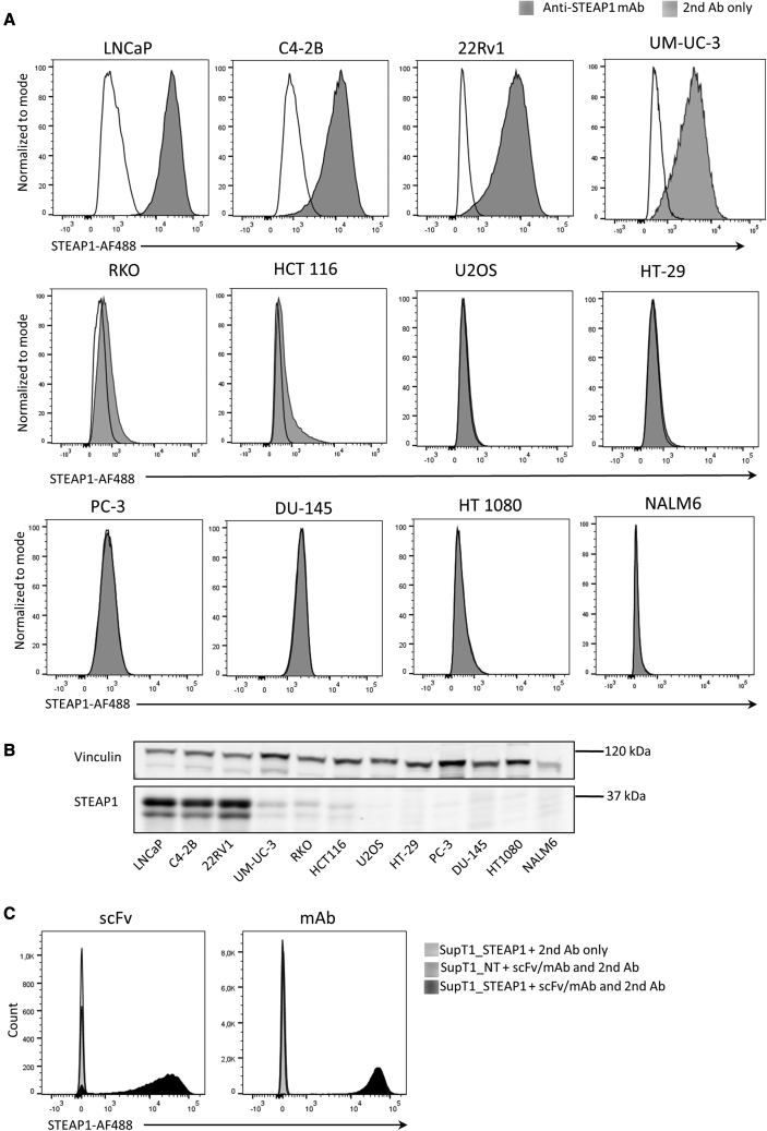
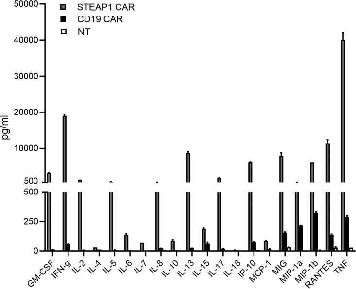
Chimeric antigen receptors (CARs) that retarget T cells against CD19 show clinical efficacy against B cell malignancies. Here, we describe the development of a CAR against the six-transmembrane epithelial antigen of prostate-1 (STEAP1), which is expressed in ∼90% of prostate cancers, and subgroups of other malignancies. STEAP1 is an attractive target, as it is associated with tumor invasiveness and progression and only expressed at low levels in normal tissues, apart from the non-vital prostate gland. We identified the antibody coding sequences from a hybridoma and designed a CAR that is efficiently expressed in primary T cells. The T cells acquired the desired anti-STEAP1 specificity, with a polyfunctional response including production of multiple cytokines, proliferation, and the killing of cancer cells. The response was observed for both CD4+ and CD8+ T cells, and against all STEAP1+ target cell lines tested. We evaluated the in vivo CAR T activity in both subcutaneous and metastatic xenograft mouse models of prostate cancer. Here, the CAR T cells infiltrated tumors and significantly inhibited tumor growth and extended survival in a STEAP1-dependent manner. We conclude that the STEAP1 CAR exhibits potent in vitro and in vivo functionality and can be further developed toward potential clinical use.

Keywords: CAR T cell; STEAP1; cancer immunotherapy; cell therapy; chimeric antigen receptor; metastatic cancer mouse model; prostate cancer.


Conflict of interest statement

J.A.K. and Yixin Jin are inventors on a patent application related to the work described in this article.

Figures

None
Graphical abstract
Figure 1
Figure 1
STEAP1-specific staining by mAb and scFv (A) A selection of six STEAP1-positive (LNCaP, C4-2B, 22Rv1, UM-UC-3, RKO, and HCT 116) and six STEAP1-negative cancer cell lines were stained with the anti-STEAP1 mAb Oslo-1 and a secondary antibody (Ab) (goat anti-mouse IgG AF488), and analyzed by flow cytometry. Gray filled histograms, anti-STEAP1 mAb + secondary (second) Ab; black open histograms, second Ab control. (B) Western blot analysis of STEAP1 expression on cell lines using a different anti-STEAP1 antibody (sc-10262). (C) STEAP1 transduced SupT1 cells (SupT1_STEAP1) and non-transduced SupT1 cells (SupT1_NT) were stained with the Oslo1 scFv (left) or the corresponding mAb (right) and analyzed by flow cytometry. Overlays display SupT1_STEAP1 cells stained with scFv/mAb +second Ab (black filled) or only with the second Ab (black open), or SupT1_NT cells stained with scFv/mAb +second Ab (light gray filled).
Figure 2
Figure 2
STEAP1 CAR design and expression (A) Schematic of STEAP1 CAR from N terminus to C terminus: signal peptide, RQR8, 2A self-cleavage peptide, signal peptide for the CAR, Oslo1 scFv targeting STEAP1, CD8a hinge domain, CD8a transmembrane domain, 4-1BB co-stimulatory domain, and CD3ζ intracellular signaling domain. (B) T cells were surface stained for CD3, CD4, and CD8 (left and middle) and analyzed by flow cytometry. CAR expression was identified by RQR8 staining. Histograms to the right show STEAP1 CAR-transduced (gray filled) CD4+ and CD8+ T cells, overlaid with NT T cells (black open). (C) The expression of the STEAP1 CAR and a CD19-specific CAR with an identical backbone was measured in transduced T cells from 10 healthy donors. Error bars indicate SD.
Figure 3
Figure 3
STEAP1-specific production of IFNγ and TNFα by CAR T cells Flow cytometry analysis of STEAP1 CAR T cells (gray bars), CD19 CAR T cells (black bars), or NT T cells (white bars) cultured with different target cells at an effector-to-target (E:T) ratio of 1:3 for 16 h. (A) IFNγ production. (B) Dot plots of T cells co-cultured with C4-2B cells, showing the applied gating. (C) IFNγ production in CD4+ (left) or CD8+ (right) T cells. (D) TNFα production. (E) Dot plots of T cells co-cultured with C4-2B cells. (F) TNFα production in CD4+ (left) or CD8+ (right) T cells. (G) Percentage of individual T cells producing both TNFα and IFNγ. The T cells were gated on RQR8+ (CAR expression), or CD3+ for the NT cells. 22Rv1, LNCaP (LN), and C4-2B are STEAP1+ prostate cancer lines. NALM6 is a STEAP1 CD19+ leukemia cell line. LNCaP and C4-2B variants with shRNA knocking down STEAP1 (shRNA1, shRNA2) or control shRNA (shGFP) were used as indicated. Data are mean of triplicates, with error bars representing SEM. The percentage of positive cells recorded for the STEAP1 CAR versus the CD19 CAR was compared by multiple t tests. Statistical significance was determined using the Holm-Sidak method, adjusting for multiple comparisons. Adjusted p values are indicated (∗p < 0.05, ∗∗p < 0.01, ∗∗∗p < 0.001, ∗∗∗∗p < 0.0001). The results are representative of two individual experiments.
Figure 4
Figure 4
Cytokine profile of T cells co-cultured with STEAP1+ 22Rv1 cells STEAP1 CAR, CD19 CAR, and NT T cells were co-cultured with 22Rv1 cells for 48 h at an E:T ratio of 1:1 and the cell culture supernatant harvested. Twenty different cytokines and chemokines were quantified using the Bio-Plex Multiplex Immunoassay system. Error bars represent SEM from duplicate cultures, each parallel kept separate through T cell stimulation and Bio-Plex assays.
Figure 5
Figure 5
STEAP1 CAR T cells specifically induce apoptosis in STEAP1+ target cells STEAP1 CAR T cells, CD19 CAR T cells, or NT T cells were cultured in triplicates for 16 h with various target cells. Apoptosis of target cells was measured by analyzing the intensity of FITC-DEVD-FMK bound to active caspase-3 by flow cytometry. Target cells cultured alone were included as controls to indicate the baseline level of active caspase-3 in each cell line. (A) Representative contour plots of 22Rv1 cells after co-culture with effector cells, showing gating strategy for identification of cells positive for active caspase-3. (B) Percentage of apoptotic 22Rv1 cells, after co-culture with effector T cells at different E:T ratios, as indicated. (C) Percentage of apoptotic cells among STEAP1-positive and STEAP1-knockdown targets, after co-culture with effector T cells (E:T ratio of 3:1). (D) Percentage of apoptotic cells among STEAP1-positive (22Rv1), STEAP1-negative (PC-3, DU-145 and HT29), or low-level STEAP1 expression (RKO) cell lines, after co-culture with effector T cells (E:T ratio of 3:1). Error bars represent SEM from triplicates. ∗p < 0.05, ∗∗p < 0.01, ∗∗∗p < 0.001. The data are representative for two independent experiments.
Figure 6
Figure 6
STEAP1 CAR T cells inhibit tumor growth in vivo NSG mice were engrafted with 2 × 10⁶ luciferase-expressing 22Rv1 prostate cancer cells subcutaneously on the hind leg. On day 9 and 14, the mice were treated with 1 × 10⁷ STEAP1 CAR T cells (N = 12), CD19 CAR T cells (N = 11), or NT T cells (N = 9) by i.v. injection, and 100 IU/g body weight rhIL-2 was given intraperitoneally twice a week. (A) Tumor growth as measured once a week by bioluminescence IVIS imaging. The data are presented as mean ± SEM in each group of mice. (B) Bioluminescence signals (mean ± SEM) 34 days after tumor engraftment. (C) Tumor size (mean ± SEM) 33 days after tumor engraftment. The length, width, and depth of the tumors were measured with a caliper. Statistical analyses in (B) and (C) were performed with Mann-Whitney U test. (D) Bioluminescence signals for each individual mouse at day 34. (E) Kaplan-Meier plot showing extended survival for the mice treated with the STEAP1 CAR T cells (red line), compared with CD19 CAR T cells (black line) or NT T cells (dotted line). The mice were euthanized when tumors reached 1,000 mm3. Statistical analysis was performed using the log rank test. (E) The body weight of the mice was measured once per week after the first treatment with T cells at day 9. Error bars indicate SEM.
Figure 7
Figure 7
STEAP1 CAR T cells expanded in vivo and infiltrated STEAP1+ tumors NSG mice were engrafted subcutaneously with 2 × 106 luciferase-expressing 22Rv1 prostate cancer cells subcutaneously on the hind leg. On day 10 and 17, the mice were treated i.v. with 1 × 107 STEAP1 CAR T cells (N = 3), CD19 CAR T cells (N = 2), or NT T cells (N = 3), and 100 IU/g body weight rhIL-2 was given intraperitoneally twice a week. Mice were sacrificed 39 days after tumor engraftment. (A) Tumor burden assessed by bioluminescence imaging (mean ± SEM) in the eight mice 38 days after tumor engraftment. (B) Peripheral blood cells were measured by flow cytometry at day 12, 14, 31, and 39, and quantified by BD Trucount beads. Left: T cells, identified by anti-human CD3. Right: CAR-expressing T cells, identified by the RQR8-specific mAb QBen10. (C and D) Tumors were fixed, paraffin-embedded, and stained for IHC with anti-huCD3 (T cells) or QBen10 (CAR/RQR8-expressing cells). (C) aCD3 and (D) aRQR8, showing representative staining from each group of mice, with 10× and 40× magnification, as indicated. For the STEAP1 CAR group, tumors from two out of three mice could be stained; the third tumor had completely regressed. T cell infiltration (C) was only observed in the STEAP1 CAR T cell-treated tumors, and corresponded to CAR T cell infiltration (D).
Figure 8
Figure 8
CAR T cells inhibit tumor growth in metastatic in vivo model in a STEAP1-dependent manner NSG mice were injected i.v. into the tail vein with 10 × 106 luciferase-expressing 22Rv1 wild-type cells, or with 22Rv1 STEAP1 knockout cells (C11). On day 19 and 26, the mice were treated i.v. with 10 × 106 STEAP1 CAR T cells (N = 5), or CD19 CAR T cells (N = 5), and 100 IU/g body weight rhIL-2 was given intraperitoneally twice a week. (A) Tumor growth was measured once or twice a week by bioluminescence IVIS imaging. The data are presented as mean ± SEM in each group of mice. (B) Bioluminescence signals 33 days after tumor injection. The signal for each mouse is indicated. Statistical analyses were performed using Mann-Whitney U test. (C) Kaplan-Meier plot showing extended survival for mice injected with wild-type 2Rv1 and treated with STEAP1 CAR T cells (red line), compared with CD19 CAR T cells (black line), and to mice injected with 22Rv1 knockout cells (C11; dotted lines). Statistical analysis was performed with log rank test. The mice were euthanized when required by animal welfare guidelines, which generally corresponded to a total photon signal of 1 × 10¹⁰ (p/s). (D and E) Tumors were fixed, paraffin-embedded and stained for IHC with anti-huCD3 (T cells) or QBen10 (CAR/RQR8-expressing cells). (D) aCD3 and (E) aRQR8, showing a representative staining from each group of mice, with 10× magnification. T cell infiltration (D) was only observed in the STEAP1 CAR T cell-treated tumors and corresponded to CAR T cell infiltration (E).

Similar articles

Cited by

References

    1. Grupp S.A., Kalos M., Barrett D., Aplenc R., Porter D.L., Rheingold S.R., Teachey D.T., Chew A., Hauck B., Wright J.F., et al. Chimeric antigen receptor-modified T cells for acute lymphoid leukemia. N. Engl. J. Med. 2013;368:1509–1518. - PMC - PubMed
    1. Brentjens R.J., Davila M.L., Riviere I., Park J., Wang X., Cowell L.G., Bartido S., Stefanski J., Taylor C., Olszewska M., et al. CD19-targeted T cells rapidly induce molecular remissions in adults with chemotherapy-refractory acute lymphoblastic leukemia. Sci. Transl. Med. 2013;5:177ra38. doi: 10.1126/scitranslmed.3005930. - DOI - PMC - PubMed
    1. Sadelain M., Brentjens R., Rivière I. The basic principles of chimeric antigen receptor design. Cancer Discov. 2013;3:388–398. - PMC - PubMed
    1. Philipson B.I., O'Connor R.S., May M.J., June C.H., Albelda S.M., Milone M.C. 4-1BB costimulation promotes CAR T cell survival through noncanonical NF-κB signaling. Sci. Signal. 2020;13 doi: 10.1126/scisignal.aay8248. - DOI - PMC - PubMed
    1. Weber E.W., Maus M.V., Mackall C.L. The emerging landscape of immune cell therapies. Cell. 2020;181:46–62. - PMC - PubMed