

The basic anatomy of the medial sural artery perforator flaps in Vietnamese adults
- PMID: 35860090
- PMCID: PMC9289320
- DOI: 10.1016/j.amsu.2022.103996
The basic anatomy of the medial sural artery perforator flaps in Vietnamese adults
Abstract
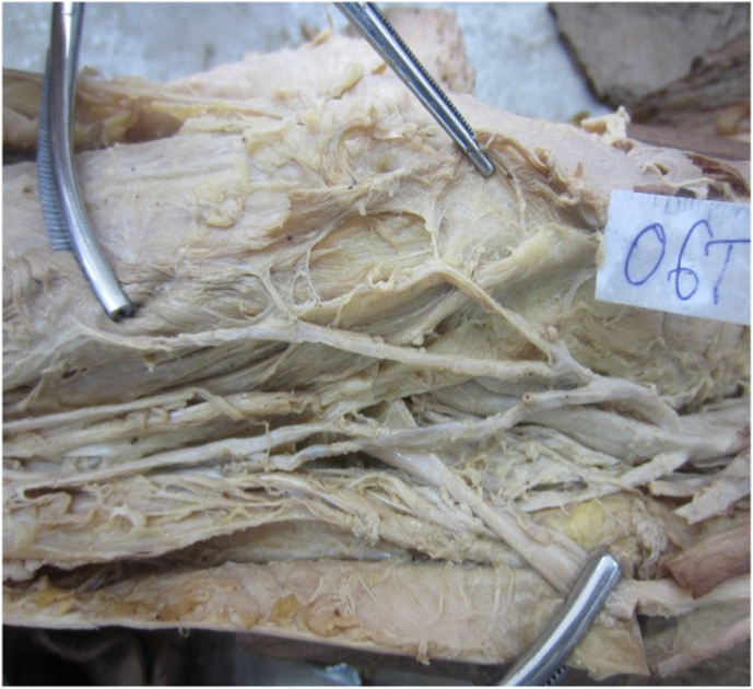
Background: Both free medial sural artery perforator flaps and pedicled medial sural artery perforator flaps have been being effectively used in treatment of body defects especially in head and neck region by plastic surgeons worldwide. However, there is a lack of comprehensive studies on the anatomy of perforating artery branches in Vietnam. This study aims to describe anatomical vascular pedicles of medial sural artery perforator flap in Vietnamese adults .
Methods: A descriptive cross-sectional study, dissected 62 lower limbs of 41 Vietnamese adult cadavers preserved by formalin in Department of Anatomy, Hanoi Medical University and Ho Chi Minh Medicine and Pharmacy University.
Results: Origin of medial sural artery was branched constantly from popliteal artery. Common stem of artery was 8.39 ± 3.5 cm in mean length. The diameter of common stem, which was measured from origin, was 2.88 ± 0.98 mm averagely. The common stem of artery, which did not have any branch (15%), divided in to 2 branches (15%), 3 branches (30%), 4 branches (40%) before entering muscle. Medial sural artery had 1 to 5 branches perforating to the skin. The distance from perforating branch to the knee joint (popliteal crease) was 10.12 ± 3.7 cm, the distance from perforator branch to middle posterior leg was 1.6 ± 0.96 cm.
Conclusions: The medial sural artery constantly originates from popliteal artery, supplies blood for medial gastrocnemius muscle. The skin area covering this muscle is nourished by one of five perforators of the medial sural artery. The perforating flaps can be created using medial sural artery perforating branches.
Keywords: Medial sural artery; Perforating branche; Perforator flap; Sural.
© 2022 The Authors.
Conflict of interest statement
No potential conflict of interest relevant to this article was reported.
Figures


References
-
- Koshima I., Soeda S. Inferior epigastric artery skin Ơflaps without rectus abdominis muscle. Br. J. Plast. Surg. 1989;42(6):645–648. - PubMed
-
- Cavadas P.C., Juan R., Rico S.G., et al. The medial sural artery perforator free flap. Plast. Reconstr. Surg. 2001;108(6):1609–1615. - PubMed
-
- Hallock G.G. Anatomic basis of the gastrocnemius perforator-based flap. Ann. Plast. Surg. 2001;47(5):517–522. - PubMed
-
- Thione A., Valdatta L., Buoro M., et al. The medial sural artery perforators: anatomic basis for a surgical plan. Ann. Plast. Surg. 2004;53(3):250–255. - PubMed
-
- Okamoto H., Sekiya I., Mizutani J., Otsuka T. Anatomical basis of the medial sural artery perforator flap in Asians. Scand. J. Plast. ReConstr. Surg. Hand Surg. 2007;41(3):125–129. - PubMed
LinkOut - more resources
Full Text Sources
