Observational Study
. 2022 Jul 4:13:940030.
doi: 10.3389/fimmu.2022.940030. eCollection 2022.

Comparison of Rapid Cytokine Immunoassays for Functional Immune Phenotyping

Affiliations
Observational Study

Comparison of Rapid Cytokine Immunoassays for Functional Immune Phenotyping

Anthony S Bonavia et al. Front Immunol. .

Abstract

Background: Cell-based functional immune-assays may allow for risk stratification of patients with complex, heterogeneous immune disorders such as sepsis. Given the heterogeneity of patient responses and the uncertain immune pathogenesis of sepsis, these assays must first be defined and calibrated in the healthy population.

Objective: Our objective was to compare the internal consistency and practicality of two immune assays that may provide data on surrogate markers of the innate and adaptive immune response. We hypothesized that a rapid turnaround, microfluidic-based immune assay (ELLA) would be comparable to a dual-color, enzyme-linked immunospot (ELISpot) assay in identifying tumor necrosis factor (TNF) and interferon (IFN)γ production following ex vivo whole blood stimulation.

Design: This was a prospective, observational cohort analysis. Whole blood samples from ten healthy, immune-competent volunteers were stimulated for either 4 hours or 18 hours with lipopolysaccharide, anti-CD3/anti-CD28 antibodies, or phorbol 12-myristate 13-acetate with ionomycin to interrogate innate and adaptive immune responses, respectively.

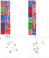
Measurements and main results: ELLA analysis produced more precise measurement of TNF and IFNγ concentrations as compared with ELISpot, as well as a four- to five-log10 dynamic range for TNF and IFNγ concentrations, as compared with a two-log10 dynamic range with ELISpot. Unsupervised clustering accurately predicted the ex vivo immune stimulant used for 90% of samples analyzed via ELLA, as compared with 72% of samples analyzed via ELISpot.

Conclusions: We describe, for the first time, a rapid and precise assay for functional interrogation of the innate and adaptive arms of the immune system in healthy volunteers. The advantages of the ELLA microfluidic platform may represent a step forward in generating a point-of-care test with clinical utility, for identifying deranged immune phenotypes in septic patients.

Keywords: cytokine; immune phenotype; immunoassay; interferon gamma; tumor necrosis factor.


Conflict of interest statement

The authors declare that the research was conducted in the absence of any commercial or financial relationships that could be construed as a potential conflict of interest.

Figures

Figure 1
Figure 1
Intra-assay variability of ELISpot immunoassay, assessed by percentage coefficient of variation (CV) for: (A) Tumor Necrosis Factor (TNF), (B) Interferon (IFN)γ, and (C) dual-staining cells (both IFNγ and TNF). (D) TNF, (E) IFNγ, and (F) dual-cytokine production following ex vivo stimulation with various immune stimulants (x-axis) for 4 hours or 18 hours. N = 2 for each measured analyte. 18 hour ELISpot data includes response to LPS and anti-CD3/anti-CD28 only, due to cell over-stimulation with PMA. SFU, spot forming units.
Figure 2
Figure 2
Intra-assay variability of ELLA microfluidic immunoassay, assessed by percentage coefficient of variation (CV) for: (A) Tumor Necrosis Factor (TNF), (B) Interferon (IFN)γ, and (C) Interleukin (IL)-6. (D) TNF, (E) IFNγ, and (F) IL-6 production following ex vivo stimulation with various immune stimulants (x-axis) for 4 hours or 18 hours. N = 3 for each measured analyte.
Figure 3
Figure 3
Correlation between cytokine concentrations, as measured by ELLA microfluidic immunoassay (x-axis), and different parameters measured by ELISpot assay at 4h post-ex vivo stimulation. Measured plasma cytokine concentration of Interferon (IFN) γ versus corresponding ELISpot spot forming units (SFU), Total Well Intensity (TWI) and Mean Spot Intensity (MSI) at (A) 4 hours and (B) 18 hours of stimulation. Measured plasma cytokine concentration of Tumor Necrosis Factor (TNF) versus corresponding ELISpot spot forming units (SFU), Total Well Intensity (TWI) and Mean Spot Intensity (MSI) at (C) 4 hours and (D) 18 hours of stimulation.
Figure 4
Figure 4
Unsupervised, hierarchical cluster analysis using Ward’s minimum variance method, to predict ex vivo stimulant by using ELLA versus ELISpot data from 10 healthy volunteers. (A) Dendrogram with heat map demonstrating hierarchical clustering by using ELISpot-derived SFU, log2-TWI, MSI and MSS, (B) Constellation plot showing data derived from Figure 4B , (C) Dendrogram with heat map demonstrating hierarchical clustering by using ELLA-derived, log2-transformed, plasma cytokine concentrations for Interferon (IFN)γ, Tumor Necrosis Factor (TNF), and interleukin (IL)-6, (D) Constellation plot showing data derived from Figure 4C .

References

    1. Singer M, Deutschman CS, Seymour CW, Shankar-Hari M, Annane D, Bauer M, et al. . The Third International Consensus Definitions for Sepsis and Septic Shock (Sepsis-3). JAMA (2016) 315(8):801–10. doi: 10.1001/jama.2016.0287 - DOI - PMC - PubMed
    1. Sweeney TE, Azad TD, Donato M, Haynes WA, Perumal TM, Henao R, et al. . Unsupervised Analysis of Transcriptomics in Bacterial Sepsis Across Multiple Datasets Reveals Three Robust Clusters. Crit Care Med (2018) 46(6):915–25. doi: 10.1097/CCM.0000000000003084 - DOI - PMC - PubMed
    1. Sweeney TE, Perumal TM, Henao R, Nichols M, Howrylak JA, Choi AM, et al. . A Community Approach to Mortality Prediction in Sepsis Via Gene Expression Analysis. Nat Commun (2018) 9(1):694. doi: 10.1038/s41467-018-03078-2 - DOI - PMC - PubMed
    1. Jacobs L, Berrens Z, Stenson EK, Zackoff MW, Danziger LA, Lahni P, et al. . The Pediatric Sepsis Biomarker Risk Model (Persevere) Biomarkers Predict Clinical Deterioration and Mortality in Immunocompromised Children Evaluated for Infection. Sci Rep (2019) 9(1):424. doi: 10.1038/s41598-018-36743-z - DOI - PMC - PubMed
    1. Wong HR, Cvijanovich NZ, Anas N, Allen GL, Thomas NJ, Bigham MT, et al. . Improved Risk Stratification in Pediatric Septic Shock Using Both Protein and Mrna Biomarkers. Persevere-Xp. Am J Respir Crit Care Med (2017) 196(4):494–501. doi: 10.1164/rccm.201701-0066OC - DOI - PMC - PubMed

Publication types