. 2022 Jul 4:16:905403.
doi: 10.3389/fnins.2022.905403. eCollection 2022.

Ceftriaxone Suppresses Group II Metabotropic Glutamate Receptor Expression Contributing to Reversal of Recognition Memory Deficits of Amyloid Precursor Protein/Presenilin 1 AD Mice

Affiliations

Ceftriaxone Suppresses Group II Metabotropic Glutamate Receptor Expression Contributing to Reversal of Recognition Memory Deficits of Amyloid Precursor Protein/Presenilin 1 AD Mice

ShuJuan Fan et al. Front Neurosci. .

Abstract

Group II metabotropic glutamate receptors (Group II mGluRs) are the peri-synaptic receptor of glutamatergic neurons and negatively regulate glutamate release from presynaptic neurons. Glutamate in the synaptic cleft is mainly taken into astrocytes by glutamate transporter-1 (GLT-1), which is primarily expressed in astrocytes. Increasing evidence showed that inhibiting or suppressing the activation of Group II mGluRs would contribute to the improvement of learning and memory deficits in Alzheimer's disease (AD) animal models. Ceftriaxone (Cef) has been reported to alleviate the spatial memory deficits in AD model mice by improving GLT-1-related clearance and metabolism of glutamate. Therefore, the present study further investigates the improving effect of Cef on recognition memory deficits and the involvement of Group II mGluRs in the process using the APP/PS1 AD mouse model. Novel object recognition tests showed that the Cef treatment significantly improved the recognition memory deficits of the AD mice. The Western blot and immunohistochemistry analysis showed that the Cef treatment significantly suppressed the upregulation of Group II mGluRs expression in APP/PS1 AD mice. The above suppression effect of Cef was blocked by dihydrokainic acid, an inhibitor of GLT-1 uptake activity. Furthermore, the Cef treatment significantly restored the downregulation in the downstream molecules of Group II mGluRs activation, including the expression of PKA and phosphorylated SNAP-25 in the APP/PS1 AD mice. The Cef treatment had no effect on the content of Aβ40 and Aβ42 in the hippocampus of APP/PS1 AD mice. The above results suggested that the suppression of Group II mGluRs contributed to the Cef-induced reversal of the recognition memory deficits in APP/PS1 AD mice.

Keywords: APP/PS1 mice; GLT-1; Group II mGluRs; SNAP-25; ceftriaxone.


Conflict of interest statement

The authors declare that the research was conducted in the absence of any commercial or financial relationships that could be construed as a potential conflict of interest.

Figures

FIGURE 1
FIGURE 1
Cef improves recognition memory deficits in 6-month and 7-month-old APP/PS1 mice. (A) Shows the protocol of novel object recognition test. (B) Shows the recognition index of the tests in 6-month and 7-month-old APP/PS1 mice in each group. The number of mice for each group are shown in Table 1. WT, wild type; Cef, ceftriaxone. The italic values upon the bars are statistical P values.
FIGURE 2
FIGURE 2
Cef suppresses the expression of mGluR2 in protein level in the hippocampus of 6 month-old APP/PS1 mice. (A) Is an mGluR2 band to indicate the expression of dimeric mGluR2 (230 kD). (B) Is a representative immunoblot of Western blot in each group, and (C) is the quantitative presentation of the immunoblot with integral optical density (IOD). (D–F) Are representative photomicrographs of immunohistochemistry staining in each group and the scale bar on them is 20 μm. (G) Is a western blot band of mGluR2 to indicate the immunohistochemistry stain is specific for mGluR2. (H) Is the quantitative presentation of the immunohistochemistry staining with mean optical density (MOD) in each group. (I–K) Are representative photomicrographs of double immunofluorescent labeling staining of mGluR2 and synapsin in WT mice. The number of mice for each test is shown in Table 1. mGluR2, metabotropic glutamate receptor 2. The italic values upon the bars are statistical P values.
FIGURE 3
FIGURE 3
DHK blocks Cef-induced improvement on recognition memory deficits and suppression on the mGluR2 protein expression in APP/PS1 mice. The histograms of (A,B) show the recognition index in test 1 (A) and test 2 (B) of the novel object recognition test. (C) Shows the expression of mGluR2 assayed by the Western blot analysis. The upper is a representative immunoblot and the lower is the quantitative presentation of the immunoblot with IOD. The number of mice for each test is shown in Table 1. The italic values upon the bars are statistical P values.
FIGURE 4
FIGURE 4
Cef upregulates the expression of PKA and phosphorylated SNAP-25 in the hippocampus of APP/PS1 mice. The upper in each of (A–C) is a representative immunoblot of Western blot and analysis, and the lower is the quantitative presentation of the immunoblot with IOD. The number of mice for each test is shown in Table 1. PKA, protein kinase A; P-SNAP25, phosphorylated synaptosomal-associated protein 25 kDa; T-SNAP25, total synaptosomal-associated protein 25 kDa. The italic values upon the bars are statistical P values.
FIGURE 5
FIGURE 5
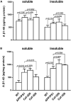
Cef has no effect on Aβ levels in the hippocampus of APP/PS1 mice. (A) Is the assay of soluble and insoluble Aβ1–40 levels in each group, and (B) is the soluble and insoluble Aβ1–42 levels. The number of mice for each test is shown in Table 1. The italic values upon the bars are statistical P values.

Similar articles

Cited by

References

    1. Ali I., Boets S., Janssens P., Van Eetveldt A., Amhaoul H., Langlois X., et al. (2016). Metabotropic glutamate receptor 2/3 density and its relation to the hippocampal neuropathology in a model of temporal lobe epilepsy in rats. Epilepsy Res. 127 55–59. 10.1016/j.eplepsyres.2016.08.010 - DOI - PubMed
    1. Anderson C. M., Swanson R. A. (2000). Astrocyte glutamate transport: review of properties, regulation, and physiological functions. Glia 32 1–14. - PubMed
    1. Arnsten A. F., Wang M. J., Paspalas C. D. (2012). Neuromodulation of thought: flexibilities and vulnerabilities in prefrontal cortical network synapses. Neuron 76 223–239. 10.1016/j.neuron.2012.08.038 - DOI - PMC - PubMed
    1. Azar O., Marcello M., Michele B., Victoria S., Tomomi A., Kohishi T., et al. (2009). Up-regulation of GLT-1 severely impairs LTD at mossy fibre–CA3 synapses. J. Physiol. 587(Pt 19) 4575–4588. 10.1113/jphysiol.2009.177881 - DOI - PMC - PubMed
    1. Bratek E., Ziembowicz A., Salinska E. (2018). Pretreatment with Group II metabotropic glutamate receptor agonist LY379268 protects neonatal rat brains from oxidative stress in an experimental model of birth asphyxia. Brain Sci. 8:48. 10.3390/brainsci8030048 - DOI - PMC - PubMed

LinkOut - more resources