

Development of a nonhuman primate model for mammalian bornavirus infection
- PMID: 35860599
- PMCID: PMC9291224
- DOI: 10.1093/pnasnexus/pgac073
Development of a nonhuman primate model for mammalian bornavirus infection
Abstract
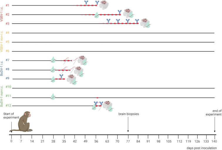
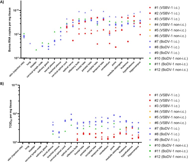
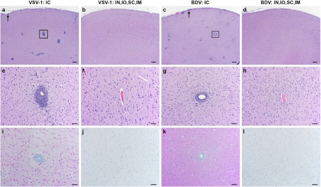
Until recently, it was assumed that members of the family Bornaviridae could not induce severe disease in humans. Today, however, Borna disease virus 1 (BoDV-1), as well as the more recently emerged variegated squirrel bornavirus 1 (VSBV-1), are known as causative agents of lethal encephalitis in humans. In order to establish animal models reflecting the pathogenesis in humans and for countermeasure efficacy testing, we infected twelve rhesus macaques (Macaca mulatta) either with VSBV-1 or with BoDV-1. For each virus, three monkeys each were inoculated with 2 × 10⁴ focus forming units by the intracerebral route or by multiple peripheral routes (intranasal, conjunctival, intramuscular, and subcutaneous; same dose in total). All BoDV-1 and VSBV-1 intracerebrally infected monkeys developed severe neurological signs around 5 to 6 or 8 to 12 weeks postinfection, respectively. Focal myoclonus and tremors were the most prominent observations in BoDV-1 and VSBV-1-infected animals. VSBV-1-infected animals also showed behavioral changes. Only one BoDV-1 peripherally infected animal developed similar disease manifestations. All animals with severe clinical disease showed high viral loads in brain tissues and displayed perivascular mononuclear cuffs with a predominance of lymphocytes and similar meningeal inflammatory infiltrates. In summary, rhesus macaques intracerebrally infected with mammalian bornaviruses develop a human-like disease and may serve as surrogate models for human bornavirus infection.
Keywords: animal model; bornavirus; nonhuman primate.
© The Author(s) 2022. Published by Oxford University Press on behalf of National Academy of Sciences.
Figures
References
-
- Hoffmann B., et al. . 2015. A variegated squirrel bornavirus associated with fatal human encephalitis. N Engl J Med. 373:154–162. - PubMed
-
- Schlottau K., et al. . 2017. Multiple detection of zoonotic variegated squirrel bornavirus 1 RNA in different squirrel species suggests a possible unknown origin for the virus. Arch Virol. 162:2747–2754.. doi: 10.1007/s00705-017-3432-z. - PubMed
LinkOut - more resources
Full Text Sources
