Case Reports
. 2022 Jul 4:9:912103.
doi: 10.3389/fmed.2022.912103. eCollection 2022.

Varying Clinical Phenotypes of Mitochondrial DNA T12811C Mutation: A Case Series Report

Affiliations
Case Reports

Varying Clinical Phenotypes of Mitochondrial DNA T12811C Mutation: A Case Series Report

Qingdan Xu et al. Front Med (Lausanne). .

Abstract

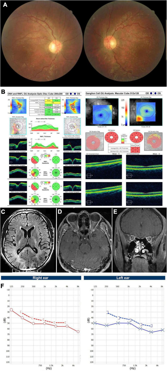
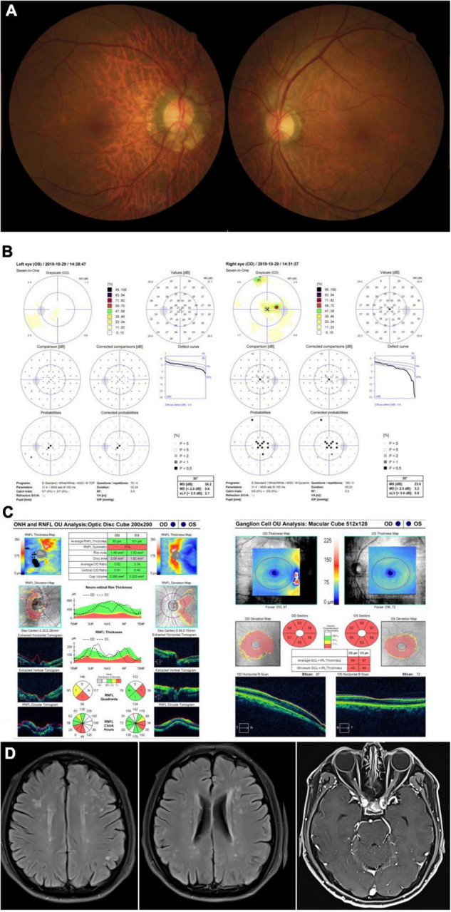
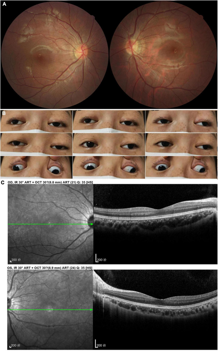
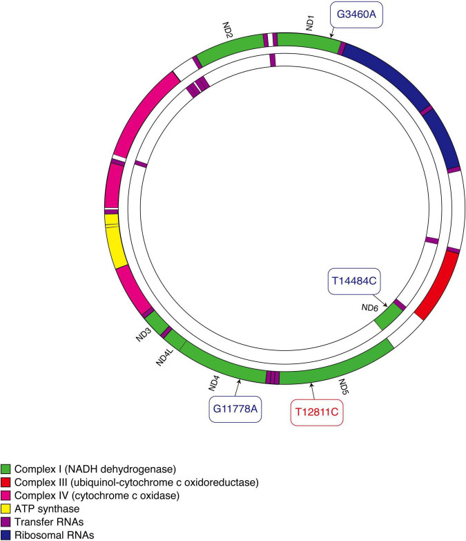
The T12811C mitochondrial DNA (mtDNA) mutation has been reported in Leber hereditary optic neuropathy (LHON) previously, with vision loss as the main manifestation. The involvement of other organ systems, including the central and peripheral nervous system, heart, and extraocular muscles, has not been well described. This case series report investigated four patients with T12811C mtDNA mutation, verified through a next generation sequencing. Two male patients presented with bilateral subacute visual decrease combined with involvement of multiple organ systems: leukoencephalopathy, hypertrophic cardiomyopathy, neurosensory deafness, spinal cord lesion and peripheral neuropathies. Two female patients presented with progressive ptosis and ophthalmoplegia, one of whom also manifested optic atrophy. This study found out that patients harboring T12811C mtDNA mutation manifested not only as vision loss, but also as a multi-system disorder affecting the nervous system, heart, and extraocular muscles.

Keywords: Leber hereditary optic neuropathy; mitochondrial DNA; mitochondrial disorder; ophthalmoplegia; optic atrophy.


Conflict of interest statement

The authors declare that the research was conducted in the absence of any commercial or financial relationships that could be construed as a potential conflict of interest.

Figures

FIGURE 1
FIGURE 1
Accessory examinations of case 1. (A) Fundus photos showed diffuse pallor in the right eye and temporal pallor in the left eye, with tortuous retinal vein bilaterally. (B) OCT showed thinning of inferior-temporal RNFL in the right eye and severe GCIPL loss bilaterally. (C) Axial T2 Flair-weighted MRI of the brain showed hyperintense signal changes involving the posterior limbs of internal capsule, occipital lobes and corpus callosum indicating leukoencephalopathy. (D) Axial and (E) coronal T1-weighted MRI of orbits with gadolinium and fat suppression showed bilateral optic nerve slight enhancement. (F) Audiography showed bilateral neurosensory deafness.
FIGURE 2
FIGURE 2
Accessory examinations of case 2. (A) Fundus photos showed temporal pallor of optic disc bilaterally. (B) Octopus visual fields showed central scotomas bilaterally. (C) OCT revealed inferior-temporal RNFL thinning in the right eye and temporal RNFL thinning in the left eye, with significant thinning of the GPICL bilaterally. (D) Brain MRI showed scattered T2 Flair-weighted hyperintense signal and orbital T1-weighted MRI imaging after contrast showed bilateral optic nerve atrophy without enhancement.
FIGURE 3
FIGURE 3
Accessory examinations of case 3. (A) Fundus photos showed temporal pallor in the right eye. (B) Nine cardinal eye positions showed impaired mobility in both eyes, including adduction, abduction, elevation, and depression. (C) OCT showed thinning of RNFL superiorly and inferiorly in the right eye, with diffuse thinning of GCIPL bilaterally. The RNFL thickness of the left eye was not available due to poor cooperation.
FIGURE 4
FIGURE 4
Accessory examinations of case 4. (A) Fundus photos showed normal optic disc and retina bilaterally. (B) Nine cardinal eye positions revealed adduction, elevation, and depression were moderately reduced bilaterally, and abduction was slightly reduced in the right eye and almost full in the left eye. (C) OCT showed normal retina thickness and macular structure bilaterally.
FIGURE 5
FIGURE 5
Schematic diagram of mitochondrial DNA. Three primary mutations causing LHON are shown in blue. The mutation that our patients carried is shown in red.

Similar articles

Cited by

References

    1. Newman NJ, Wallace DC. Mitochondria and Leber’s hereditary optic neuropathy. Am J Ophthalmol. (1990) 109:726–30. - PubMed
    1. Yu-Wai-Man P, Griffiths PG, Hudson G, Chinnery PF. Inherited mitochondrial optic neuropathies. J Med Genet. (2009) 46:145–58. 10.1136/jmg.2007.054270 - DOI - PMC - PubMed
    1. Carelli V, La Morgia C, Valentino ML, Barboni P, Ross-Cisneros FN, Sadun AA. Retinal ganglion cell neurodegeneration in mitochondrial inherited disorders. Biochim Biophys Acta. (2009) 1787:518–28. 10.1016/j.bbabio.2009.02.024 - DOI - PubMed
    1. Man PYW, Turnbull DM, Chinnery PF. Leber hereditary optic neuropathy. J Med Genet. (2002) 39: 162–9. 10.1136/jmg.39.3.162 - DOI - PMC - PubMed
    1. Sadun AA, La Morgia C, Carelli V. Leber’s hereditary optic neuropathy. Curr Treat Option Neurol. (2011) 13:109–17. 10.1007/s11940-010-0100-y - DOI - PubMed

Publication types

LinkOut - more resources