. 2022 Sep 28;190(2):1474-1489.
doi: 10.1093/plphys/kiac334.

A secreted fungal subtilase interferes with rice immunity via degradation of SUPPRESSOR OF G2 ALLELE OF skp1

Affiliations

A secreted fungal subtilase interferes with rice immunity via degradation of SUPPRESSOR OF G2 ALLELE OF skp1

Xiaoyang Chen et al. Plant Physiol. .

Abstract

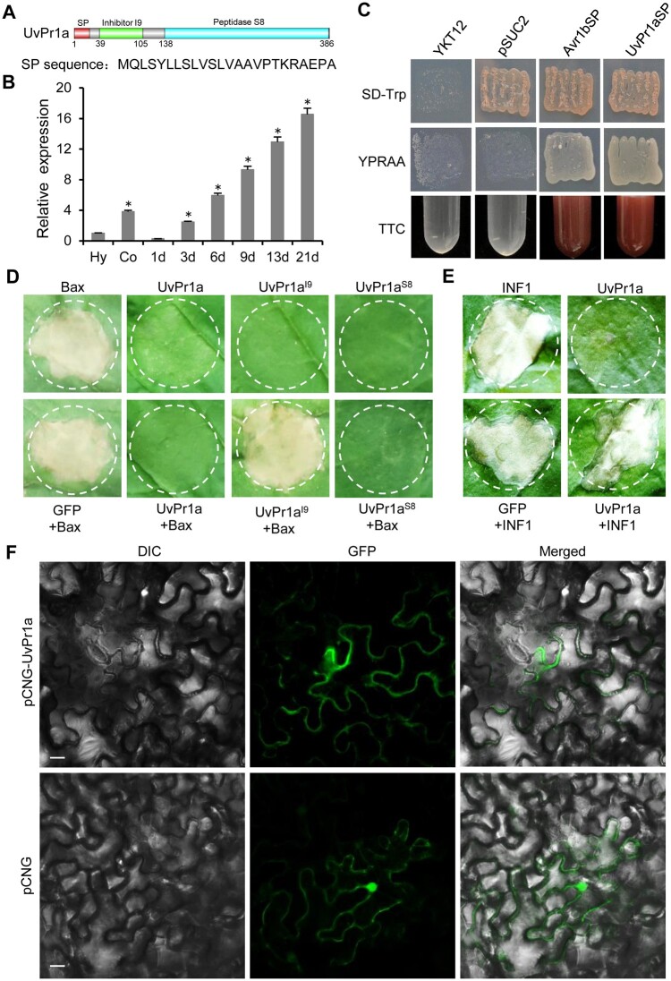
Serine protease subtilase, found widely in both eukaryotes and prokaryotes, participates in various biological processes. However, how fungal subtilase regulates plant immunity is a major concern. Here, we identified a secreted fungal subtilase, UvPr1a, from the rice false smut (RFS) fungus Ustilaginoidea virens. We characterized UvPr1a as a virulence effector localized to the plant cytoplasm that inhibits plant cell death induced by Bax. Heterologous expression of UvPr1a in rice (Oryza sativa) enhanced plant susceptibility to rice pathogens. UvPr1a interacted with the important rice protein SUPPRESSOR OF G2 ALLELE OF skp1 (OsSGT1), a positive regulator of innate immunity against multiple rice pathogens, degrading OsSGT1 in a protease activity-dependent manner. Furthermore, host-induced gene silencing of UvPr1a compromised disease resistance of rice plants. Our work reveals a previously uncharacterized fungal virulence strategy in which a fungal pathogen secretes a subtilase to interfere with rice immunity through degradation of OsSGT1, thereby promoting infection. These genetic resources provide tools for introducing RFS resistance and further our understanding of plant-pathogen interactions.


Figures

Figure 1
Figure 1
UvPr1a is a cytoplasmic effector. A, Predicted Pfam domain of UvPr1a. B, RT-qPCR detects the transcription level of UvPr1a relative to β-tubulin in conidia (Co) and hyphae (Hy) in PSB and at different infection stages (1–21 dpi) of U. virens. Error bars represent the standard deviation. Asterisks represent significant differences from Hy using the test of LSD at P < 0.05. C, Yeast secretion assay of UvPr1a signal peptide. Yeast strain YTK12 was transformed with the empty vector pSUC2 (negative control), pSUC2-UvPr1aSP, or pSUC2-Avr1SP (positive control) and tested for growth on SD–Trp or YPRAA medium and for invertase activity with a colorimetric TTC assay. D, Transient expression of UvPr1a, UvPr1aI9, and UvPr1aS8 suppresses PCD triggered by Bax in N. benthamiana leaves. Representative leaves were photographed 5 days after infiltration. E, Transient expression of UvPr1a did not suppresses PCD triggered INF1 in N. benthamiana leaves. Representative leaves were photographed 5 days after infiltration. F, UvPr1a-GFP localization in N. benthamiana leaves. DIC, differential interference contrast; GFP, green fluorescent protein. Scale bar = 20 μm.
Figure 2
Figure 2
UvPr1a is a key virulence effector. A, Colony morphology of U. virens wild-type HWD-2, ΔUvPr1a-9, ΔUvPr1a-24, CΔUvPr1a, CΔUvPr1aΔS8, and CΔUvPr1aΔI9 strains on PSA after 14 days of darkness at 28°C. B, Colony diameters of mutant strains on PSA after 14 days at 28°C. C, Conidial production of mutant strains grown in PSB medium at 180 rpm for 7 days. D, Virulence assays of mutant strains on rice spikelets at 21 dpi. E, Mean number of rice smut balls per panicle. Data were collected from three independent experiments for each treatment. Error bars represent standard deviation, and asterisks represent significant difference from HWD-2 using the test of LSD at P = 0.05.
Figure 3
Figure 3
Heterologous expression of UvPr1a in rice increases susceptibility to rice pathogens. A, Representative images of 35S-UvPr1a transgenic rice plants and the wild-type Nip rice plants at the mature stage following growth in field conditions. B, Left: Resistance assays of 35S-UvPr1a transgenic rice plants and the wild-type Nip rice plants to U. virens strain HWD-2 infection at 25 dpi. Right: Mean number of rice smut balls measured in resistance assays. C, Left: Disease symptoms at 21 dpi of 35S-UvPr1a transgenic rice plants and the wild-type Nip rice plants after inoculation with Xoo PXO99A. Right: Mean lesion lengths at 21 dpi on the leaves of 35S-UvPr1a transgenic rice plants and the wild-type Nip rice plants after inoculation with Xoo PXO99A. D, Left: Leaves of 35S-UvPr1a transgenic rice plants and the wild-type Nip rice plants at 5 dpi following spot-inoculation with a spore suspension of M. oryzae Guy11. Right: Relative fungal biomass was determined using qPCR for the M. oryzae Pot2 gene normalized to rice OsUBQ1. E, Left: Disease symptoms at 3 dpi of 35S-UvPr1a transgenic rice plants and the wild-type Nip rice plants after inoculation with R. solani HG81. Right: Leaf lesion area of 35S-UvPr1a transgenic rice plants and the wild-type Nip rice plants after inoculation with R. solani HG81. All inoculation experiments were repeated 3 times. Asterisks represent significant differences using the test of LSD at P < 0.05 between Nip and 35S-UvPr1a transgenic rice plants, and standard errors are shown in bar graphs.
Figure 4
Figure 4
UvPr1a physically interacts with rice OsSGT1 in vitro and in vivo. A, Y2H analysis of the interaction between UvPr1a and OsSGT1. SD-3, SD–Trp–Leu–His; BD, pGBKT7; AD, pGADT7. BD-53 + AD-T served as the positive control. BD-UvPr1a + AD or BD + AD-OsSGT1 served as the negative control. B, GST pull-down assay showing the interaction between OsSGT1-His and UvPr1a-GST. Recombinant UvPr1a-GST bound resin was incubated with E. coli crude extracts containing OsSGT1-His and analyzed by immunoblotting. C, Co-IP assay showing that UvPr1a interacts with OsSGT1 in vivo. Co-IP assay was performed on protein extracts from N. benthamiana leaves co-expressed with OsSGT1-Flag and UvPr1a-GFP. D, BiFC assays of the interaction between UvPr1a and OsSGT1. Nicotiana benthamiana leaves were co-infiltrated with UvPr1a-nYFP and OsSGT1-cYFP constructs. YFP signals were observed 2 days after infiltration. Co-infiltration with UvPr1a-nYFP and cYFP or nYFP and OsSGT1-cYFP constructs were used as negative control. Scale bar = 20 µm.
Figure 5
Figure 5
OsSGT1 positively regulates rice disease resistance against multiple rice pathogens. A, Representative images of Nip, OsSGT1-OE, and ossgt1 rice plants at the mature stage following growth under field conditions. B, Left: Resistance assays of Nip, OsSGT1-OE and ossgt1 rice plants against infection by U. virens strain HWD-2 at 25 dpi. Right: Mean number of rice smut balls seen in resistance assays. C, Left: Leaf lesions on Nip, OsSGT1-OE, and ossgt1 rice plants after inoculated with Xoo PXO99 at 21 dpi. Right: Lesion lengths on rice leaves at 21 dpi after inoculation with Xoo PXO99A. D, Left: Disease symptoms on the leaves of Nip, OsSGT1-OE, and ossgt1 rice plants. Leaves were spot inoculated with a spore suspension of M. oryzae Guy11 and photographed at 5 dpi. Right: Relative fungal biomass was determined using qPCR for M. oryzae Pot2 and normalized to rice OsUBQ1. E, Left: Disease symptoms at 3 dpi of Nip, OsSGT1-OE, and ossgt1 rice plants after inoculation with R. solani HG81. Right: Leaf lesions on Nip, OsSGT1-OE and ossgt1 rice plants after inoculation with R. solani HG81 at 3 dpi. All inoculation experiments were repeated 3 times. Asterisks represent significant differences using the test of LSD at P < 0.05, and standard errors are shown in bar graphs.
Figure 6
Figure 6
UvPr1a mediates protein degradation of OsSGT1. A, Transient expression of UvPr1a suppresses PCD triggered by OsSGT1 in N. benthamiana leaves. pVX-UvPr1 and pVX-OsSGT1 fusion constructs were co-infiltrated in N. benthamiana leaves. Representative leaves were photographed 5 days after infiltration. B, UvPr1a degrades OsSGT1 protein in vitro, as determined by immunoblotting using 5 µL of purified OsSGT1-His, with 1, 2, 5, and 10 µL of recombinant UvPr1a-GST or GST incubated at 30°C for 15 min. The reaction products were analyzed by immunoblotting with anti-His and anti-GST antibodies. C, OsSGT1-Flag co-expression with UvPr1a-GFP or GFP in N. benthamiana leaves. The relative protein level of OsSGT1-Flag was detected by Western blotting. D, Relative protein levels of OsSGT1 in Nip and 35S-UvPr1a transgenic rice plants as detected with anti-SGT1.
Figure 7
Figure 7
HIGS of UvPr1a increases rice resistance to RFS. A, Resistance assays of T2 and T3 HIGS transgenic rice plants harboring the UvPr1aHIGS construct against U. virens at 25 dpi. B, Mean number of rice smut balls in T2 and T3 transgenic rice plants from resistance assays. The experiment was repeated 3 times, and 30 rice panicles were used per inoculation. Error bars represent the standard deviation. C, Relative UvPr1a transcript levels of T3 transgenic lines at 6 dpi. Levels of the U. virens β-tubulin gene were used for normalization in RT-qPCR. The experiment was repeated 3 times and error bars represent the standard deviation. D, Visualization of siRNAs targeting UvPr1a in infected transgenic rice spikelets at 6 dpi by FISH using a specific probe. An, anther; hy, U. virens hyphae. Scale bar = 20 μm. Asterisks indicate statistically significant differences compared to Nip using the test of LSD at P < 0.05.
Figure 8
Figure 8
A working model illustrating how UvPr1a degrades OsSGT1 to suppress rice immunity during U. virens infection. During infection, the U. virens effector UvPr1a is secreted by HWD-2 and translocated into host cells, and then physically interacts with OsSGT1. UvPr1a directly degraded OsSGT1, thereby inhibiting plant immunity and showed susceptibility to pathogens. However, in the ΔUvPr1a mutants, the OsSGT1 protein was expressed normally, and the rice plants had normal immune responses and showed resistance to pathogens.

Similar articles

Cited by

References

    1. Aarts N, Metz M, Holub E, Staskawicz BJ, Daniels MJ, Parker JE (1998) Different requirements for EDS1 and NDR1 by disease resistance genes define at least two R gene-mediated signaling pathways in Arabidopsis. Proc Natl Acad Sci USA 95: 10306–10311 - PMC - PubMed
    1. Azevedo C, Sadanandom A, Kitagawa K, Freialdenhoven A, Shirasu K, Schulze-Lefert P (2002) The RAR1 interactor SGT1, an essential component of R gene-triggered disease resistance. Science 295: 2073–2076 - PubMed
    1. Azevedo C, Betsuyaku S, Peart J, Takahashi A, Noel L, Sadanandom A, Casais C, Parker J, Shirasu K (2006) Role of SGT1 in resistance protein accumulation in plant immunity. EMBO J 25: 2007–2016 - PMC - PubMed
    1. Baker D, Shiau AK, Agard DA (1993) The role of pro regions in protein folding. Curr Opin Cell Biol 5: 966–970 - PubMed
    1. Bryan PN (2002) Prodomains and protein folding catalysis. Chem Rev 102: 4805–4816 - PubMed

Publication types

MeSH terms

Substances