. 2022 Aug 23;16(8):11792-11801.
doi: 10.1021/acsnano.2c04962. Epub 2022 Jul 21.

Stretchable Sponge Electrodes for Long-Term and Motion-Artifact-Tolerant Recording of High-Quality Electrophysiologic Signals

Affiliations

Stretchable Sponge Electrodes for Long-Term and Motion-Artifact-Tolerant Recording of High-Quality Electrophysiologic Signals

Li-Wei Lo et al. ACS Nano. .

Abstract

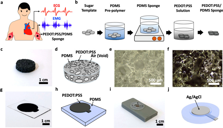
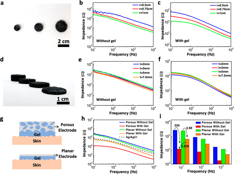
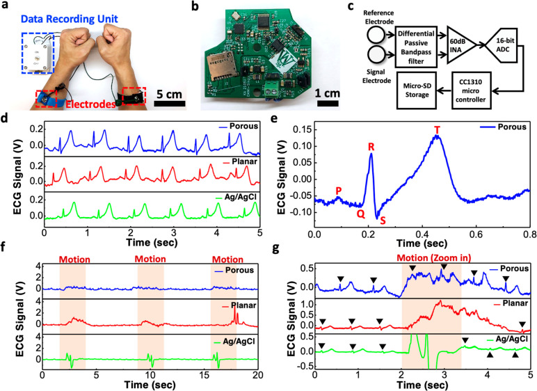
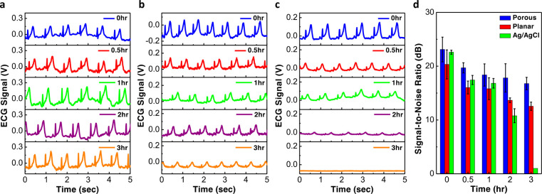
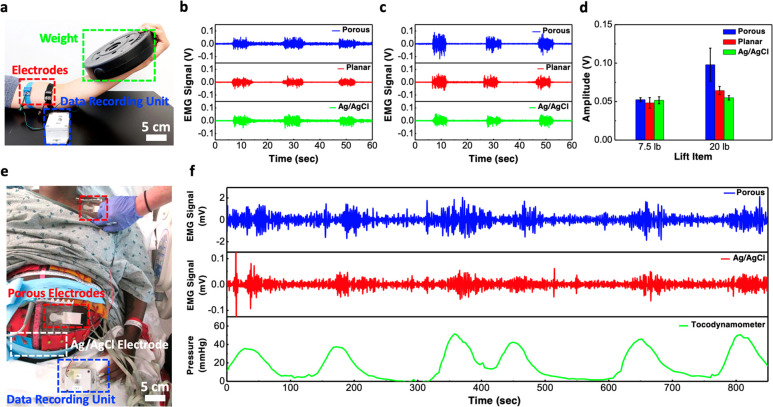
Soft electronic devices and sensors have shown great potential for wearable and ambulatory electrophysiologic signal monitoring applications due to their light weight, ability to conform to human skin, and improved wearing comfort, and they may replace the conventional rigid electrodes and bulky recording devices widely used nowadays in clinical settings. Herein, we report an elastomeric sponge electrode that offers greatly reduced electrode-skin contact impedance, an improved signal-to-noise ratio (SNR), and is ideally suited for long-term and motion-artifact-tolerant recording of high-quality biopotential signals. The sponge electrode utilizes a porous polydimethylsiloxane sponge made from a sacrificial template of sugar cubes, and it is subsequently coated with a poly(3,4-ethylenedioxythiophene) polystyrenesulfonate (PEDOT:PSS) conductive polymer using a simple dip-coating process. The sponge electrode contains numerous micropores that greatly increase the skin-electrode contact area and help lower the contact impedance by a factor of 5.25 or 6.7 compared to planar PEDOT:PSS electrodes or gold-standard Ag/AgCl electrodes, respectively. The lowering of contact impedance resulted in high-quality electrocardiogram (ECG) and electromyogram (EMG) recordings with improved SNR. Furthermore, the porous structure also allows the sponge electrode to hold significantly more conductive gel compared to conventional planar electrodes, thereby allowing them to be used for long recording sessions with minimal signal degradation. The conductive gel absorbed into the micropores also serves as a buffer layer to help mitigate motion artifacts, which is crucial for recording on ambulatory patients. Lastly, to demonstrate its feasibility and potential for clinical usage, we have shown that the sponge electrode can be used to monitor uterine contraction activities from a patient in labor. With its low-cost fabrication, softness, and ability to record high SNR biopotential signals, the sponge electrode is a promising platform for long-term wearable health monitoring applications.

Keywords: electrocardiography; electromyography; porous elastomer; porous electrode; stretchable electronics; uterine contraction monitoring.


Conflict of interest statement

The authors declare no competing financial interest.

Figures

Figure 1
Figure 1
Concept of the soft PEDOT:PSS/PDMS sponge electrode. (a) Schematics illustrating the use of the PEDOT:PSS/PDMS sponge electrode for ECG and EMG recording applications. (b) Schematic diagrams illustrating the fabrication steps used to make the sponge electrodes. (c) Photograph of a sponge electrode. (d) Schematic illustrating the structure of the sponge electrode. (e, f) Optical micrographs showing the microstructures of the PDMS sponge (e) before and (f) after the PEDOT:PSS coating. (g, h) Photograph and schematic diagram of the PEDOT:PSS thin-film directly printed on a piece of planar PDMS substrate. (i, j) Photograph and schematic diagram illustrating the structure of a commercial Ag/AgCl electrode used as a gold-standard reference in this study.
Figure 2
Figure 2
Skin-electrode impedance characterization of the porous PEDOT:PSS/PDMS electrode. (a) Photograph showing three sponge electrodes with the same thickness of 2 mm and different radii of 0.5, 0.75, and 1 cm. (b, c) Impedance spectra measured using the sponge electrodes of different radii (b) without and (c) with the use of conductive hydrogel. (d) Photograph showing four sponge electrodes with the same radius of 1 cm and different thicknesses of 2, 3, 5, and 7.5 mm. (e, f) Impedance spectra measured using the sponge electrodes of different thicknesses (e) without and (f) with the use of conductive hydrogel. (g) Schematic diagrams illustrating the difference in the electrode-gel-skin contact area between the sponge electrode and planar electrode. (h) Comparison of impedance spectra measured using the sponge electrode, planar electrode, and commercial Ag/AgCl electrode. (i) Impedance values at 10, 100, and 1000 Hz for the porous electrode and planar electrode with and without the use of conductive hydrogel.
Figure 3
Figure 3
ECG recording and the effect of motion artifacts. (a) Photograph showing the ECG recording setup. (b) Photograph showing the PCB board of our in-house built biopotential data recorder. (c) Block diagram of the portable data recording unit. (d) ECG signals measured using the porous PEDOT:PSS/PDMS electrode, planar PEDOT:PSS electrode, and commercial Ag/AgCl electrode. (e) Representative cycle of the ECG waveform acquired by the sponge electrode showing clear P wave, QRS complex, and T wave. (f) ECG signals measured under the presence of motion artifacts caused by periodic body movement. (g) Zoomed-in view of the data in (f) showing the ECG peaks along with the motion artifacts.
Figure 4
Figure 4
Long-term ECG signal recording. (a–c) ECG signals measured after various amount of time using the (a) porous PEDOT:PSS/PDMS electrodes; (b) planar PEDOT:PSS electrodes; and (c) commercial Ag/AgCl electrodes. (d) Comparison of the SNR between the three different types of electrodes.
Figure 5
Figure 5
EMG signal recording from skeletal muscle and smooth muscle. (a) Photograph showing the setup for measuring EMG signal from the contraction of biceps. (b, c) EMG signals measured using various kinds of electrodes when the subject was lifting a (b) 7.5-lb or (c) 20-lb weight. (d) Comparison of the EMG signal amplitude measured with sponge, planar, and commercial Ag/AgCl electrodes. (e) Photograph showing the setup for recording EMG signals from uterine contraction activities in a clinical setting. (f) Comparison of EMG waveforms recorded from our porous electrodes and in-house built data recorder, the commercial BioSemi active Ag/AgCl electrodes and BioSemi biopotential measurement system and the corresponding uterine contractions recorded from a tocodynamometer.

Similar articles

Cited by

References

    1. Zamarayeva A. M.; Ostfeld A. E.; Wang M.; Duey J. K.; Deckman I.; Lechêne B. P.; Davies G.; Steingart D. A.; Arias A. C. Flexible and Stretchable Power Sources for Wearable Electronics. Sci. Adv. 2017, 3, e160205110.1126/sciadv.1602051. - DOI - PMC - PubMed
    1. Hua Q.; Sun J.; Liu H.; Bao R.; Yu R.; Zhai J.; Pan C.; Wang Z. L. Skin-Inspired Highly Stretchable and Conformable Matrix Networks for Multifunctional Sensing. Nat. Commun. 2018, 9, 244.10.1038/s41467-017-02685-9. - DOI - PMC - PubMed
    1. Yuk H.; Lu B.; Lin S.; Qu K.; Xu J.; Luo J.; Zhao X. 3D Printing of Conducting Polymers. Nat. Commun. 2020, 11, 1604.10.1038/s41467-020-15316-7. - DOI - PMC - PubMed
    1. Wang Y.; Lee S.; Yokota T.; Wang H.; Jiang Z.; Wang J.; Koizumi M.; Someya T. A Durable Nanomesh On-Skin Strain Gauge for Natural Skin Motion Monitoring with Minimum Mechanical Constraints. Sci. Adv. 2020, 6, eabb704310.1126/sciadv.abb7043. - DOI - PMC - PubMed
    1. Wang C.; Qi B.; Lin M.; Zhang Z.; Makihata M.; Liu B.; Zhou S.; Huang Y. hsi; Hu H.; Gu Y.; Chen Y.; Lei Y.; Lee T.; Chien S.; Jang K. I.; Kistler E. B.; Xu S. Continuous Monitoring of Deep-Tissue Haemodynamics with Stretchable Ultrasonic Phased Arrays. Nat. Biomed. Eng. 2021, 5, 749–758. 10.1038/s41551-021-00763-4. - DOI - PubMed

Publication types