. 2022 Aug 10;96(15):e0078622.
doi: 10.1128/jvi.00786-22. Epub 2022 Jul 21.

PSMD12-Mediated M1 Ubiquitination of Influenza A Virus at K102 Regulates Viral Replication

Affiliations

PSMD12-Mediated M1 Ubiquitination of Influenza A Virus at K102 Regulates Viral Replication

Xianfeng Hui et al. J Virol. .

Abstract

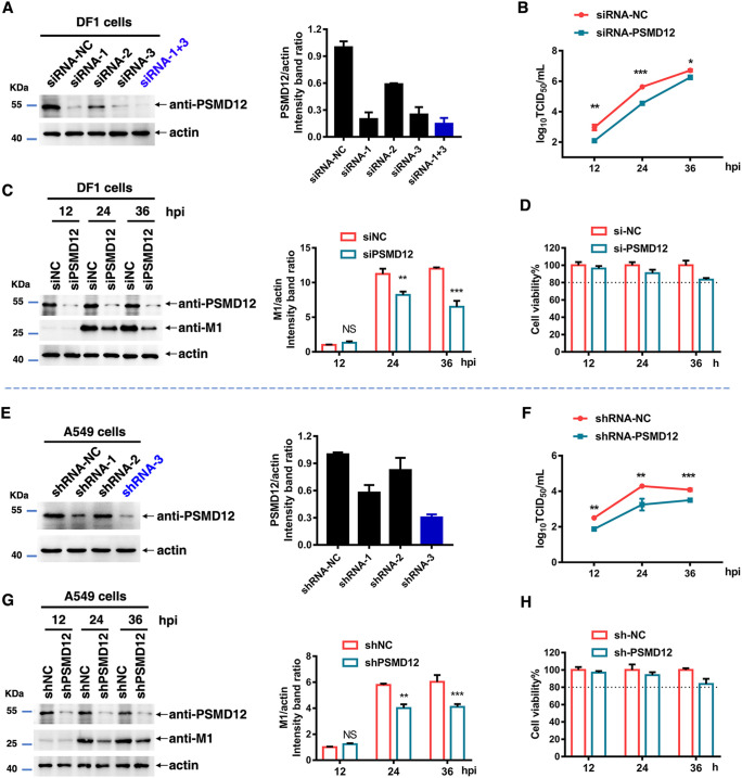
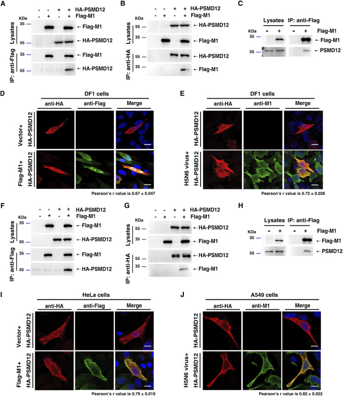
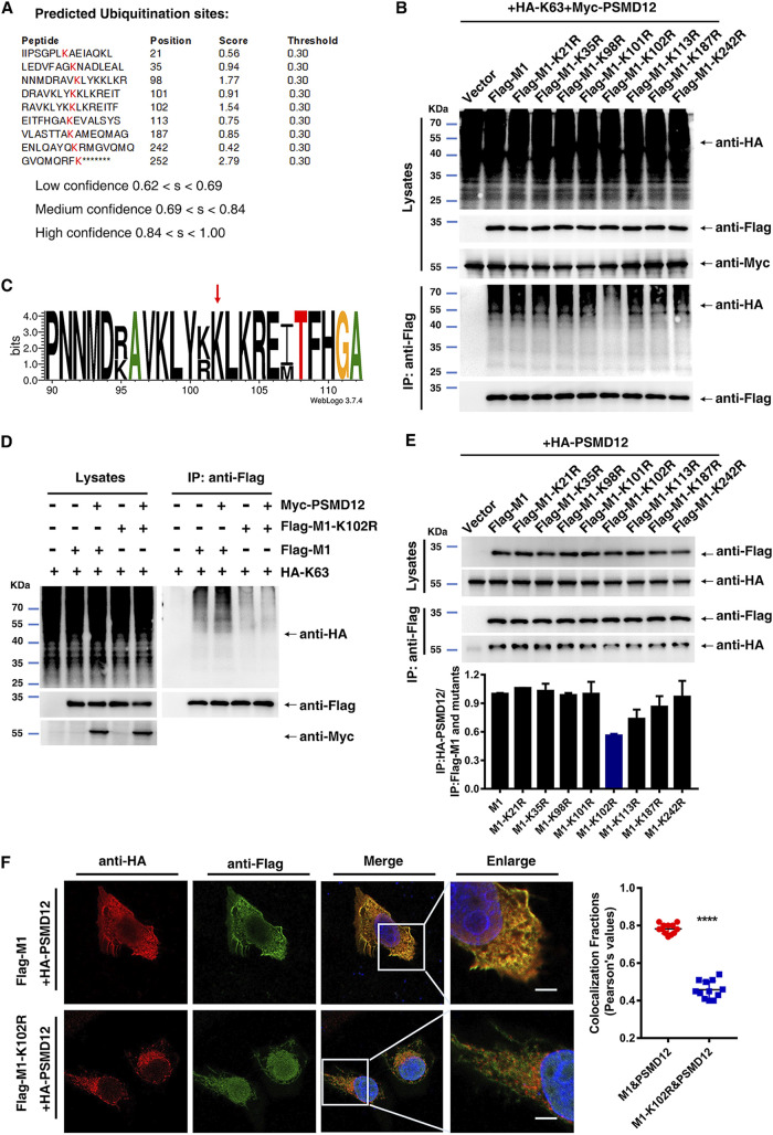
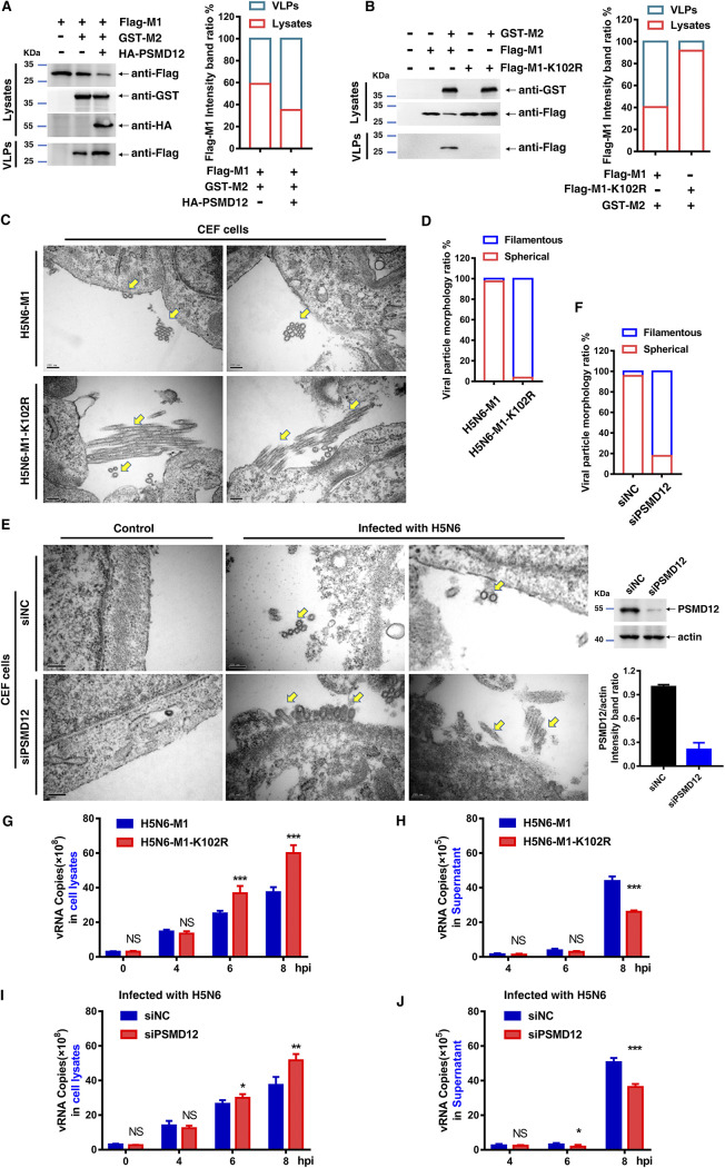
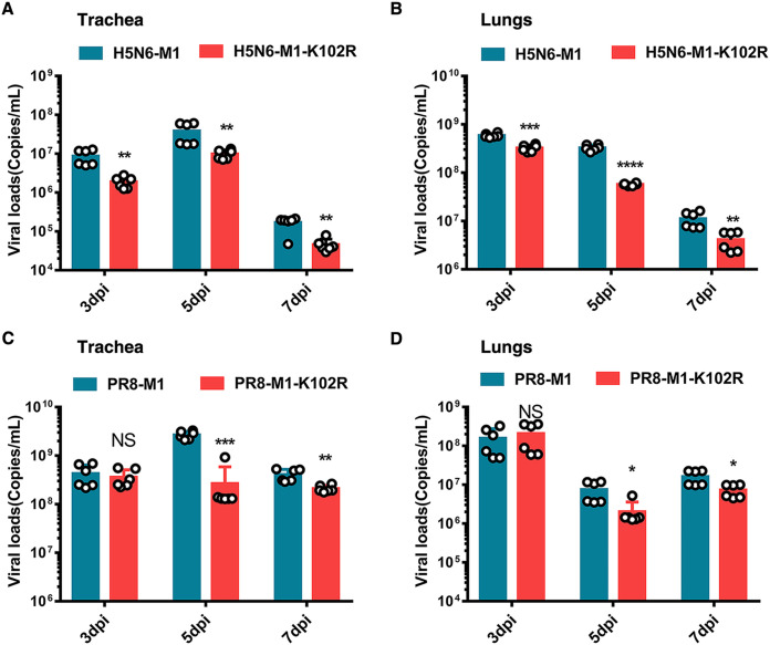
The M1 of influenza A virus (IAV) is important for the virus life cycle, especially for the assembly and budding of viruses, which is a multistep process that requires host factors. Identifying novel host proteins that interact with M1 and understanding their functions in IAV replication are of great interest in antiviral drug development. In this study, we identified 19 host proteins in DF1 cells suspected to interact with the M1 protein of an H5N6 virus through immunoprecipitation (IP)/mass spectrometry. Among them, PSMD12, a 26S proteasome regulatory subunit, was shown to interact with influenza M1, acting as a positive host factor in IAV replication in avian and human cells. The data showed that PSMD12 promoted K63-linked ubiquitination of M1 at the K102 site. H5N6 and PR8 with an M1-K102 site mutant displayed a significantly weaker replication ability than the wild-type viruses. Mechanistically, PSMD12 promoted M1-M2 virus-like particle (VLP) release, and an M1-K102 mutation disrupted the formation of supernatant M1-M2 VLPs. An H5N6 M1-K102 site mutation or knockdown PSMD12 disrupted the budding release of the virus in chicken embryo fibroblast (CEF) cells, which was confirmed by transmission electron microscopy. Further study confirmed that M1-K102 site mutation significantly affected the virulence of H5N6 and PR8 viruses in mice. In conclusion, we report the novel host factor PSMD12 which affects the replication of influenza virus by mediating K63-linked ubiquitination of M1 at K102. These findings provide novel insight into the interactions between IAV and host cells, while suggesting an important target for anti-influenza virus drug research. IMPORTANCE M1 is proposed to play multiple biologically important roles in the life cycle of IAV, which relies largely on host factors. This study is the first one to identify that PSMD12 interacts with M1, mediates K63-linked ubiquitination of M1 at the K102 site, and thus positively regulates influenza virus proliferation. PSMD12 promoted M1-M2 VLP egress, and an M1-K102 mutation affected the M1-M2 VLP formation. Furthermore, we demonstrate the importance of this site to the morphology and budding of influenza viruses by obtaining mutant viruses, and the M1 ubiquitination regulator PSMD12 has a similar function to the M1 K102 mutation in regulating virus release and virus morphology. Additionally, we confirm the reduced virulence of H5N6 and PR8 (H1N1) viruses carrying the M1-K102 site mutation in mice. These findings provide novel insights into IAV interactions with host cells and suggest a valid and highly conserved candidate target for antiviral drug development.

Keywords: K102 site; K63-linked ubiquitination; M1 protein; PSMD12; influenza A virus; replication.


Conflict of interest statement

The authors declare no conflict of interest.

Figures

FIG 1
FIG 1
PSMD12 was identified to positively regulate the proliferation of the H5N6 influenza virus. (A) Schematic diagram of the identification of host proteins that coprecipitated with influenza A viral protein M1. (i) Mass spectrometry was used to identify host proteins that coimmunoprecipitated with HA-tagged influenza viral protein M1. (ii) To identify host factors that affected viral replication, cells were transfected with plasmids to overexpress each of the candidate host genes and then infected with influenza virus. Virus titers and cell viability were then determined. (iii) Based on the screening results, the molecular mechanisms of the candidate host proteins were further explored. (B) DF1 cells were transfected with the indicated plasmids, infected with H5N6 virus at an MOI of 0.05, and the supernatant was collected for viral plaque measurement. (C) Viability of DF1 cells transfected with the indicated plasmids.
FIG 2
FIG 2
Knockdown of PSMD12 inhibits H5N6 virus replication. (A) DF1 cells were transfected with siRNA-PSMD12 or siRNA-negative control (NC), and cells were harvested and analyzed via Western blotting (WB). (B, C) DF1 cells were infected with H5N6 virus 24 h after transfection with siRNAs. Supernatant was collected at different time points for TCID50 measurement (B), and the cells were harvested and analyzed via WB (C). (D) Cell viability was determined at the indicated time points. (E) The effect of knockdown by different shRNAs in A549 cells was analyzed via WB. (F, G) PSMD12-knockdown A549 cells were infected with H5N6 virus, and the supernatant was collected at different time points for TCID50 measurement (F); cells were harvested and analyzed via WB (G). (H) Cell viability was determined at the indicated time points. Error bars, mean ± SD of three experiments. All comparisons were tested by two-tailed Student's t test; *, P < 0.05; **, P < 0.01; ***, P < 0.001.
FIG 3
FIG 3
PSMD12 interacted with M1. (A) DF1 cells were transfected with plasmids encoding HA-PSMD12 and Flag-M1. The lysates were subjected to anti-Flag IP and analyzed via Western blotting (WB). (B) DF1 cells were transfected with plasmids encoding HA-PSMD12 and Flag-M1. The lysates were subjected to anti-HA IP and analyzed via WB. (C) DF1 cells were transfected with a plasmid encoding Flag-M1, and the lysate was subjected to IP and analyzed for endogenous PSMD12 via WB. (D, E) DF1 cells were transfected with plasmids encoding Flag-M1 (H5N6 virus) and HA-PSMD12 (avian source) and analyzed for colocalization of Flag-M1 and HA-PSMD12. Scale bar, 10 μm (D). DF1 cells were transfected with HA-PSMD12 plasmid, infected with H5N6 virus, and analyzed for colocalization of HA-PSMD12 and M1 of H5N6 virus. Scale bar, 10 μm (E). (F) HEK293T cells were transfected with plasmids encoding HA-PSMD12 (human source) and Flag-M1 (H5N6 virus). The lysates were subjected to anti-Flag IP and analyzed via WB. (G) HEK293T cells were transfected with plasmids encoding HA-PSMD12 and Flag-M1. The lysates were subjected to anti-HA IP and analyzed via WB. (H) HEK293T cells were transfected with a plasmid encoding Flag-M1, and the lysate was subjected to IP and analyzed for endogenous PSMD12 via WB. (I, J) HeLa cells were transfected with plasmids encoding Flag-M1 (H5N6 virus) and HA-PSMD12 (human source) and analyzed for colocalization of Flag-M1 and HA-PSMD12. Scale bar, 10 μm (I). A549 cells were transfected with an HA-PSMD12 plasmid, infected with H5N6 virus, and analyzed for colocalization of HA-PSMD12 and M1 of H5N6 virus. Scale bar, 10 μm (J). Pearson correlation coefficients were determined for molecular colocalization in confocal microscopy images using Coloc 2 of Fiji (regions of interest were chosen within 3 to 5 cells each group, n = 12). Fig. S3 contains scanned complete images of Western blots reference to this figure.
FIG 4
FIG 4
Host protein PSMD12 triggers K63-linked M1 ubiquitination. (A) HEK293T cells were transfected with the indicated plasmid combinations to measure ubiquitination of M1. (B) HEK293T cells overexpressing PSMD12 were transfected with the indicated plasmid combinations to measure ubiquitination of 1. (C) HEK293T cells overexpressing PSMD12 were transfected with the indicated plasmid combinations to measure K6/K11/K27/K29/K33/K48/K63-linked ubiquitination of M1. (D) HEK293T cells overexpressing PSMD12 were transfected with the indicated plasmid combinations to measure K6-linked ubiquitination of M1. (E) HEK293T cells overexpressing PSMD12 were transfected with the indicated plasmid combinations to measure K11-linked ubiquitination of M1. (F) HEK293T cells overexpressing PSMD12 were transfected with the indicated plasmid combinations to measure K29-linked ubiquitination of M1. (G) HEK293T cells overexpressing PSMD12 were transfected with the indicated plasmid combinations to measure K33-linked ubiquitination of M1. (H) HEK293T cells overexpressing PSMD12 were transfected with the indicated plasmid combinations to measure K48-linked ubiquitination of M1. (I) HEK293T cells overexpressing PSMD12 were transfected with the indicated plasmid combinations to measure K63-linked ubiquitination of M1.
FIG 5
FIG 5
The K102 site is critical for K63-linked ubiquitination of M1 and K102R impairs the interaction between M1 and PSMD12. (A) The ubiquitination sites of M1 were predicted by BDM-PUB and UBPRED. (B) HEK293T cells were transfected with the indicated plasmid combinations to measure K63-linked ubiquitination of M1. (C) WebLogo output for amino acid sequences of M1 from H1N1, H2N2, H3N2, H5N1, H5N6, H7N9, and H9N2 influenza viruses. (D) HEK293T cells overexpressing Myc-PSMD12 and HA-K63 were transfected with the indicated plasmid combinations to measure the ubiquitination of PR8 Flag-M1/M1-K102R. (E) Interaction between HA-PSMD12 and wild-type and mutant M1. (F) HeLa cells were transfected with plasmids encoding Flag-M1/M1-K102R and HA-PSMD12, followed by analysis for colocalization of Flag-M1/M1-K102R and HA-PSMD12. Scale bar, 10 μm. Pearson correlation coefficients were determined for molecular colocalization in confocal microscopy images using Coloc 2 of Fiji (regions of interest were chosen within 3 to 5 cells each group, n = 12).
FIG 6
FIG 6
Mutation at the M1 K102 site alters viral replication in cells and PSMD12 fails to regulate M1 K102 mutant H5N6 proliferation. (A, B) CEF cells were infected with H5N6-M1 or H5N6-M1-K102R virus, the supernatant was collected at different time points for TCID50 measurement, and the cell lysates were analyzed by WB. (C, D) A549 cells were infected with PR8-M1 or PR8-M1-K102R virus, the supernatant was collected at different time points for TCID50 measurement, and the cell lysates were analyzed by WB. (E, F) DF1 cells were transfected with the indicated plasmids and infected with H5N6-M1 or H5N6-M1-K102R virus at an MOI of 0.05. The supernatant was collected at 24 h postinfection for TCID50 measurement (E), and cells were harvested and analyzed via WB (F). (G, H) DF1 cells were infected with H5N6-M1 or H5N6-M1-K102R virus at an MOI of 0.05 after transfection with siRNAs. The supernatant was collected at 24 h postinfection for TCID50 measurement (G), and cells were harvested and analyzed via WB (H). Error bars, mean ± SD of three experiments. All comparisons were tested by two-tailed Student’s t test; *, P < 0.05; **, P < 0.01; ***, P < 0.001.
FIG 7
FIG 7
M1 K102 mutation or knockdown PSMD12 restricts virus release. (A, B) HEK293T cells were transfected with the indicated plasmid combinations to detect the influence on the egress of M1. (C) CEF cells were infected with H5N6-M1 or H5N6-M1-K102R virus at an MOI of 0.1 for 10 h followed by transmission electron microscopy. The yellow arrows indicate virus particles. Scale bar, 200 nm. (D) Quantification of virion morphology under 10 random fields. Virus particle length greater than 2-fold the virion diameter are defined as filamentous phenotype. (E) CEF cells (siNC and siPSMD12) were infected with H5N6 virus at an MOI of 0.1 for 10 h followed by transmission electron microscopy. The yellow arrows indicate virus particles. Scale bar, 200 nm. The efficiency of siRNA knockdown was evaluated by Western blotting. (F) Quantification of virion morphology under 10 random fields. Virus particles length greater than 2-fold the virion diameter are defined as filamentous phenotype. (G, H) DF1 cells were infected with H5N6-M1 or H5N6-M1-K102R virus (MOI of 5); the cell lysates (G) and supernatant (H) were collected at different time points, and vRNA of M1 was analyzed by qRT-PCR. (I, J) DF1 cells were infected with H5N6-M1 virus at an MOI of 5 after transfection with siRNAs. The cell lysates (I) and supernatant (J) were collected at different time points, and vRNA of M1 was analyzed by qRT-PCR. Error bars, mean ± SD of three experiments. All comparisons were done by two-tailed Student’s t test; *, P < 0.05; **, P < 0.01; ***, P < 0.001.
FIG 8
FIG 8
M1-K102 site mutation of influenza A virus affects its pathogenicity in mice. (A, B) Six-week-old C57/BL6 mice were infected with the same doses of H5N6-M1 and H5N6-M1-K102R viruses. Body weight (A) and survival (B) were monitored daily for 2 weeks (n = 10). (C, D) Six-week-old C57/BL6 mice were infected with the same doses of PR8-M1 and PR8-M1-K102R viruses. Body weight (C) and survival (D) were monitored daily for 2 weeks (n = 10).
FIG 9
FIG 9
Viral loads of C57/BL6 mice after influenza virus infection. Six-week-old C57/BL6 mice were infected intranasally with WT and M1-K102R mutant H5N6 or PR8 viruses at a dose of 2× MLD50. (A, B) Viral RNA copies were determined by qRT-PCR at 3, 5, and 7 dpi in the trachea (A) and lung (B) of mice infected with H5N6-M1 or H5N6-M1-K102R virus. (C, D) Viral RNA copies were determined by qRT-PCR at 3, 5, and 7 dpi in the trachea (C) and lung (D) of mice infected with PR8-M1 or PR8-M1-K102R virus. Error bars, mean ± SD of three experiments. All comparisons were done by two-tailed Student’s t test; *, P < 0.05; **, P < 0.01; ***, P < 0.001.

References

    1. Gong W, He X, Huang K, Zhang Y, Li C, Yang Y, Zou Z, Jin M. 2021. Interaction of nuclear export protein with G protein pathway suppressor 2 (GPS2) facilitates influenza A virus replication by weakening the inhibition of GPS2 to RNA synthesis and ribonucleoprotein assembly. J Virol 95:e00008-21. 10.1128/JVI.00008-21. - DOI - PMC - PubMed
    1. Ali A, Avalos RT, Ponimaskin E, Nayak DP. 2000. Influenza virus assembly: effect of influenza virus glycoproteins on the membrane association of M1 protein. J Virol 74:8709–8719. 10.1128/jvi.74.18.8709-8719.2000. - DOI - PMC - PubMed
    1. Watanabe T, Kawakami E, Shoemaker JE, Lopes TJS, Matsuoka Y, Tomita Y, Kozuka-Hata H, Gorai T, Kuwahara T, Takeda E, Nagata A, Takano R, Kiso M, Yamashita M, Sakai-Tagawa Y, Katsura H, Nonaka N, Fujii H, Fujii K, Sugita Y, Noda T, Goto H, Fukuyama S, Watanabe S, Neumann G, Oyama M, Kitano H, Kawaoka Y. 2014. Influenza virus-host interactome screen as a platform for antiviral drug development. Cell Host Microbe 16:795–805. 10.1016/j.chom.2014.11.002. - DOI - PMC - PubMed
    1. Gannage M, Dormann D, Albrecht R, Dengjel J, Torossi T, Rämer PC, Lee M, Strowig T, Arrey F, Conenello G, Pypaert M, Andersen J, García-Sastre A, Münz C. 2009. Matrix protein 2 of influenza A virus blocks autophagosome fusion with lysosomes. Cell Host Microbe 6:367–380. 10.1016/j.chom.2009.09.005. - DOI - PMC - PubMed
    1. Stauffer S, Feng Y, Nebioglu F, Heilig R, Picotti P, Helenius A. 2014. Stepwise priming by acidic pH and a High K+ concentration is required for efficient uncoating of influenza A virus cores after penetration. J Virol 88:13029–13046. 10.1128/JVI.01430-14. - DOI - PMC - PubMed

MeSH terms

Substances

LinkOut - more resources