. 2022 Aug;12(8):e2714.
doi: 10.1002/brb3.2714. Epub 2022 Jul 21.

A window into eye movement dysfunction following mTBI: A scoping review of magnetic resonance imaging and eye tracking findings

Affiliations

A window into eye movement dysfunction following mTBI: A scoping review of magnetic resonance imaging and eye tracking findings

Matthew A McDonald et al. Brain Behav. 2022 Aug.

Abstract

Mild traumatic brain injury (mTBI), commonly known as concussion, is a complex neurobehavioral phenomenon affecting six in 1000 people globally each year. Symptoms last between days and years as microstructural damage to axons and neurometabolic changes result in brain network disruption. There is no clinically available objective biomarker to diagnose the severity of injury or monitor recovery. However, emerging evidence suggests eye movement dysfunction (e.g., saccades and smooth pursuits) in patients with mTBI. Patients with a higher symptom burden and prolonged recovery time following injury may show higher degrees of eye movement dysfunction. Likewise, recent advances in magnetic resonance imaging (MRI) have revealed both white matter tract damage and functional network alterations in mTBI patients, which involve areas responsible for the ocular motor control. This scoping review is presented in three sections: Section 1 explores the anatomical control of eye movements to aid the reader with interpreting the discussion in subsequent sections. Section 2 examines the relationship between abnormal MRI findings and eye tracking after mTBI based on the available evidence. Finally, Section 3 communicates gaps in our knowledge about MRI and eye tracking, which should be addressed in order to substantiate this emerging field.

Keywords: DTI; MRI; concussion; eye tracking; fMRI; mTBI; ocular motor; oculomotor; saccades; smooth pursuit; white matter tracts.


Conflict of interest statement

The authors declare that there is no conflict of interest.

Figures

FIGURE 1
FIGURE 1
(a) T1‐weighted, (b) T2‐weighted, and (c) T2 FLAIR MRI sequences acquired on a healthy 17‐year‐old male, representing key sequences of a clinical MRI protocol
FIGURE 2
FIGURE 2
Diffusion MRI in a healthy 17‐year‐old male. To derive neural tract direction, DTI scans use six or more gradient directions, sufficient to compute the diffusion tensor. The level of diffusion weighting is indicated by the b‐value, a parameter that reflects the length and strength of the magnetic field gradients; slow moving water molecules across shorter diffusion distances require a higher b‐value (Stejskal & Tanner, 1965). (a) Six direction gradients used as input (directions in square brackets) which is combined with (b) to calculate diffusion parameters. (c) Apparent diffusion coefficient (ADC) map which requires a minimum of three directions. By collecting images with at least two different values, a pure parametric image of the ADC can be calculated (Le Bihan, 2013), where the ADC represents the magnitude of diffusion of water molecules within the tissue as the sole source of contrast. (d) Axial diffusivity (AD). (e) Radial diffusivity (RD) map as a quantitative measurement of diffusion, removing T2 effects. (f) Fractional anisotropy (FA) map. (g) Color FA map. (h) Diffusion tractography map of corpus callosum with 54 diffusion gradients. Note: (d)–(g) require six or more directional gradients
FIGURE 3
FIGURE 3
Values, which are eigenvalues (“pointiness” and “size” of diffusion) and eigenvectors (describing orientation in space), of the diffusion tensor are used to give directional information of water diffusion. (a)–(e) The five main diffusion MRI ellipsoid parameters to quantify water diffusion along a trajectory
FIGURE 4
FIGURE 4
Default mode network BOLD signal activation in a 17‐year‐old male acquired on a resting state fMRI sequence while lying quietly awake with eyes closed. Resolution: 1.5 mm × 1.5 mm × 3 mm, acquired over 5 min. Ninety slices per location were recorded in a multiband sequence
FIGURE 5
FIGURE 5
Corpus callosum (CC) illustrated with deterministic tractography with cut‐off value of 0.15 as a tensor FA threshold for terminating tracts. Twenty thousand streamlines (tracts) are generated for each bundle of tracts. Scanning was acquired on a 3‐Tesla MRI (GE Signa™ Premier) with a 48‐channel head coil and 54 diffusion gradients. Voxel size = 2 mm isotropic with three b‐values = 1000, 2000, and 3000 s/mm2, 15, 15, and 20 directions respectively, and 4× b = 0
FIGURE 6
FIGURE 6
Anterior, superior, and posterior thalamic radiations illustrated with deterministic tractography with cut‐off value of 0.15 as a tensor FA threshold for terminating tracts. Twenty thousand streamlines (tracts) are generated for each bundle of tracts. Scanning was acquired on a 3‐Tesla MRI (GE Signa™ Premier) with a 48‐channel head coil and 54 diffusion gradients. Voxel size = 2 mm isotropic with three b‐values = 1000, 2000, and 3000 s/mm2, 15, 15, and 20 directions respectively, and 4× b = 0
FIGURE 7
FIGURE 7
Corona radiata illustrated with deterministic tractography with a cut‐off value of 0.15 as a tensor FA threshold for terminating tracts. Twenty thousand streamlines (tracts) are generated for each bundle of tracts. Scanning was acquired on a 3‐Tesla MRI (GE Signa™ Premier) with a 48‐channel head coil and 54 diffusion gradients. Voxel size = 2 mm isotropic with three b‐values = 1000, 2000, and 3000 s/mm2, 15, 15, and 20 directions respectively, and 4× b = 0
FIGURE 8
FIGURE 8
Inferior fronto‐occipital fasciculus (IFOF) illustrated with deterministic tractography with a cut‐off value of 0.15 as a tensor FA threshold for terminating tracts. Twenty thousand streamlines (tracts) are generated for each bundle of tracts. Scanning was acquired on a 3‐Tesla MRI (GE Signa™ Premier) with a 48‐channel head coil and 54 diffusion gradients. Voxel size = 2 mm isotropic with three b‐values = 1000, 2000, and 3000 s/mm2, 15, 15, and 20 directions respectively, and 4× b = 0
FIGURE 9
FIGURE 9
Superior longitudinal fasciculus (SLF) illustrated with deterministic tractography with a cut‐off value of 0.15 as a tensor FA threshold for terminating tracts. Twenty thousand streamlines (tracts) are generated for each bundle of tracts. Scanning was acquired on a 3‐Tesla MRI (GE Signa™ Premier) with a 48‐channel head coil and 54 diffusion gradients. Voxel size = 2 mm isotropic with three b‐values = 1000, 2000, and 3000 s/mm2, 15, 15, and 20 directions respectively, and 4× b = 0
FIGURE 10
FIGURE 10
Arcuate fasciculus (AF) illustrated with deterministic tractography with a cut‐off value of 0.15 as a tensor FA threshold for terminating tracts. Twenty thousand streamlines (tracts) are generated for each bundle of tracts. Scanning was acquired on a 3‐Tesla MRI (GE Signa™ Premier) with a 48‐channel head coil and 54 diffusion gradients. Voxel size = 2 mm isotropic with three b‐values = 1000, 2000, and 3000 s/mm2, 15, 15, and 20 directions respectively, and 4× b = 0
FIGURE 11
FIGURE 11
Uncinate fasciculus (UF) illustrated with deterministic tractography with a cut‐off value of 0.15 as a tensor FA threshold for terminating tracts. Twenty thousand streamlines (tracts) are generated for each bundle of tracts. Scanning was acquired on a 3‐Tesla MRI (GE Signa™ Premier) with a 48‐channel head coil and 54 diffusion gradients. Voxel size = 2 mm isotropic with three b‐values = 1000, 2000, and 3000 s/mm2, 15, 15, and 20 directions respectively, and 4× b = 0
FIGURE 12
FIGURE 12
Cingulum bundle (CB) illustrated with deterministic tractography with a cut‐off value of 0.15 as a tensor FA threshold for terminating tracts. Twenty thousand streamlines (tracts) are generated for each bundle of tracts. Scanning was acquired on a 3‐Tesla MRI (GE Signa™ Premier) with a 48‐channel head coil and 54 diffusion gradients. Voxel size = 2 mm isotropic with three b‐values = 1000, 2000, and 3000 s/mm2, 15, 15, and 20 directions respectively, and 4× b = 0
FIGURE 13
FIGURE 13
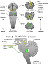
Anterior, axial, and sagittal views of the anatomical origins of cranial nerves III, IV, and VI
FIGURE 14
FIGURE 14
Smooth pursuit gaze trajectory (orange line) along a circular target path (blue line) of a healthy participant at 200 frames per second. Horizontal and vertical coordinates are separated above and below, respectively. High amplitude vertical orange lines represent blink events, which are typically excluded from analysis

Similar articles

Cited by

References

    1. Arfanakis, K. , Haughton, V. M. , Carew, J. D. , Rogers, B. P. , Dempsey, R. J. , & Meyerand, M. E. (2002). Diffusion tensor MR imaging in diffuse axonal injury. AJNR. American Journal of Neuroradiology, 23(5), 794–802. - PMC - PubMed
    1. Asanuma, C. , Thach, W. T. , & Jones, E. G. (1983). Distribution of cerebellar terminations and their relation to other afferent terminations in the ventral lateral thalamic region of the monkey. Brain Research, 286(3), 237–265. 10.1016/0165-0173(83)90015-2 - DOI - PubMed
    1. Asken, B. M. , DeKosky, S. T. , Clugston, J. R. , Jaffee, M. S. , & Bauer, R. M. (2018). Diffusion tensor imaging (DTI) findings in adult civilian, military, and sport‐related mild traumatic brain injury (mTBI): A systematic critical review. Brain Imaging and Behavior, 12(2), 585–612. 10.1007/s11682-017-9708-9 - DOI - PubMed
    1. Astafiev, S. V. , Shulman, G. L. , Metcalf, N. V. , Rengachary, J. , MacDonald, C. L. , Harrington, D. L. , & Corbetta, M. (2015). Abnormal white matter blood‐oxygen‐level‐dependent signals in chronic mild traumatic brain injury. Journal of Neurotrauma, 32(16), 1254–1271. 10.1089/neu.2014.3547 - DOI - PMC - PubMed
    1. Bahrami, N. , Sharma, D. , Rosenthal, S. , Davenport, E. M. , Urban, J. E. , Wagner, B. , & Maldjian, J. A. (2016). Subconcussive head impact exposure and white matter tract changes over a single season of youth football. Radiology, 281(3), 919–926. 10.1148/radiol.2016160564 - DOI - PMC - PubMed

Publication types