. 2022 Oct;32(5):391-400.
doi: 10.1089/nat.2021.0101. Epub 2022 Jul 20.

Inhibition of Epidermal Growth Factor Receptor Signaling by Antisense Oligonucleotides as a Novel Approach to Epidermal Growth Factor Receptor Inhibition

Affiliations

Inhibition of Epidermal Growth Factor Receptor Signaling by Antisense Oligonucleotides as a Novel Approach to Epidermal Growth Factor Receptor Inhibition

Thushara W Madanayake et al. Nucleic Acid Ther. 2022 Oct.

Abstract

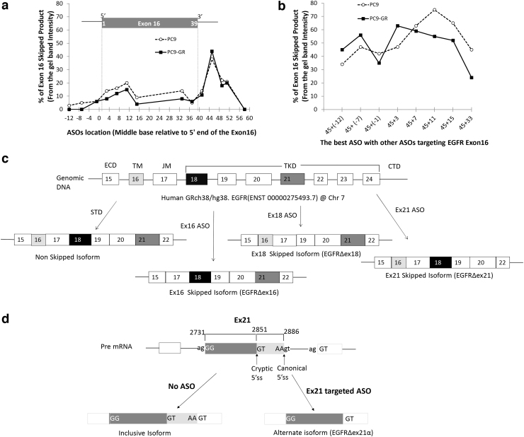
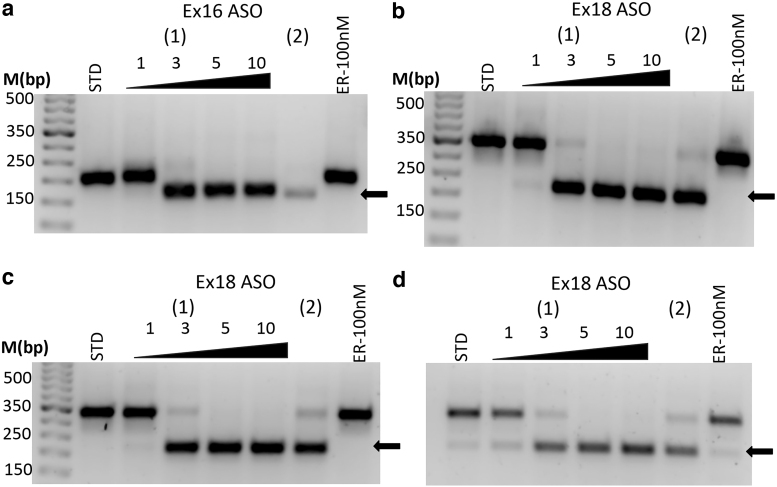
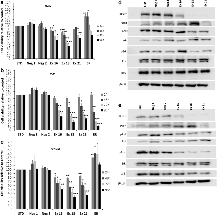
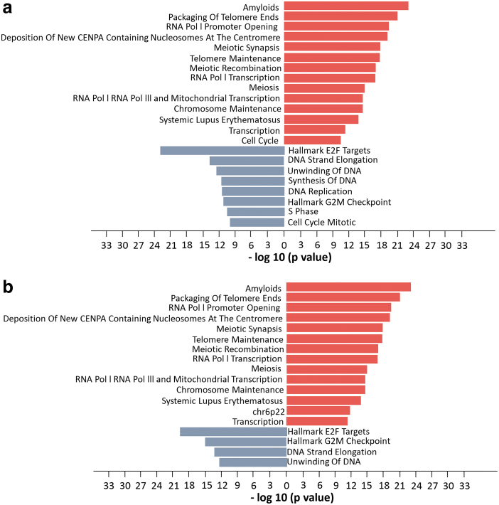
We report a novel method to inhibit epidermal growth factor receptor (EGFR) signaling using custom morpholino antisense oligonucleotides (ASOs) to drive expression of dominant negative mRNA isoforms of EGFR by ASO-induced exon skipping within the transmembrane (16) or tyrosine kinase domains (18 and 21). In vivo ASO formulations induced >95% exon skipping in several models of nonsmall cell lung cancer (NSCLC) and were comparable in efficacy to erlotinib in reducing colony formation, cell viability, and migration in EGFR mutant NSCLC (PC9). However, unlike erlotinib, ASOs maintained their efficacy in both erlotinib-resistant subclones (PC9-GR) and wild-type overexpressing EGFR models (H292), in which erlotinib had no significant effect. The most dramatic ASO-induced phenotype resulted from targeting the EGFR kinase domain directly, which resulted in maximal inhibition of phosphorylation of EGFR, Akt, and Erk in both PC9 and PC9GR cells. Phosphoproteomic mass spectrometry confirmed highly congruent impacts of exon 16-, 18-, and 21-directed ASOs compared with erlotinib on PC9 genome-wide cell signaling. Furthermore, EGFR-directed ASOs had no impact in EGFR-independent NSCLC models, confirming an EGFR-specific therapeutic mechanism. Further exploration of synergy of ASOs with existing tyrosine kinase inhibitors may offer novel clinical models to improve EGFR-targeted therapies for both mutant and wild-type NSCLC patients.

Keywords: antisense oligonucleotides; epidermal growth factor receptor; exon skipping; receptor tyrosine kinase; tyrosine kinase inhibitors.


Conflict of interest statement

T.R. holds a patent related to the therapeutic use of antisense oligonucleotides.

Figures

FIG. 1.
FIG. 1.
Optimization of ASO target sequence and schematic diagram of human EGFR with its expected splice variants. To optimize exon 16 skipping, we systematically designed and tested in vitro ASOs centered on every fourth base pair near the 5′ and 3′ ends of exon 16. The x-axis shows the base pair location of the center of each ASO target relative to the start of exon 16. PC9 (dashed line) and PC9-GR (solid line) cells were exposed to 5 μM of EGFR ASOs for 48 h and quantified with respect to percent EGFR exon 16 skipped product (skipped/total) relative to the wild-type isoform by RT-PCR. We first examined (a) single ASOs, which revealed a distinct peak in efficacy for both cell lines centered on exon base pair 45 located with the 5′ splice site of intron 16 and a secondary peak within the 5′ exon centered on base pair 11. We then examined whether the ASO centered on base pair 45 could be combined with a second ASO to provide a synergistic effect (b). Synergy was detected using a second ASO when it targeted the 5′ end of the exon (nonoverlapping) and a decreased efficacy when the second ASO was located near the original ASO (base pair 33) thought to be capable of sterically competing with the first ASO. (c) ASO targeting of exons 16 and 18 resulted in mRNA expression of the expected isoforms. In contrast, ASO targeting of exon 21 resulted in the use of a cryptic 5′ splice site formed within exon 21 that results in persistent inclusion of the 5′ portion of exon 21 (d). ASO, antisense oligonucleotide; EGFR, epidermal growth factor receptor; RT-PCR, reverse transcriptase–polymerase chain reaction.
FIG. 2.
FIG. 2.
In vivo ASOs induce dose-dependent complete skipping of targeted exons in multiple NSCLC cell lines. The in vivo ASO formulation showed significantly improved efficacy compared to the in vitro formulation (Fig. 1) and was able to induce complete targeted exon skipping in H292 cells of exon 16 (a) and exon 18 (b) and exon 18 in both PC9 (c) and PC9-GR (d). ASOs were used as 0, 1, 3, 5, and 10 μM concentrations for 48 h. (1) Represents the optimized ASO and (2) is a secondary ASO at 5 μM. Arrow (lower bands) represents the skipped product. No effect was observed with erlotinib (ER). NSCLC, nonsmall cell lung cancer.
FIG. 3.
FIG. 3.
ASO-induced skipping of exons 16, 18, and 21 results in reduced in vitro cell viability and EGFR signaling inhibition in H292, PC9, and PC9-GR cells. Cell viability was assessed in (a) H292, (b) PC9, and (c) PC9-GR cell lines measured using Cell Titer Glo Assay (Promega) and revealed that ASO targeting of exon 18 resulted in the largest reduction in cell viability across all three cell lines, whereas erlotinib only reduced viability in PC9 cells. Western blot analysis of (d) PC9 cells and (e) PC9-GR cells revealed that ASOs directed to the tyrosine kinase domain (exons 18 and 21) resulted in more profound suppression of EGFR signaling than targeting the transmembrane domain (exon 16). Blots show results for phosphorylation of EGFR on tyrosine (Y) 1,068, Akt at serine (S) 473, Erk targets at threonine (T) 202/tyrosine (Y) 204, and S6 at serine (S) 235/236. Cells were treated with 5 μM concentrations of the indicated ASOs and 100 nM erlotinib (ER). Neg1 and Neg2 are custom negative controls, and STD refers to the company-provided standard negative control. All the treatments lasted 48 h. Asterisks indicate two-tailed t-test significance using thresholds of *P < 0.05, **P < 0.01, and ***P < 0.001.
FIG. 4.
FIG. 4.
Pathway enrichment analysis of (a) combined proteomic analysis following EGFR ASO 16, 18, and 21 treatment compared with standard ASO control (STD) in PC9 cells. Comparable pathway enrichment was observed following (b) direct inhibition of EGFR using erlotinib treated relative to STD. Analysis was performed using the DE proteins. Figures represent pathways that enriched for increased (red) and decreased (blue) protein expression. All pathways have negative log10 P values >10. DE, differentially expressed.
FIG. 5.
FIG. 5.
Pathway enrichment analysis of (a) combined phosphoproteomic analysis following EGFR ASO 16, 18, and 21 treatment compared with standard ASO control (STD) in PC9 cells reveals inhibition of cell cycle and transcription and upregulation of apoptosis and EGFR (Erbb1) downstream pathways. Comparable pathway enrichment was observed following (b) direct inhibition of EGFR using erlotinib treated relative to STD. Analysis was performed using the DE proteins. Figures represent pathways that are formed only using upregulated DE proteins (red) and downregulated DE proteins (blue). All pathways were selected based on negative log10 P values >10.

Similar articles

Cited by

References

    1. Manning G, Whyte DB, Martinez R, Hunter T and Sudarsanam S. (2002). The protein kinase complement of the human genome. Science 298:1912–1934. - PubMed
    1. Blume-Jensen P and Hunter T. (2001). Oncogenic kinase signalling. Nature 411:355–365. - PubMed
    1. Hubbard SR. (1999). Structural analysis of receptor tyrosine kinases. Prog Biophys Mol Bio 71:343–358. - PubMed
    1. Yarden Y and Pines G. (2012). The ERBB network: at last, cancer therapy meets systems biology. Nat Rev Cancer 12:553–563. - PubMed
    1. Li SQ, Schmitz KR, Jeffrey PD, Wiltzius JJW, Kussie P and Ferguson KM. (2005). Structural basis for inhibition of the epidermal growth factor receptor by cetuximab. Cancer Cell 7:301–311. - PubMed

Publication types

MeSH terms