. 2022 Aug 30;146(9):699-714.
doi: 10.1161/CIRCULATIONAHA.121.058017. Epub 2022 Jul 18.

Cardiomyocyte-Specific Long Noncoding RNA Regulates Alternative Splicing of the Triadin Gene in the Heart

Affiliations

Cardiomyocyte-Specific Long Noncoding RNA Regulates Alternative Splicing of the Triadin Gene in the Heart

Yuanbiao Zhao et al. Circulation. .

Abstract

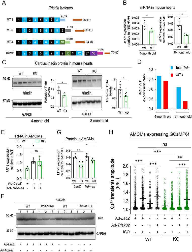
Background: Abnormalities in Ca2+ homeostasis are associated with cardiac arrhythmias and heart failure. Triadin plays an important role in Ca2+ homeostasis in cardiomyocytes. Alternative splicing of a single triadin gene produces multiple triadin isoforms. The cardiac-predominant isoform, mouse MT-1 or human Trisk32, is encoded by triadin exons 1 to 8. In humans, mutations in the triadin gene that lead to a reduction in Trisk32 levels in the heart can cause cardiac dysfunction and arrhythmias. Decreased levels of Trisk32 in the heart are also common in patients with heart failure. However, mechanisms that maintain triadin isoform composition in the heart remain elusive.

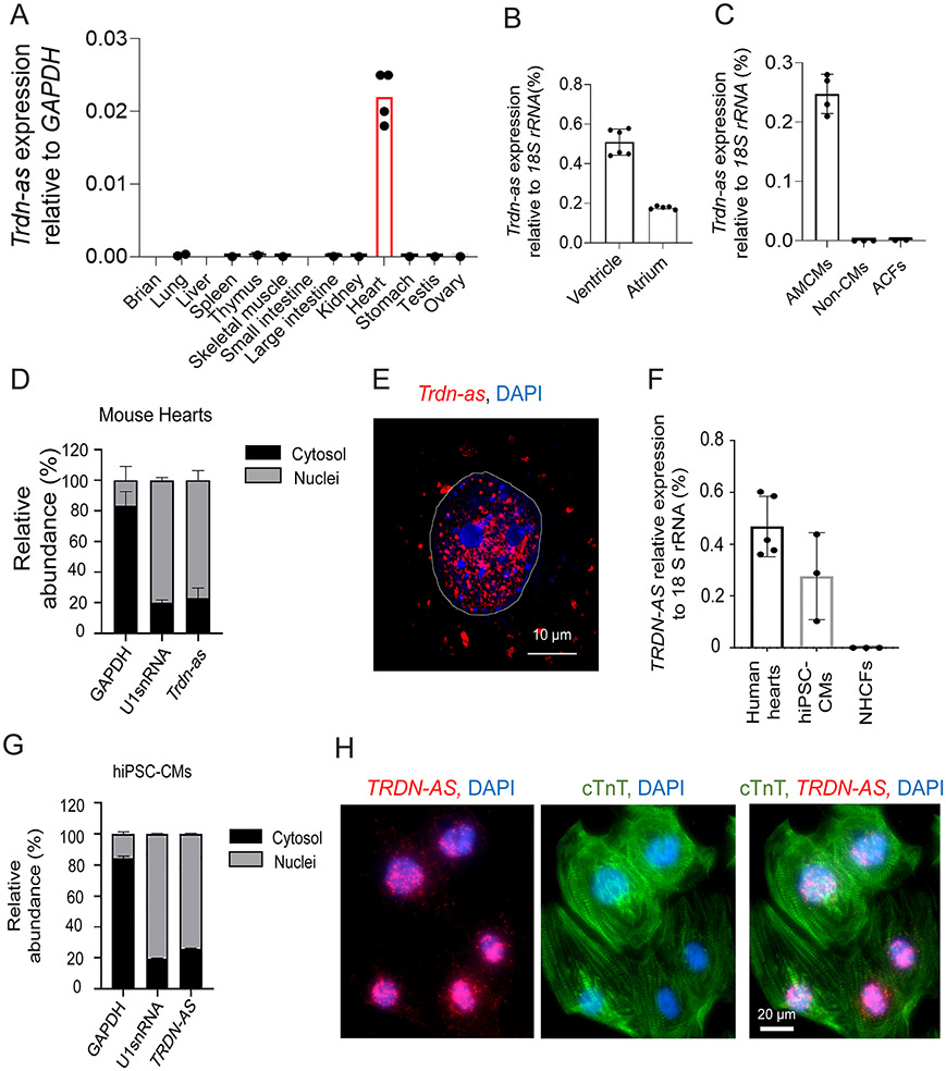
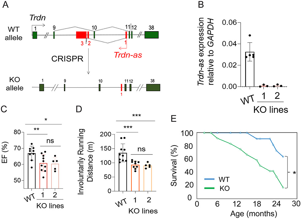
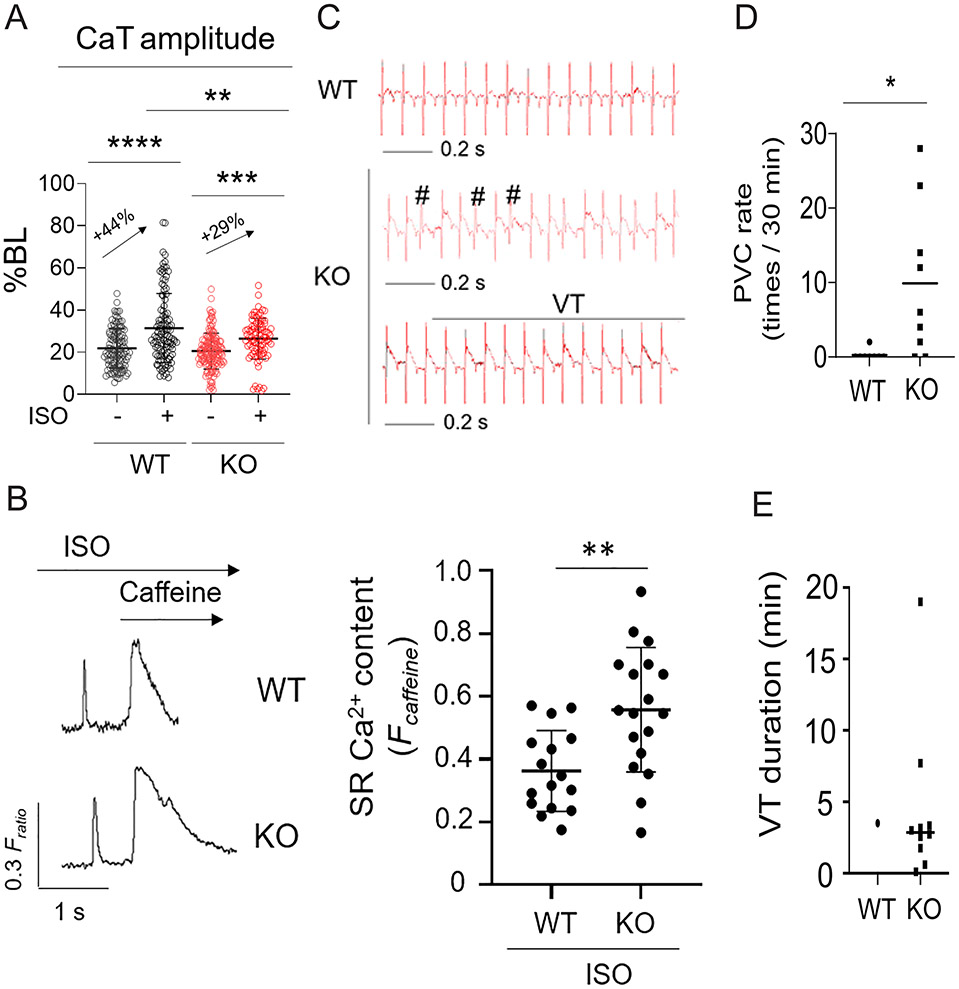
Methods: We analyzed triadin expression in heart explants from patients with heart failure and cardiac arrhythmias and in hearts from mice carrying a knockout allele for Trdn-as, a cardiomyocyte-specific long noncoding RNA encoded by the antisense strand of the triadin gene, between exons 9 and 11. Catecholamine challenge with isoproterenol was performed on Trdn-as knockout mice to assess the role of Trdn-as in cardiac arrhythmogenesis, as assessed by ECG. Ca2+ transients in adult mouse cardiomyocytes were measured with the IonOptix platform or the GCaMP system. Biochemistry assays, single-molecule fluorescence in situ hybridization, subcellular localization imaging, RNA sequencing, and molecular rescue assays were used to investigate the mechanisms by which Trdn-as regulates cardiac function and triadin levels in the heart.

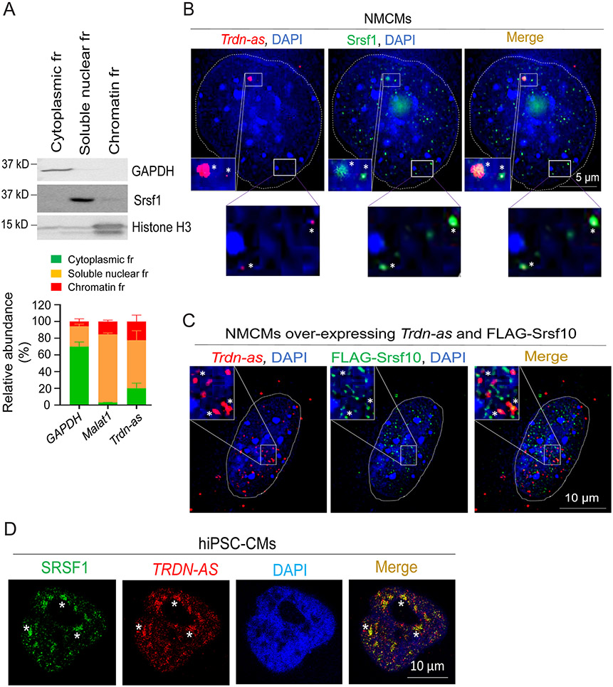
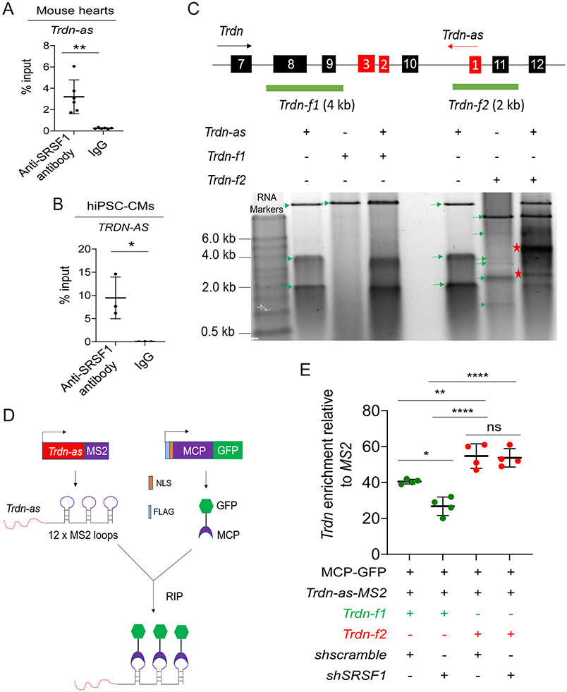
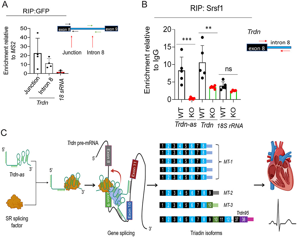
Results: We report that Trdn-as maintains cardiac function, at least in part, by regulating alternative splicing of the triadin gene. Knockout of Trdn-as in mice downregulates cardiac triadin, impairs Ca2+ handling, and causes premature death. Trdn-as knockout mice are susceptible to cardiac arrhythmias in response to catecholamine challenge. Normalization of cardiac triadin levels in Trdn-as knockout cardiomyocytes is sufficient to restore Ca2+ handling. Last, Trdn-as colocalizes and interacts with serine/arginine splicing factors in cardiomyocyte nuclei and is essential for efficient recruitment of splicing factors to triadin precursor mRNA.

Conclusions: These findings reveal regulation of alternative splicing as a novel mechanism by which a long noncoding RNA controls cardiac function. This study indicates potential therapeutics for heart disease by targeting the long noncoding RNA or pathways regulating alternative splicing.

Keywords: RNA, long noncoding; arrhythmias, cardiac; heart failure; myocytes, cardiac; splicing, alternative.


Figures

Figure 1.
Figure 1.. Human TRDN-AS and mouse Trdn-as are exclusively expressed in cardiomyocytes (CMs) and enriched in nuclei.
A-C) Expression of Trdn-as in mouse tissues (A), adult mouse heart ventricle and atria (B), CMs (AMCMs), non-CMs, and cardiac fibroblasts (ACFs) isolated from adult mouse hearts (C) by qPCR. Tissues or cells were harvested from at least 2 mice. Each filled circle represents one animal. Data are presented as mean in A or mean ± SD in B and C. D) qPCR analysis of nuclear and cytoplasmic RNAs in adult mouse hearts. GAPDH and U1 snRNA are used as markers of cytoplasmic and nuclear fractions, respectively. N = 3 mouse hearts. E) Cellular distributions of Trdn-as in neonatal mouse cardiomyocytes (NMCMs) by smFISH. Trdn-as was overexpressed in NMCMs by adenoviral delivery with a MOI of 5. smFISH assays were performed 72 hours post-transduction. The dotted line surrounds a CM nucleus. F) Expression of TRDN-AS in healthy human hearts, hiPSC-CMs, and normal human cardiac fibroblasts (NHCFs) by qPCR. Each filled circle represents an independent induction for hiPSC-CMs or an individual human heart explant. G) qPCR analysis of nuclear and cytoplasmic RNAs in hiPSC-CMs. GAPDH and U1 snRNA are used as markers of cytoplasmic and nuclear fractions, respectively. N = 3 independent inductions. H) Cellular distributions of human TRDN-AS in hiPSC-CMs by FISH. TRDN-AS was over-expressed in hiPSC-CMs by adenoviral delivery with a MOI of 5. FISH was performed 48 hours post-infection. hiPSC-CMs are marked by cardiac troponin T (cTnT).
Figure 2.
Figure 2.. Knockout of Trdn-as impaired cardiac function and caused premature death.
A) Knockout (KO) of Trdn-as by CRISPR/Cas9 genome editing. Trdn exons are shown in green and Trdn-as in red. B) Deletion of Trdn-as in hearts of two KO lines, line 1 and line 2 confirmed by qPCR. Each filled circle represents one mouse. Data are presented as mean ± SD. C-D) Left ventricular ejection fraction (EF) by echocardiography and running distance of indicated mice. Mice ran at speeds of 15, 25, 28, 31, 34, 37, and 40 m/min, each for 1 minute until mice were exhausted. Each filled circle represents one mouse. Data are presented as mean ± SD. One-way ANOVA with Tukey’s post test; *p = 0.05, **p < 0.05, ***p < 0.01, ns: not significant. E) Kaplan-Meier survival curve for WT (n = 11) and Trdn-as KO (n = 12) mice by OASIS2. Log-Rank test; *p<0.05. Both males and females were used for these assays.
Figure 3.
Figure 3.. Trdn-as KO mice are susceptible to cardiac arrhythmias in response to catecholamine challenge.
A) Ca2+ transients (CaT) of isolated AMCMs were field-stimulated at 1 Hz and recorded by the IonOptix system. AMCMs were treated with 0.1 μM ISO for 10 min prior to recording. 10 to 15 CMs per mouse, and 3 mice for each group were recorded. Each open circle represents an individual AMCM. Data are presented as mean ± SD. One-way ANOVA with Tukey’s post test, **p<0.01, ***p<0.001, ****p<0.0001. B) Modulation of SR Ca2+ load by Trdn-as under adrenergic stimulation. Representative IonOptix traces (left panel) of WT and Trdn-as KO AMCMS, which were isolated, loaded with Fura2-AM, pretreated with 500 nM Isoproterenol, and paced at 0.5 Hz. Where indicated, caffeine (10 mM) was added to the solution to induce Ca2+ release from SR, visible as high, extended peak. SR Ca2+ content was measured (right panel) by assessing the height of this peak , . 3 to 5 AMCMs per mouse, and 5 mice for each group were assayed. Each filled circle represents an individual AMCM. Data are presented as mean ± SD. P-values were calculated by Student’s t-test, **p<0.01. C) ECG records showing typical traces of WT mice and representative examples of PVC (#) and VT in KO mice after i.p. injection of ISO (0.1 mg/kg). D-E) Rate of PVC (D) and Duration of individual VT during a 30-min period after ISO injection (E). N = 8 WT mice and 9 KO mice. Each filled circle or square represents an individual WT or KO mouse. Data are presented as mean. P-values were calculated by Student’s t-test, *p<0.05.
Figure 4.
Figure 4.. Trdn-as is essential and sufficient for maintenance of cardiac triadin levels in mouse hearts.
A) Triadin isoforms are alternative splicing products of a single gene, Trdn. Three short isoforms, MT-1 (ENSMUST00000219982.2), MT-2 (ENSMUST00000217779.2), and MT-3 (ENSMUST00000219931.2) are expressed in the heart, while full-length triadin, Trdn95 (ENSMUST00000095761.5), is mainly expressed in skeletal muscle. Trdn exons are numbered. 3’ UTR sequence for each isoform is unique. B) Expression of triadin MT-1 in WT or Trdn-as KO hearts by qPCR. Total RNA was extracted from adult mouse hearts at indicated ages. Expression of triadin isoforms were quantified by qPCR using paired primers amplifying specific isoforms of triadin. N = 4 or 5 animals per each group. Each filled circle represents a mouse. Data are presented as mean ± SD. C) Immunoblotting analysis of triadin in WT or Trdn-as KO hearts at indicated ages. Lysates of adult mouse hearts were subjected to western blot. A predominant triadin band of ~32 kD was quantified on the right. Trdn95 protein was not detected in heart lysates. N = 4 or 8 animals per each group. Each filled circle represents a mouse. Data are presented as mean ± SD. D) Mean expression of total Trdn and MT-1 in Trdn-as KO vs WT (KO / WT) hearts at indicated ages. Expression of Trdn exons 7-8 was quantified using qPCR to represent total Trdn mRNA levels. Mean expression of total Trdn and MT-1 in the heart was calculated from 4 mice per group at the age of 4 months or 8 mice per group at the age of 8 months. E-G) Re-expression of Trdn-as in Trdn-as KO AMCMs rescued MT-1 expression at mRNA and protein levels. LacZ or Trdn-as was delivered into AMCMs by adenovirus (Ad). Gene expression was analyzed by qPCR for MT-1 RNA in E 48 hours post-infection or by immunoblotting for triadin protein in F-G 72 hours post-infection. Levels of indicated protein in AMCMs isolated from three WT or KO hearts are shown in F. In E and G, each filled circle represents a mouse. Data are presented as mean ± SD. H) Overexpression of human cardiac triadin (Trisk32) rescued the defect in Ca2+ transients in Trdn-as KO AMCMs. LacZ or Trisk32, and GCaMP6f were delivered into AMCMs by adenovirus (Ad). 48 hours later, AMCMs were treated with 0.1 μM ISO for 15 mins. Ca2+ transients were imaged and analyzed using the GCaMP6-based system. AMCMs were isolated from 3 WT or 3 Trdn-as KO mice. Each open circle represents one AMCM. Data are presented as mean + SD. One-way ANOVA with Tukey’s post test (E, G, H) or Student’s t-test (B, C), *p<0.05, **p<0.01, ***p<0.001, ns, not significant.
Figure 5.
Figure 5.. TRDN-AS was down-regulated in patients with heart failure.
A) Expression of TRDN-AS in hearts of healthy donors (Non-failing; NF) or patients with HF. Each open circle represents an individual human subject. Data are presented as mean + SD. B) Expression of cardiac triadin in NF or HF human hearts. Multiple bands with molecular weights of 30 to 50 kD appeared due to multiple isoforms and/or glycosylation of triadin in the heart, as shown in previous studies , . Trdn95 was not detectable in any samples by immunoblotting. Calnexin serves as a loading control. Non-glycosylated triadin, denoted by an arrow, was quantified. Each filled circle represents an individual human subject. Data are presented as mean ± SD. C) Expression correlation between TRDN-AS and cardiac triadin TRISK32 was analyzed using a simple linear regression model in GraphPad Prism 9.3. Each filled circle represents an individual human subject. Student’s t-test (A, B), **p<0.01.
Figure 6.
Figure 6.. Trdn-as and TRDN-AS colocalize with SR splicing factors in CMs.
A) qPCR analysis of the relative abundance of transcripts from subcellular fractions (fr) of AMCMs. Immunoblotting for GAPDH as a marker for cytoplasm, Srsf1 for soluble nuclear, and histone H3 for chromatin fractions are shown in the top panel. GAPDH and Malat1 RNAs are used as markers for cytoplasmic and soluble nuclear fractions, respectively. N = 3 independent experiments. Data are presented as mean + SD. B-C) Confocal imaging analysis of Trdn-as and Srsf1 in B and over-expressed Trdn-as and FLAG-Srsf10 in C in NMCMs. Endogenous (in B) or over-expressed Trdn-as (in C) were detected by smFISH. In C, NMCMs were infected with adenovirus vectors carrying Trdn-as or FLAG-Srsf10 with a MOI of 5. 48 hours later, over-expressed Trdn-as and FLAG-Srsf10 were analyzed using smFISH and immunostaining, respectively. Nuclei are surrounded by white lines. Insets are enlarged on the right corner. Colocalizations are indicated by stars. D) Confocal imaging analysis of TRDN-AS and SRSF1 in a hiPSC-CM by FISH. TRDN-AS was over-expressed in hiPSC-CMs by adenovirus with a MOI of 5. 48 hours later, TRDN-AS and SRSF1 were analyzed using FISH and immunostaining, respectively. Colocalizations are indicated by stars.
Figure 7.
Figure 7.. Trdn-as interact with SR splicing factors and Trdn pre-mRNA.
A) RIP-qPCR analysis of the interaction of Trdn-as with Srsf1 in adult mouse hearts. n=6 independent experiments. Data are presented as mean ± SD. Student’s t-test, **p<0.01. B) RIP-qPCR analysis of the interaction of TRDN-AS with SRSF1 in hiPSC-CMs. n=3 independent experiments. Data are presented as mean ± SD. Student’s t-test, *p<0.05. C) Analysis of interactions between Trdn-as and Trdn pre-mRNA by native agarose gel electrophoresis. Trdn and Trdn-as genes schematics are shown on top. Exons of Trdn and Trdn-as are numbered in black and red boxes, respectively. The black and red arrows indicate the direction of Trdn or Trdn-as transcription, respectively. Full-length Trdn-as and two partial fragments of Trdn pre-mRNA, Trdn-f1 and Trdn-f2 were in vitro transcribed. RNA was purified after treatment with proteinase K and RNase-free DNase I. The mixture of Trdn-as and either Trdn-f1 or Trdn-f2 was resolved in native agarose gel electrophoresis. Green arrows indicate individual RNAs. Asterisks indicate RNA-RNA duplex. D) A schematic diagram shows pulldown of lncRNA-associated complexes using the MS2-MCP system. MS2 RNA stem loops strongly interact with the MS2 coat protein (MCP). Trdn-as was fused to 12 x MS2 stem loops (Trdn-as-MS2). Nuclear-localized, FLAG-tagged MCP was fused to GFP (FLAG-NLS-MCP-GFP). Trdn-as associated complexes are precipitated using a GFP antibody. E) qPCR analysis of Trdn pre-mRNA levels in complexes precipitated by the GFP antibody. HEK293 cells were transfected with various combinations of expression plasmids encoding MCP-GFP, shscramble, shSRSF1, MS2, or Trdn-as-MS2. 48 hours later, nuclear extracts of HEK293 cells were incubated with in vitro transcribed Trdn pre-mRNA Trdn-f1 or Trdn-f2. MS2 or Trdn-as-MS2 associated complexes were precipitated by anti-GFP antibody. Trdn pre-mRNA was analyzed using qPCR to amplify Trdn exon 8 for Trdn-f1 or Trdn exon 11 for Trdn-f2, respectively. N = 4 independent experiments. Data are presented as mean ± SD. One-way ANOVA with Tukey’s post hoc test. *p<0.05, **p<0.01, ****p<0.0001, ns, not significant.
Figure 8.
Figure 8.. Trdn-as is required for recruitment of SR splicing factors to Trdn transcripts in the heart.
A) RIP of Trdn-as associated complexes in AMCMs using the MS2-MCP system in Figure 7D. AMCMs were infected by adenoviruses carrying either MS2 or Trdn-as-MS2 and MCP-GFP. RIP with an anti-GFP antibody was performed 48 hours post-infection. Junction of Trdn exon 8 and intron 8, Trdn intron 8 denoting Trdn pre-mRNA, and 18s rRNA were then quantified by qPCR. N = 4 under each condition. Relative enrichment was calculated by normalization of RNA levels in AMCMs expressing Trdn-as-MS2 to those in AMCMs expressing MS2 post-RIP with the antibody against GFP. Data are presented as mean + SD. B) RIP with anti-SRSF1 antibody followed by qPCR analysis of RNAs in Srsf1-associated complexes in WT or Trdn-as KO mouse hearts. Trdn was quantified by qPCR to amplify the exon 8/intron 8 junction. N = 4-6 mouse hearts under each condition. Data are presented as mean + SD. One-way ANOVA with Tukey’s post test, ***p<0.001, ns, not significant. C) Model of Trdn-as to modulate triadin isoform composition in the heart. Trdn-as colocalizes and interacts with SR splicing factors in nuclear speckles. Trdn-as forms an RNA/RNA duplex with Trdn pre-mRNA and facilitates the recruitment of SR splicing factors to Trdn pre-mRNA to produce the predominant triadin isoform MT-1 in the heart. Knockout of Trdn-as impairs the recruitment of splicing factors to Trdn pre-mRNA, leading to abnormal composition of triadin isoforms in the heart, Ca2+ mishandling, and susceptibility to cardiac arrhythmias.

Comment in

References

    1. Hill JA, Olson EN. Cardiac plasticity. N Engl J Med. 2008;358:1370–1380. doi:10.1056/NEJMra072139. - DOI - PubMed
    1. Smith GL, Eisner DA. Calcium Buffering in the Heart in Health and Disease. Circulation. 2019;139:2358–2371. doi:10.1161/CIRCULATIONAHA.118.039329 - DOI - PMC - PubMed
    1. Bers DM. Cardiac sarcoplasmic reticulum calcium leak: basis and roles in cardiac dysfunction. Annu Rev Physiol. 2014;76:107–127. doi:10.1146/annurev-physiol-020911-153308 - DOI - PubMed
    1. Guo W, Campbell KP. Association of triadin with the ryanodine receptor and calsequestrin in the lumen of the sarcoplasmic reticulum. J Biol Chem. 1995;270:9027–30. doi: 10.1074/jbc.270.16.9027. - DOI - PubMed
    1. Kobayashi YM, Alseikhan BA, Jones LR. Localization and characterization of the calsequestrin-binding domain of triadin 1. Evidence for a charged beta-strand in mediating the protein-protein interaction. J Biol Chem. 2000;275:17639–46. doi: 10.1074/jbc.M002091200. - DOI - PubMed

Publication types

MeSH terms