

Neuronal Alterations in Secondary Thalamic Degeneration Due to Cerebral Infarction: A 11C-Flumazenil Positron Emission Tomography Study
- PMID: 35862203
- PMCID: PMC9508960
- DOI: 10.1161/STROKEAHA.122.038846
Neuronal Alterations in Secondary Thalamic Degeneration Due to Cerebral Infarction: A 11C-Flumazenil Positron Emission Tomography Study
Abstract
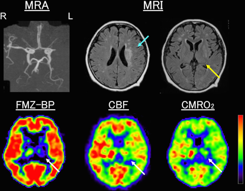
Background: Studies using animal experiments have shown secondary neuronal degeneration in the thalamus after cerebral infarction. Neuroimaging studies in humans have revealed changes in imaging parameters in the thalamus, remote to the infarction. However, few studies have directly demonstrated neuronal changes in the thalamus in vivo. The purpose of this study was to determine whether secondary thalamic neuronal damage may manifest as a decrease in central benzodiazepine receptors in patients with cerebral infarction and internal carotid artery or middle cerebral artery disease.
Methods: We retrospectively analyzed the data of 140 patients with unilateral cerebral infarction ipsilateral to internal carotid artery or middle cerebral artery disease. All patients had quantitative measurements of 11C-flumazenil binding potential (FMZ-BP), cerebral blood flow, and cerebral metabolic rate of oxygen using positron emission tomography in the chronic stage. Region of interest analysis was performed using NeuroFlexer-an automated region of interest analysis software using NEUROSTAT.
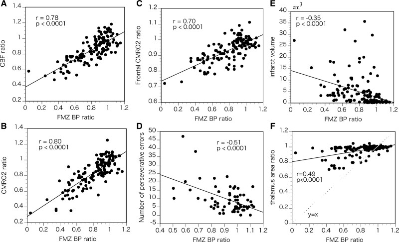
Results: In the thalamus ipsilateral to the infarcts, the values of FMZ-BP, cerebral blood flow, and cerebral metabolic rate of oxygen were significantly lower than those in the contralateral thalamus. Significant correlations were found between the ipsilateral-to-contralateral ratio of FMZ-BP and the ipsilateral-to-contralateral ratio of cerebral blood flow or cerebral metabolic rate of oxygen in the thalamus. Patients with corona radiata infarcts and striatocapsular infarcts had significantly decreased ipsilateral-to-contralateral FMZ-BP ratio in the thalamus compared with those without. The ipsilateral-to-contralateral ratio of FMZ-BP in the thalamus was significantly correlated with the ipsilateral-to-contralateral cerebral metabolic rate of oxygen ratio in the frontal cortex and showed a significant negative correlation with the number of perseverative errors on the Wisconsin Card Sorting Test.
Conclusions: Secondary thalamic neuronal damage may manifest as a decrease in central benzodiazepine receptors in patients with cerebral infarction and internal carotid artery or middle cerebral artery disease, which may be associated with frontal lobe dysfunction.
Keywords: cerebral infarction; cerebrovascular disorders; positron emission tomography; receptors, GABA-A; thalamus.
Figures
Similar articles
-
Permanent cortical damage detected by flumazenil positron emission tomography in acute stroke.Stroke. 1998 Feb;29(2):454-61. doi: 10.1161/01.str.29.2.454. Stroke. 1998. PMID: 9472889
-
Selective neuronal damage and borderzone infarction in carotid artery occlusive disease: a 11C-flumazenil PET study.J Nucl Med. 2005 Dec;46(12):1973-9. J Nucl Med. 2005. PMID: 16330559
-
Early detection of irreversibly damaged ischemic tissue by flumazenil positron emission tomography in cats.Stroke. 1997 Oct;28(10):2045-51; discussion 2051-2. doi: 10.1161/01.str.28.10.2045. Stroke. 1997. PMID: 9341717
-
Glucose and [11C]flumazenil positron emission tomography abnormalities of thalamic nuclei in temporal lobe epilepsy.Neurology. 1999 Dec 10;53(9):2037-45. doi: 10.1212/wnl.53.9.2037. Neurology. 1999. PMID: 10599778
-
Imaging the ischemic penumbra and treatment effects by PET.Keio J Med. 2001 Dec;50(4):249-56. doi: 10.2302/kjm.50.249. Keio J Med. 2001. PMID: 11806502 Review.
Cited by
-
Early histopathological changes of secondary degeneration in the spinal cord after total MCA territory stroke.Sci Rep. 2023 Dec 11;13(1):21934. doi: 10.1038/s41598-023-49230-x. Sci Rep. 2023. PMID: 38082027 Free PMC article.
-
Secondary thalamic dysfunction underlies abnormal large-scale neural dynamics in chronic stroke.Proc Natl Acad Sci U S A. 2024 Nov 12;121(46):e2409345121. doi: 10.1073/pnas.2409345121. Epub 2024 Nov 6. Proc Natl Acad Sci U S A. 2024. PMID: 39503890 Free PMC article.
-
The p75 neurotrophin receptor attenuates secondary thalamic damage after cortical infarction by promoting angiogenesis.CNS Neurosci Ther. 2024 Jul;30(7):e14875. doi: 10.1111/cns.14875. CNS Neurosci Ther. 2024. PMID: 39072998 Free PMC article.
-
Alterations in the functional connectivity of thalamic subregions after basal ganglia stroke.Front Neurol. 2025 Jun 6;16:1584290. doi: 10.3389/fneur.2025.1584290. eCollection 2025. Front Neurol. 2025. PMID: 40546256 Free PMC article.
References
-
- Feeney DM, Baron JC. Diaschisis. Stroke. 1986;17:817–830. doi: 10.1161/01.str.17.5.817 - PubMed
-
- Zhang J, Zhang Y, Xing S, Liang Z, Zeng J. Secondary neurodegeneration in remote regions after focal cerebral infarction: a new target for stroke management? Stroke. 2012;43:1700–1705. doi: 10.1161/STROKEAHA.111.632448 - PubMed
-
- Fujie W, Kirino T, Tomukai N, Iwasawa T, Tamura A. Progressive shrinkage of the thalamus following middle cerebral artery occlusion in rats. Stroke. 1990;21:1485–1488. doi: 10.1161/01.str.21.10.1485 - PubMed
-
- Iizuka H, Sakatani K, Young W. Neural damage in the rat thalamus after cortical infarcts. Stroke. 1990;21:790–794. doi: 10.1161/01.str.21.5.790 - PubMed
-
- De Reuck J, Decoo D, Lemahieu I, Strijckmans K, Goethals P, Van Maele G. Ipsilateral thalamic diaschisis after middle cerebral artery infarction. J Neurol Sci. 1995;134:130–135. doi: 10.1016/0022-510x(95)00229-2 - PubMed
Publication types
MeSH terms
Substances
LinkOut - more resources
Full Text Sources
