. 2022 Sep 22;7(18):e135407.
doi: 10.1172/jci.insight.135407.

Spatiotemporal regulation of human IFN-ε and innate immunity in the female reproductive tract

Affiliations

Spatiotemporal regulation of human IFN-ε and innate immunity in the female reproductive tract

Nollaig M Bourke et al. JCI Insight. .

Abstract

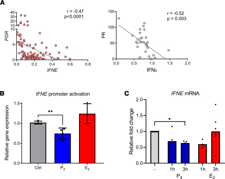
Although published studies have demonstrated that IFN-ε has a crucial role in regulating protective immunity in the mouse female reproductive tract, expression and regulation of IFN-ε in the human female reproductive tract (hFRT) have not been characterized to our knowledge. We obtained hFRT samples from a well-characterized cohort of women to enable us to comprehensively assess ex vivo IFN-ε expression in the hFRT at various stages of the menstrual cycle. We found that among the various types of IFNs, IFN-ε was uniquely, selectively, and constitutively expressed in the hFRT epithelium. It had distinct expression patterns in the surface and glandular epithelia of the upper hFRT compared with basal layers of the stratified squamous epithelia of the lower hFRT. There was cyclical variation of IFN-ε expression in the endometrial epithelium of the upper hFRT and not in the distal FRT, consistent with selective endometrial expression of the progesterone receptor and regulation of the IFNE promoter by progesterone. Because we showed IFN-ε stimulated important protective IFN-regulated genes in FRT epithelium, this characterization is a key element in understanding the mechanisms of hormonal control of mucosal immunity.

Keywords: Cytokines; Immunology; Reproductive Biology.


Conflict of interest statement

Conflict of interest: SLA has received consulting fees from Mayne Pharma and Merck. Magee-Womens Research Institute receives research funding from Estetra SRL, EvoFem, and Merck.

Figures

Figure 1
Figure 1. Distinct expression patterns of IFN-ε in upper and lower hFRT mucosa.
(AC) Representative images of IFN-ε expression in sections from matched biopsy samples from 33 women. Sections from the (A) endometrium, (B) ectocervix, and (C) vagina were stained for expression of IFN-ε (brown staining, highlighted with red arrows) or IgG control. Scale bar, 200 μm. EP, epithelium, GE, glandular epithelium; LE, luminal epithelium; ST, stroma.
Figure 2
Figure 2. Cyclic variation of IFN-ε expression only in the upper hFRT.
(A) IFNE mRNA expression, as determined by quantitative PCR (qPCR), in vaginal, ectocervical, and endometrial biopsy samples stratified into follicular (n = 16) and luteal (n = 16) stages of the menstrual cycle. Quantification (B) and representative IHC images (C) of endometrial epithelial IFN-ε staining intensity in women in the follicular or luteal stage of the menstrual cycle, using the Aperio positive pixel–count algorithm to generate intensity values for staining. IFN-ε staining is highlighted with red arrows. Significance was determined using either Kruskal-Wallis testing with Dunn’s multiple-comparison analysis (A) or Mann-Whitney U test (B). **P < 0.01; ***P < 0.001. GE, glandular epithelium; LE, luminal epithelium; ST, stroma.
Figure 3
Figure 3. Expression of PR selective and cyclic changes only in upper hFRT.
(A)PR gene (PGR) mRNA expression, as determined by qPCR, in vaginal, ectocervical, and endometrial biopsy samples. (B) Representative images of PR expression in sections from matched biopsy samples from 33 women. Sections from the endometrium, ectocervix, and vagina were stained for expression of PR (brown) or IgG control. Scale bar, 200 μm. (C) PGR mRNA expression, as determined by qPCR, in vaginal, ectocervical, and endometrial biopsy samples stratified into follicular (n = 16) and luteal (n = 16) stages of the menstrual cycle. (D) Quantification and (E) representative IHC images of cytoplasmic PR staining intensity in endometrial epithelial cells from women in the follicular or luteal stage of the menstrual cycle. H-scores for staining were generated using the Aperio cytoplasm algorithm, which classifies cytoplasmic staining intensity scoring as 0, none; 1+, weak; 2+ moderate; or 3+, strong, and uses this to generate an H-score using the following formula: 1 × (%1+) + 2 × (%2+) + 3 × (%3+). PR staining is highlighted with red arrows. Significance determined using either Kruskal-Wallis testing with Dunn’s multiple-comparison analysis (A and C) or Mann-Whitney U test (D). **P < 0.01, ****P < 0.0001. EP, epithelium, GE, glandular epithelium; LE, luminal epithelium; ST, stroma.
Figure 4
Figure 4. Regulation of IFN-ε by PR.
(A) Negative correlation of both mRNA (left) and protein expression (right) of IFN-ε and PR in hFRT cells. Spearman correlation analysis. (B) Luciferase reporter assay measuring activation of the human IFNE promoter in ECC-1 cells after treatment with either 10 nM progesterone or 10 nM estrogen for 4 hours. Data are from 4 independent biological replicates, each in technical triplicate, shown as mean +SEM and analyzed using Student’s 2-tailed t test. **P < 0.01. (C) Primary uterine epithelial cells were isolated from endometrial biopsy specimens (from up to 6 donors) and cultured for 3 days prior to stimulation for either 1 or 3 hours with 10 nM progesterone or 10 nM estrogen. IFNE expression was quantified using qPCR, expressed relative to expression of 18S and fold change relative to unstimulated control. Significance was determined using Kruskal-Wallis testing with Dunn’s multiple-comparison analysis. *P < 0.05.
Figure 5
Figure 5. Exclusive expression of IFN-ε in hFRT regulates immune-protective IRGs.
(A) Spearman’s correlation analysis of the expression of IFNE with the IRGs MX1, CXCL10, and OAS2 across hFRT biopsy samples. (B) Primary uterine epithelial cells were isolated from endometrial biopsy specimens and cultured for 3 days prior to stimulation with IFN-β (n = 9) or IFN-ε (n = 20). (C) Expression of type I IFN (IFNA1, IFNA2, IFNA4, IFNB, IFNE), type II IFN (IFNG), and type III IFN (IL28A, IL28B, IL29) was quantified by qPCR in matched vaginal, ectocervical, and endometrial biopsy samples from 32 women regardless of phase of menses. In vaginal samples, IFNA1 was not detectable (N/D) in 10, IFNA2 was N/D in 24, IFNA4 was N/D in 31, IFNB was N/D in 13, IFNG was N/D in 2, IFNL1and IFNL2 were N/D in 31, and IFNL3 was N/D in 30 specimens. In ectocervical samples, IFNA1 was N/D in 6, IFNA2 was N/D in 24, IFNA4 was N/D in 28, IFNB was N/D in 7, IFNG was N/D in 1, IFNL1 was N/D in 30, IFNL2 was N/D in 31, and was IFNL3 was N/D in 27 specimens. In endometrial samples, IFNA1 was N/D in 10, IFNA2 was N/D in 24, IFNA4 was N/D in 31, IFNB was N/D in 13, IFNG was N/D in 2, IFNL1 and IFNL2 was N/D in 31, and IFNL3 was N/D in 30 specimens. Gene expression was quantified using qPCR, normalized to 18S expression, and expressed relative to untreated control cells. The box plots depict the minimum and maximum values (whiskers), the upper and lower quartiles, and the median. The length of the box represents the interquartile range. Data were analyzed using Kruskal-Wallis testing with Dunn’s multiple-comparison analysis: ****P < 0.0001; or Mann-Whitney U test: *P < 0.05, **P < 0.01, ***P < 0.001. US, unstimulated.
Figure 6
Figure 6. IFN-ε protein is expressed in CVL.
(A) Concentrations of IFN-ε, IL-15, and IL-6 were quantified in CVL fluid (n = 32) using either laboratory-developed (IFN-ε) or commercially available (IL-15, IL-6) immunoassays. There was undetectable cytokine expression in 4 samples for IFN-ε and 2 samples for IL-15. (B) CVL IFN-ε, IL-15, and IL-6 expression was stratified by cycle stage into follicular stage samples (n = 16) and luteal stage samples (n = 16). There was undetectable cytokine expression for IFN-ε in 1 follicular and 3 luteal stage samples and IL-15 in 1 follicular and 1 luteal stage sample. (C) IFNE, IL15, and IL6 mRNA expression, as determined by qPCR, in endometrial, ectocervical, and vaginal biopsy samples stratified into follicular (n = 16) and luteal (n = 16) stages of menstrual cycle. Mann-Whitney U tests were applied to determine cyclic differences for each gene or protein of interest. *P < 0.05, **P < 0.01,***P < 0.001.

References

    1. Roy A, Matzuk MM. Reproductive tract function and dysfunction in women. Nat Rev Endocrinol. 2011;7(9):517–525. doi: 10.1038/nrendo.2011.79. - DOI - PubMed
    1. Wira CR, et al. Sex hormone regulation of innate immunity in the female reproductive tract: the role of epithelial cells in balancing reproductive potential with protection against sexually transmitted pathogens. Am J Reprod Immunol. 2010;63(6):544–565. doi: 10.1111/j.1600-0897.2010.00842.x. - DOI - PMC - PubMed
    1. Wira CR, et al. The role of sex hormones in immune protection of the female reproductive tract. Nat Rev Immunol. 2015;15(4):217–230. doi: 10.1038/nri3819. - DOI - PMC - PubMed
    1. Kaushic C, et al. Effects of estradiol and progesterone on susceptibility and early immune responses to Chlamydia trachomatis infection in the female reproductive tract. Infect Immun. 2000;68(7):4207–4216. doi: 10.1128/IAI.68.7.4207-4216.2000. - DOI - PMC - PubMed
    1. Kaushic C, et al. Progesterone increases susceptibility and decreases immune responses to genital herpes infection. J Virol. 2003;77(8):4558–4565. doi: 10.1128/JVI.77.8.4558-4565.2003. - DOI - PMC - PubMed