. 2022 Jul 26;119(30):e2202125119.
doi: 10.1073/pnas.2202125119. Epub 2022 Jul 21.

LRBA is essential for urinary concentration and body water homeostasis

Affiliations

LRBA is essential for urinary concentration and body water homeostasis

Yu Hara et al. Proc Natl Acad Sci U S A. .

Abstract

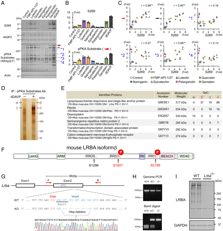
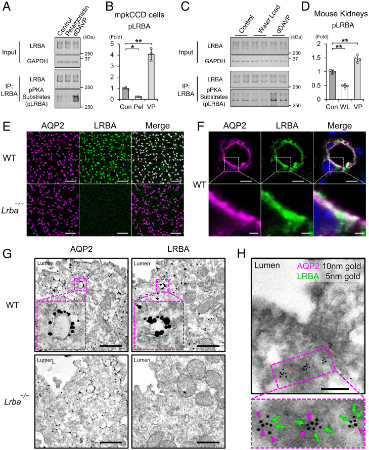
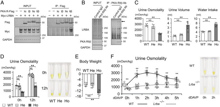
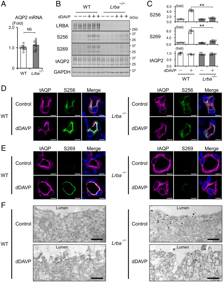
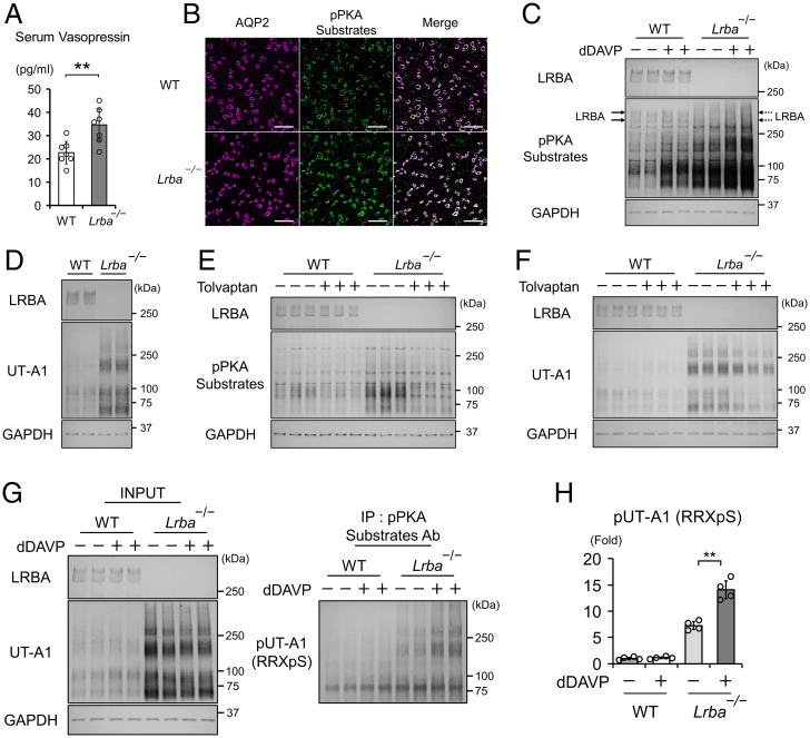
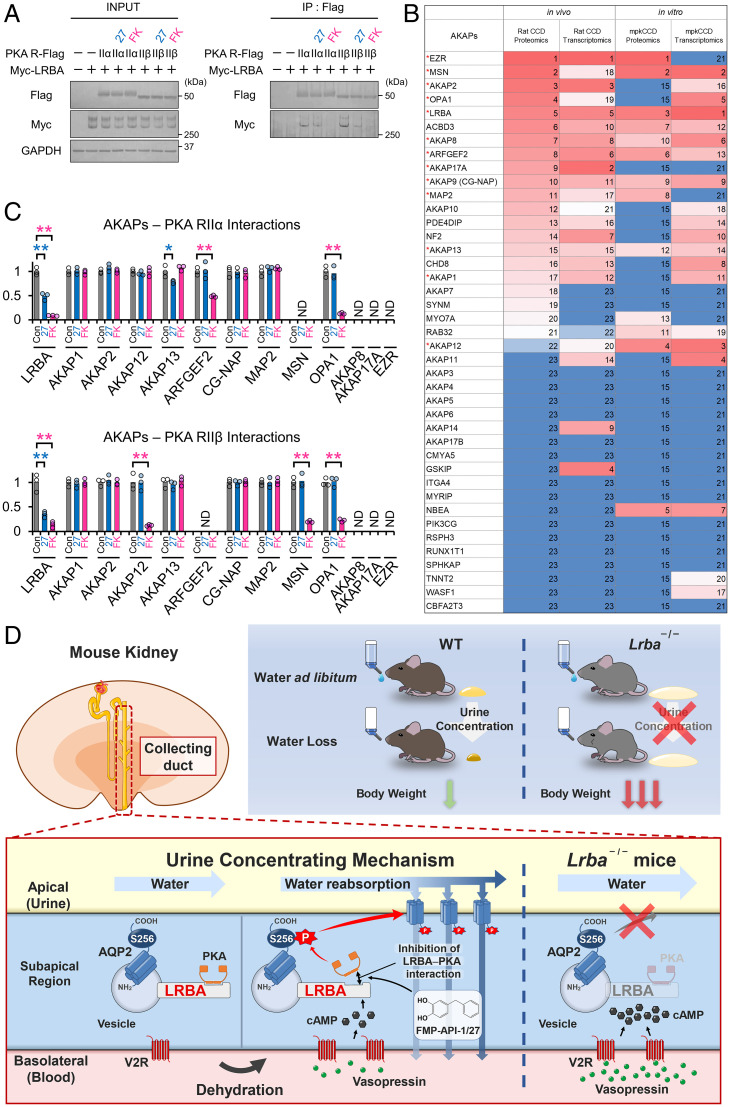
Protein kinase A (PKA) directly phosphorylates aquaporin-2 (AQP2) water channels in renal collecting ducts to reabsorb water from urine for the maintenance of systemic water homeostasis. More than 50 functionally distinct PKA-anchoring proteins (AKAPs) respectively create compartmentalized PKA signaling to determine the substrate specificity of PKA. Identification of an AKAP responsible for AQP2 phosphorylation is an essential step toward elucidating the molecular mechanisms of urinary concentration. PKA activation by several compounds is a novel screening strategy to uncover PKA substrates whose phosphorylation levels were nearly perfectly correlated with that of AQP2. The leading candidate in this assay proved to be an AKAP termed lipopolysaccharide-responsive and beige-like anchor protein (LRBA). We found that LRBA colocalized with AQP2 in vivo, and Lrba knockout mice displayed a polyuric phenotype with severely impaired AQP2 phosphorylation. Most of the PKA substrates other than AQP2 were adequately phosphorylated by PKA in the absence of LRBA, demonstrating that LRBA-anchored PKA preferentially phosphorylated AQP2 in renal collecting ducts. Furthermore, the LRBA-PKA interaction, rather than other AKAP-PKA interactions, was robustly dissociated by PKA activation. AKAP-PKA interaction inhibitors have attracted attention for their ability to directly phosphorylate AQP2. Therefore, the LRBA-PKA interaction is a promising drug target for the development of anti-aquaretics.

Keywords: AKAP; AQP2; LRBA; PKA; urinary concentration.


Conflict of interest statement

The authors declare no competing interest.

Figures

Fig. 1.
Fig. 1.
FMP-API-1/27 and flavonoids phosphorylate LRBA and AQP2 in a correlated manner. (A–C) Screening for PKA substrates whose phosphorylation are well correlated with pAQP2-S269. (A) Representative blots of pAQP2-S269 and pPKA substrates. (B) Phosphorylation levels of AQP2-S269 and the PKA substrate (indicated by red arrowhead) are quantified by densitometric analysis (n = 3). (C) Phosphorylation levels of AQP2-S269 and PKA substrates (indicated by arrowheads) are strongly correlated in scatterplots. (D and E) Immunoprecipitated PKA substrates (indicated by β and δ) are identified as LRBA by liquid chromatography–tandem mass spectrometry (LC-MS/MS). (D) Representative silver staining of pPKA substrates immunoprecipitated by pPKA substrate antibody. (E) Identified proteins by LC-MS/MS. (F) PTM analysis reveals the RRXS sites of LRBA phosphorylated by dDAVP. (G and H) Lrba knockout mice are generated by CRISPR/Cas9 genome-editing technology. (G) The target sequence for Lrba gene editing. (H) Genotyping of Lrba knockout mice after BanII digestion of the PCR products from genomic DNA. (I) Anti-LRBA antibody detects renal LRBA in WT mice. (C) Pearson correlation coefficient r value and two-sided Student's t test. **P < 0.01. Ab, antibody; IP, immunoprecipitation; PAM, protospacer adjacent motif; SpC, spectral count.
Fig. 2.
Fig. 2.
LRBA colocalized with AQP2 at vesicles in the subapical region. (A and B) The effects of pelargonidin and vasopressin on LRBA phosphorylation in mpkCCD cells (n = 3). (C and D). The effects of water load and vasopressin on LRBA phosphorylation in vivo (n = 4). (E and F) Immunofluorescence staining showing that AQP2 (magenta) and LRBA (green) are colocalized in the kidneys of WT mice. (E) Scale bars, 100 μm. (F) Scale bars: top, 5 μm; bottom, 1 μm. (G) Immuno-electron microscopy showing that AQP2 and LRBA are localized at vesicles in renal collecting ducts of WT mice. Scale bars, 500 nm. (H) Colocalization of AQP2 and LRBA at the same intracellular vesicles. Double-label immuno-electron microscopy of AQP2 (10-nm gold indicated by magenta arrowheads) and LRBA (5-nm gold indicated by green arrows) in renal collecting ducts of WT mice. Scale bars, 200 nm. Data are reported as mean ± SD. *P < 0.05, **p < 0.01 by Dunnett's test. IP, immunoprecipitation; Pel, pelargonidin; VP, dDAVP; WL, water load.
Fig. 3.
Fig. 3.
Lrba knockout mice display the NDI phenotype. (A) LRBA interacts with PKA RIIα and RIIβ in vitro. FLAG-tagged PKA regulatory subunits and Myc-tagged LRBA were overexpressed in HEK293T cells. Anti-FLAG beads were used to perform coimmunoprecipitations. (B) LRBA interacts with PKA RIIβ in vivo. Membrane fraction of mouse kidneys was prepared. Anti-RIIβ antibody was used to perform a coimmunoprecipitation assay. (C) The baseline urine-concentrating ability is impaired in Lrba−/− mice. Metabolic cages were used to monitor urine osmolality, urine volume, and water intake for 24 h. (n = 5 WT mice; n = 4, He mice; n = 7 Ho mice). (D and E) The maximal urine-concentrating ability is impaired in Lrba−/− mice. Mice were deprived of water for 12 h. (D) Left, urine osmolality at the beginning and end of the experiments (n = 6). Right, representative urine samples. (E) Percentage of weight loss after water deprivation test (n = 6). (F) Urine osmolality of Lrba−/− mice is unresponsive to vasopressin. Left, changes in urine osmolality of WT and Lrba−/− mice after the treatment of dDAVP (0.4 μg/kg) (n = 7). Right, representative urine samples. (C–E) Data are reported as mean ± SD. **P < 0.01, NS, not significant by Dunnett's test. (F) **P < 0.01 by two-sided Student's t test. Ab, antibody; He, Lrba+/−; Ho, Lrba−/−; IP, immunoprecipitation.
Fig. 4.
Fig. 4.
Lrba knockout impairs AQP2 phosphorylation and trafficking. (A) AQP2 mRNA levels are not different between WT and Lrba−/− mice. AQP2 mRNA levels were quantified by qPCR (n = 10). (B and C) AQP2 phosphorylation is impaired in Lrba−/− mice. dDAVP (0.4 μg/kg) was intraperitoneally injected into WT and Lrba−/− mice for 1 h (n = 6). (D and E) Enhanced signal intensity of pAQP2-S256 and pAQP2-S269 by dDAVP is impaired in Lrba−/− mice. Immunofluorescence staining of AQP2 (magenta) and pAQP2-S256 (green) (D) or pAQP2-S269 (green) (E) in the kidneys of WT and Lrba−/− mice. dDAVP was administered as in B. Scale bars, 5 μm. (F) Immuno-electron microscopy showing that accumulation of AQP2 at the apical plasma membrane by dDAVP is impaired in Lrba−/− mice. dDAVP was administered as in B. Scale bars, 500 nm. Data are reported as mean ± SD. (A) Two-sided Student's t test. (C) **P < 0.01 by Dunnett's test. NS, not significant.
Fig. 5.
Fig. 5.
Vasopressin phosphorylates most of the renal PKA substrates in Lrba−/− mice. (A) Serum vasopressin levels are elevated in Lrba−/− mice (n = 3 WT mice; n = 4 Lrba−/− mice). (B) pPKA substrates are localized at renal collecting ducts. Immunofluorescence staining of AQP2 (magenta) and pPKA substrates (green) in WT and Lrba−/− mice. Scale bars, 100 μm. (C) Vasopressin enhances phosphorylation of PKA substrates in Lrba−/− mice. dDAVP (0.4 μg/kg) was intraperitoneally administered into WT and Lrba−/− mice for 1 h (n = 6). (D) Protein expression of UT-A1 is increased in Lrba−/− mice (n = 6). (E) Tolvaptan suppresses PKA activity in Lrba−/− mice. WT and Lrba−/− mice were subcutaneously infused with tolvaptan (25 mg/kg/d) for 48 h using osmotic minipumps (n = 6). (F) Tolvaptan decreases protein expressions of UT-A1. Mice were administered tolvaptan as in E (n = 5). (G) UT-A1 is phosphorylated by vasopressin in Lrba−/− mice. At 1 h after the administration of dDAVP (0.4 μg/kg), UT-A1 was immunoprecipitated by a pPKA-substrate antibody and probed with anti–UT-A1 antibody. (H) Densitometric analysis of pUT-A1 (n = 4). Data are reported as mean ± SD. (A) **P < 0.01 by two-sided Student's t test. (H) **P < 0.01 by Tukey test. (CH) The inner medulla was dissected from the kidneys. Ab, antibody; IP, immunoprecipitation.
Fig. 6.
Fig. 6.
The LRBA–PKA interaction is the drug target of FMP-API-1/27. (A) FMP-API-1/27 dissociates the LRBA–PKA RIIβ interaction. PKA RII subunits (RIIα, RIIβ)-Flag and Myc-LRBA were overexpressed in HEK293T cells. FMP-API-1/27 (200 μM) or forskolin (10 μM) was added to the cells 1 h before coimmunoprecipitation. Representative blots of coimmunoprecipitated Myc-LRBA are shown (n = 3). (B) Highly expressed AKAP proteins and mRNA in renal collecting ducts are listed based on recent omics data (–43). Expression levels are indicated by a color scale, with red representing relative high expression levels and blue indicating relative low expression levels. The numbers in the table indicate rank in sequential order. Highly expressed AKAPs, which are evaluated in C and SI Appendix, Fig. S10, are indicated by red asterisks. (C) FMP-API-1/27 and forskolin inhibit specific AKAP–PKA interactions. A coimmunoprecipitation assay for measuring AKAP–PKA interactions (SI Appendix, Fig. S10) was performed as in A. Densitometric analysis of coimmunoprecipitated AKAPs (n = 3). (D) Schematic summary of the physiological role of LRBA in the urine-concentrating system. Lrba−/− mice exhibited polyuria, and their urine-concentrating ability did not respond to vasopressin. As a result of diluted urine, serum vasopressin levels were elevated to compensate for water loss in Lrba−/− mice. Exclusion of PKA from the LRBA compartment by Lrba knockout caused a failure of PKA-induced AQP2 phosphorylation. The strength of LRBA–PKA interaction was dynamically changed by AQP2 activators. Vasopressin and FMP-API-1/27 dissociated PKA from LRBA, presumably to enhance AQP2 phosphorylation at S256. (C) Mean data are reported. **P < 0.05, *P < 0.01 by Tukey test. IP, immunoprecipitation; ND, AKAP–PKA interactions not detected.

Similar articles

Cited by

References

    1. Sands J. M., Bichet D. G., Physicians A. C., Society A. P.; American College of Physicians; American Physiological Society, Nephrogenic diabetes insipidus. Ann. Intern. Med. 144, 186–194 (2006). - PubMed
    1. Moeller H. B., Rittig S., Fenton R. A., Nephrogenic diabetes insipidus: Essential insights into the molecular background and potential therapies for treatment. Endocr. Rev. 34, 278–301 (2013). - PMC - PubMed
    1. Fujimoto M., et al. , Clinical overview of nephrogenic diabetes insipidus based on a nationwide survey in Japan. Yonago Acta Med. 57, 85–91 (2014). - PMC - PubMed
    1. Ando F., Activation of AQP2 water channels by protein kinase A: Therapeutic strategies for congenital nephrogenic diabetes insipidus. Clin. Exp. Nephrol. 25, 1051–1056 (2021). - PMC - PubMed
    1. Smith F. D., et al. , Local protein kinase A action proceeds through intact holoenzymes. Science 356, 1288–1293 (2017). - PMC - PubMed

Publication types

MeSH terms