. 2022 Jul 21;18(7):e1010691.
doi: 10.1371/journal.ppat.1010691. eCollection 2022 Jul.

Potent universal beta-coronavirus therapeutic activity mediated by direct respiratory administration of a Spike S2 domain-specific human neutralizing monoclonal antibody

Affiliations

Potent universal beta-coronavirus therapeutic activity mediated by direct respiratory administration of a Spike S2 domain-specific human neutralizing monoclonal antibody

Michael S Piepenbrink et al. PLoS Pathog. .

Abstract

Severe Acute Respiratory Syndrome Coronavirus-2 (SARS-CoV-2) marks the third novel β-coronavirus to cause significant human mortality in the last two decades. Although vaccines are available, too few have been administered worldwide to keep the virus in check and to prevent mutations leading to immune escape. To determine if antibodies could be identified with universal coronavirus activity, plasma from convalescent subjects was screened for IgG against a stabilized pre-fusion SARS-CoV-2 spike S2 domain, which is highly conserved between human β-coronavirus. From these subjects, several S2-specific human monoclonal antibodies (hmAbs) were developed that neutralized SARS-CoV-2 with recognition of all variants of concern (VoC) tested (Beta, Gamma, Delta, Epsilon, and Omicron). The hmAb 1249A8 emerged as the most potent and broad hmAb, able to recognize all human β-coronavirus and neutralize SARS-CoV and MERS-CoV. 1249A8 demonstrated significant prophylactic activity in K18 hACE2 mice infected with SARS-CoV-2 lineage A and lineage B Beta, and Omicron VoC. 1249A8 delivered as a single 4 mg/kg intranasal (i.n.) dose to hamsters 12 hours following infection with SARS-CoV-2 Delta protected them from weight loss, with therapeutic activity further enhanced when combined with 1213H7, an S1-specific neutralizing hmAb. As little as 2 mg/kg of 1249A8 i.n. dose 12 hours following infection with SARS-CoV Urbani strain, protected hamsters from weight loss and significantly reduced upper and lower respiratory viral burden. These results indicate in vivo cooperativity between S1 and S2 specific neutralizing hmAbs and that potent universal coronavirus neutralizing mAbs with therapeutic potential can be induced in humans and can guide universal coronavirus vaccine development.


Conflict of interest statement

I have read the journal’s policy and the authors of this manuscript have the following competing interests: M.S.P., J.-G.P., A.D., F.S.O., M.B., S.S., N.B.E., P.A.G., M.R.W., L.M.-S., and J.J.K. are co-inventors on patents that include claims related to the hmAbs described. A.L., D.C., J.W., P.L., and V.L.T. are employees of Aridis Pharmaceuticals.

Figures

Fig 1
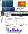
Fig 1. Isolation of SARS-CoV-2 S2-specific human monoclonal antibodies (hmAbs).
(A) Schematic representation of the S2-STBL and S1/S2 chimera proteins used as baits for ELISA and flow cytometry. (B) Human plasma from either convalescent or healthy subjects was diluted 1:1000 in PBS and tested in duplicate in an ELISA against indicated proteins; Absorbance at 450 nM is shown. Each row is an individual subject. (C) Representative gating strategy for S2+ B cell isolation. Initial plots are gated on live CD3−CD4−CD14-annexinV-CD19+CD27+ B cells. (D) hmAbs were tested at 10 and 1 μg/ml in duplicate by ELISA for binding to indicated protein; area under the curve (AUC) is indicated.
Fig 2
Fig 2. In vitro neutralization and ADCP of SARS-CoV-2 by S2- specific hmAbs.
(A) SARS-CoV-2 neutralization of S2 hmAbs. Vero E6 cells were infected with SARS-CoV-2 WA-1 or SARS-CoV-2 Delta for 1 h. After 1 h of viral adsorption, the indicated concentrations of S2 hmAbs were added and at 24 h.p.i., infected cells were fixed for virus titration by immunostaining assay. Data was expressed as mean and SD of quadruplicates. (B) Summary of viral neutralization (NT50) using either pseudovirus representing SARS-CoV-2 D614G mutation, or live SARS-CoV-2 WA-1 or Delta. (C) Ab-dependent cellular phagocytosis (ADCP) assay. SARS-CoV-2 Wuhan-Hu-1 S-coated and BSA-coated beads were incubated with 5 μg/ml hmAb for 2 h and then added to THP-1 cells. After incubation for 3 h at 37°C, cells were assayed for fluorescent bead uptake by flow cytometry. The ADCP score of each mAb was calculated by multiplying the percentage of bead positive cells (frequency of phagocytosis) by the mean fluorescence intensity (MFI) of the beads (degree of phagocytosis) and dividing by 106. (D) Binding to S2 protein fragments by hmAbs (5 μg/ml) determined by ELISA. (E). SPR competition assays were performed by capturing S2-Frag4 to the chip surface, followed by sequential injections of 50 nM of 1249A8 (Inject1) and the various S2 Abs at 50nM concentration (Inject 2). (Inset) Summary of the competition sensorgram data, where Ab binding levels (RU), measured after Inject1 (black), were normalized to 100, and compared to Ab binding levels after the second injection (Inject 2, red), which occurred after 1249A8 binding.
Fig 3
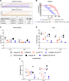
Fig 3. Prophylactic activity of 1249A8 hmAb against rSARS-CoV-2 WA1-Venus and rSARS-CoV-2 Beta-mCherry in K18 ACE2 transgenic mice model.
Female K18 hACE2 transgenic mice were treated i.p. with 1249A8 (10 mg/kg or 40 mg/kg), 1213H7 (5 mg/kg), alone or in combination, or isotype control hmAb (40 mg/kg), followed by infection with both rSARS-CoV-2 Venus and rSARS-CoV-2 Beta/mCherry Beta. Body weight (A) and survival (B) were evaluated at the indicated days post-infection (n = 5 mice/group). Mice that loss >25% of their body weight were humanely euthanized. Error bars represent standard deviations (SEM) of the mean for each group of mice. Viral titers in the nasal turbinate (C) and lung (D) at 2 and 4 DPI were determined by plaque assay in Vero E6 cells (n = 4 mice/group/day). Symbols represent individual mice, bars indicate the mean and SD of lung virus titers. Dotted lines indicate limit of detection, titers below limit of detection are presented at limit of detection. Proportion of rSARS-CoV-2 WA-1/Venus and rSARS-CoV-2 Beta/mCherry determined by fluorescence in nasal turbinate (E) and lung (F). (G) At 2 and 4 DPI, lungs were collected to determine Venus and mCherry fluorescence expression using an Ami HT imaging system. BF, bright field. (H) Venus (Green) and (I) mCherry (Red) radiance values were quantified based on the mean values for the regions of interest in mouse lungs. Mean values were normalized to the autofluorescence in mock-infected mice at each time point and were used to calculate fold induction. (J) Gross pathological scores in the lungs of mock-infected and rSARS-CoV-2-infected K18 hACE2 transgenic mice were calculated based on the percentage of area of the lungs affected by infection. Dotted line indicates limit of detection. * indicates p<0.05 as compared to isotype control hmAb as determined by one-way ANOVA.
Fig 4
Fig 4. Universal β-coronavirus in vitro activity of 1249A8 hmAb.
(A) Confluent monolayers of Vero E6 cells were infected (MOI 0.1) with SARS-CoV-2 WA-1, Beta (B.1.351), Gamma (P.1), or Epsilon (B.1.427/B.1.429). Mock-infected cells (bottom) were included as control. In a separate experiment, confluent monolayers of Vero E6 cells were infected (MOI 0.1) with SARS-CoV (Urbani v2163) or MERS-CoV (recombinant MERS-CoV-RFP delta ORF5 ic). Cells were incubated with the indicated primary S2 hmAb (1 μg/ml) and developed with a FITC-conjugated secondary anti-human Ab. 4,6-Diamidino-2-phenylindole (DAPI; blue) was used for nuclear stain. As internal control, mock and SARS-CoV-2 WA-1 infected cells were stained with a SARS-CoV cross-reactive NP mAb (1C7C7) or a ⍺-MERS NP pAb. Scale bars indicate 50 μm. (B) Binding of hmAb 1249A8 and other known S2-specific mAbs at 5, 0.5, and 0.05 μg/ml to the Spike proteins of β-coronaviruses as determined by ELISA in the presence or absence of 8M urea. (C) Summary table of Surface Plasmon Resonance (SPR) and Biolayer Interferometry (BLI) of 1249A8 against the Spike protein of SARS-CoV-2, MERS-CoV, and SARS-CoV. (D) Viral neutralization by 1249A8 and CV3-25. Vero E6 cells were infected with 100 PFU of SARS-CoV (Urbani v2163) or MERS-CoV (recombinant MERS-CoV-RFP delta ORF5 ic) for 1 h. After 1 h of viral adsorption, the indicated concentrations of S2 hmAbs were added. At 24 h.p.i., infected cells were fixed for virus titration by immunostaining assay. (E) ADCP of S2-specific mAb against MERS-CoV Spike coated beads.
Fig 5
Fig 5. Neutralization and prophylactic in vivo activity of 1249A8 and 1213H7 against SARS-CoV-2 Omicron.
(A) Binding of 1249A8 (60, 40, 27, 18, 12 nM) to SARS-CoV-2 Wuhan and Omicron Spike protein determined by BLI. (B) Vero AT cells were infected with 600 pfu SARS-CoV-2 Omicron (BEIR) and after 1 h of viral adsorption, the indicated mAb(s) was added and at 24 h.p.i infected cells were fixed for virus titration by immunostaining assay. 1213H7 and 1249A8 were tested alone (open symbols) and together keeping 1213H7 constant (C) (50 ng/ml) or 1249A8 constant (2 μg/ml) and titrating the reciprocal mAb (closed symbols). Resulting NT50 (ng/ml) are indicated. K18 hACE2 mice were treated with 1249A8 (40 mg/kg), 1213H7 (10 mg/kg), or isotype control mAb (40 mg/kg) either alone or in combination i.p. or i.n. as indicated and 24 h later challenged i.n. with 105 PFU SARS-CoV-2 Omicron (BEIR) and virus titer in nasal turbinates (C) and lungs (D) determined at 2 and 4 dpi by plaque assay and gross lung pathology measured (E) (n = 4 mice/group/day). Each symbol represents an individual animal. Dotted line indicates limit of detection, titers below limit of detection are presented at limit of detection. *indicates p<0.05 compared to isotype control group as determined by one-way ANOVA.
Fig 6
Fig 6. Therapeutic activity of intranasal 1249A8 and 1213H7 in hamsters infected with SARS-CoV-2 Delta.
Golden Syrian hamsters were infected i.n. with 104 CCID50 SARS-CoV-2 Delta and 12 h p.i. treated i.n. with a single dose of indicated mAb(s). n = 4–8 per group. (A) Body weight was measured daily. Mean ± SEM indicated. Nasal turbinate (B), cranial lung (C), and caudal lung (D) viral titers were measured at 3 d p.i. by plaque assay. Each symbol represents an individual animal. Dotted line indicates limit of detection, titers below limit of detection are presented at limit of detection. *indicates p<0.05 compared to isotype control group as determined by one-way ANOVA.
Fig 7
Fig 7. Therapeutic activity of intranasal 1249A8 and 1213H7 in hamsters infected with SARS-CoV.
Golden Syrian hamsters were infected i.n. with 104 pfu SARS-CoV (SARS-Urbani) and 12 h p.i. treated i.n. with a single dose of indicated mAb(s). n = 4–8 per group. (A) Body weight was measured daily. Mean ± SEM indicated. (B) Oropharyngeal swabs were collected days 1, 2, and 3 p.i. and sum of daily virus titer for each animal indicated. Nasal turbinate (C), cranial lung (C), and caudal lung (E) viral titers were measured at 3 d p.i. by plaque assay. Each symbol represents an individual animal. *indicates p<0.05 compared to isotype control group as determined by one-way ANOVA.

Update of

References

    1. Focosi D, Maggi F, Franchini M, McConnell S, Casadevall A. Analysis of Immune Escape Variants from Antibody-Based Therapeutics against COVID-19: A Systematic Review. Int J Mol Sci. 2021;23(1). Epub 20211221. doi: 10.3390/ijms23010029 . - DOI - PMC - PubMed
    1. Cele S, Jackson L, Khoury DS, Khan K, Moyo-Gwete T, Tegally H, et al. Omicron extensively but incompletely escapes Pfizer BNT162b2 neutralization. Nature. 2021. Epub 20211223. doi: 10.1038/s41586-021-04387-1 . - DOI - PMC - PubMed
    1. Papanikolaou V, Chrysovergis A, Ragos V, Tsiambas E, Katsinis S, Manoli A, et al. From delta to Omicron: S1-RBD/S2 mutation/deletion equilibrium in SARS-CoV-2 defined variants. Gene. 2022;814:146134. Epub 20220104. doi: 10.1016/j.gene.2021.146134 ; PubMed Central PMCID: PMC8725615. - DOI - PMC - PubMed
    1. Thakur V, Bhola S, Thakur P, Patel SKS, Kulshrestha S, Ratho RK, et al. Waves and variants of SARS-CoV-2: understanding the causes and effect of the COVID-19 catastrophe. Infection. 2021. Epub 20211216. doi: 10.1007/s15010-021-01734-2 ; PubMed Central PMCID: PMC8675301. - DOI - PMC - PubMed
    1. Shah M, Woo HG. Omicron: A Heavily Mutated SARS-CoV-2 Variant Exhibits Stronger Binding to ACE2 and Potently Escapes Approved COVID-19 Therapeutic Antibodies. Front Immunol. 2021;12:830527. Epub 20220124. doi: 10.3389/fimmu.2021.830527 ; PubMed Central PMCID: PMC8819067. - DOI - PMC - PubMed

Publication types