. 2022 Aug 9;88(15):e0096822.
doi: 10.1128/aem.00968-22. Epub 2022 Jul 12.

Chitin-Active Lytic Polysaccharide Monooxygenases Are Rare in Cellulomonas Species

Affiliations

Chitin-Active Lytic Polysaccharide Monooxygenases Are Rare in Cellulomonas Species

James Li et al. Appl Environ Microbiol. .

Abstract

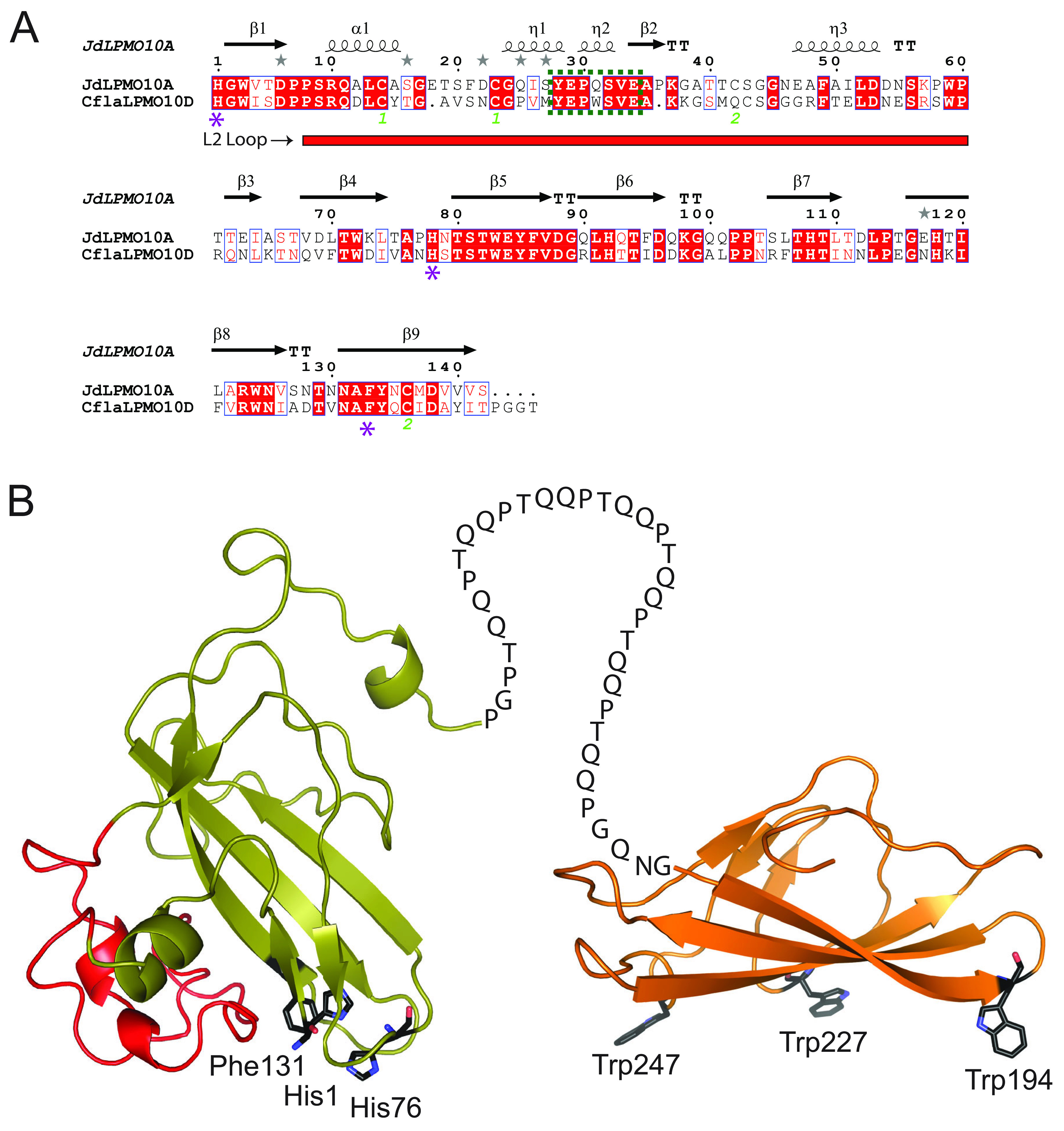
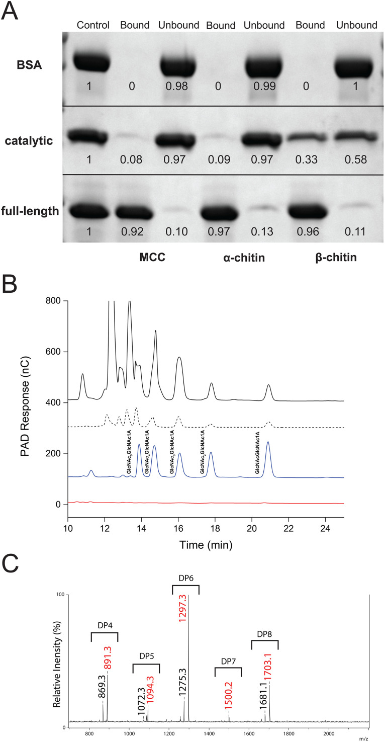
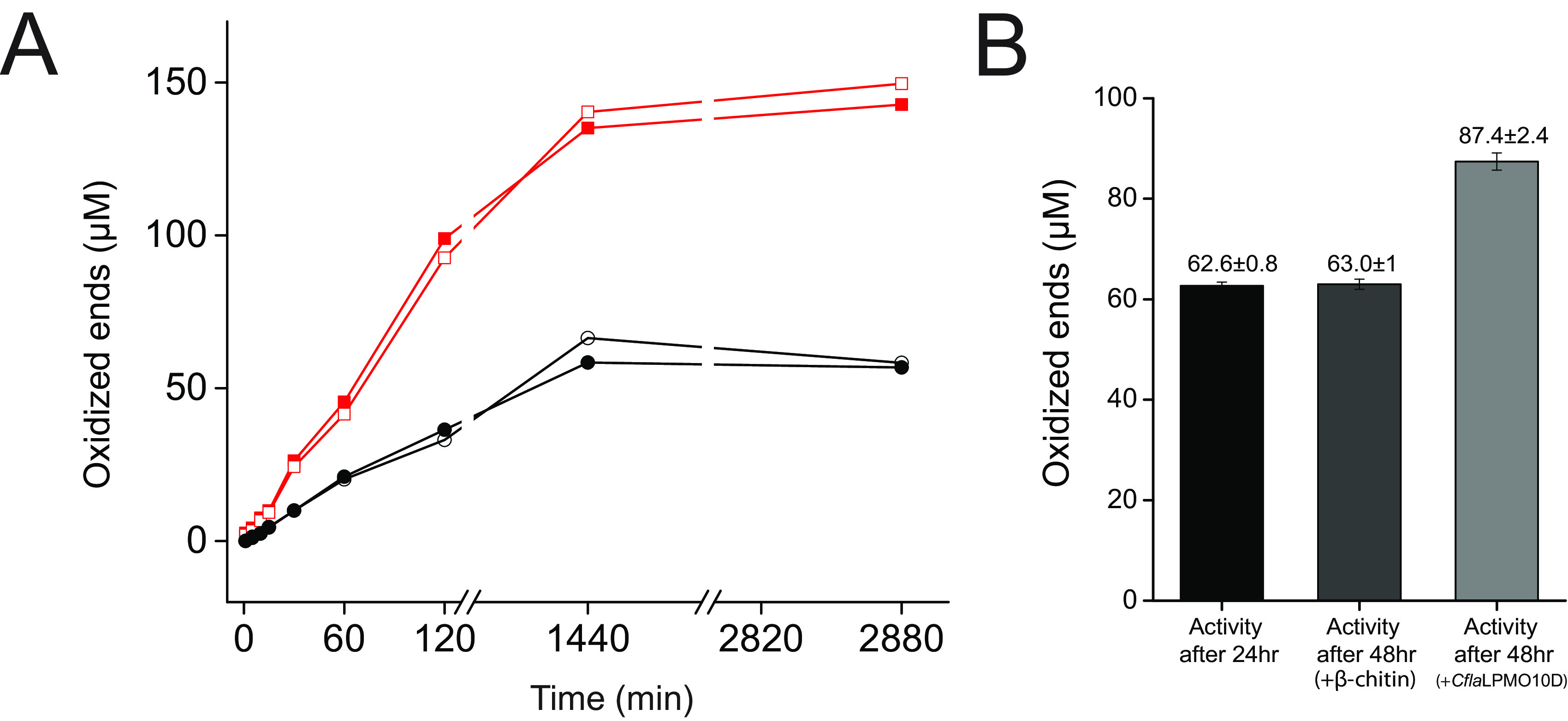
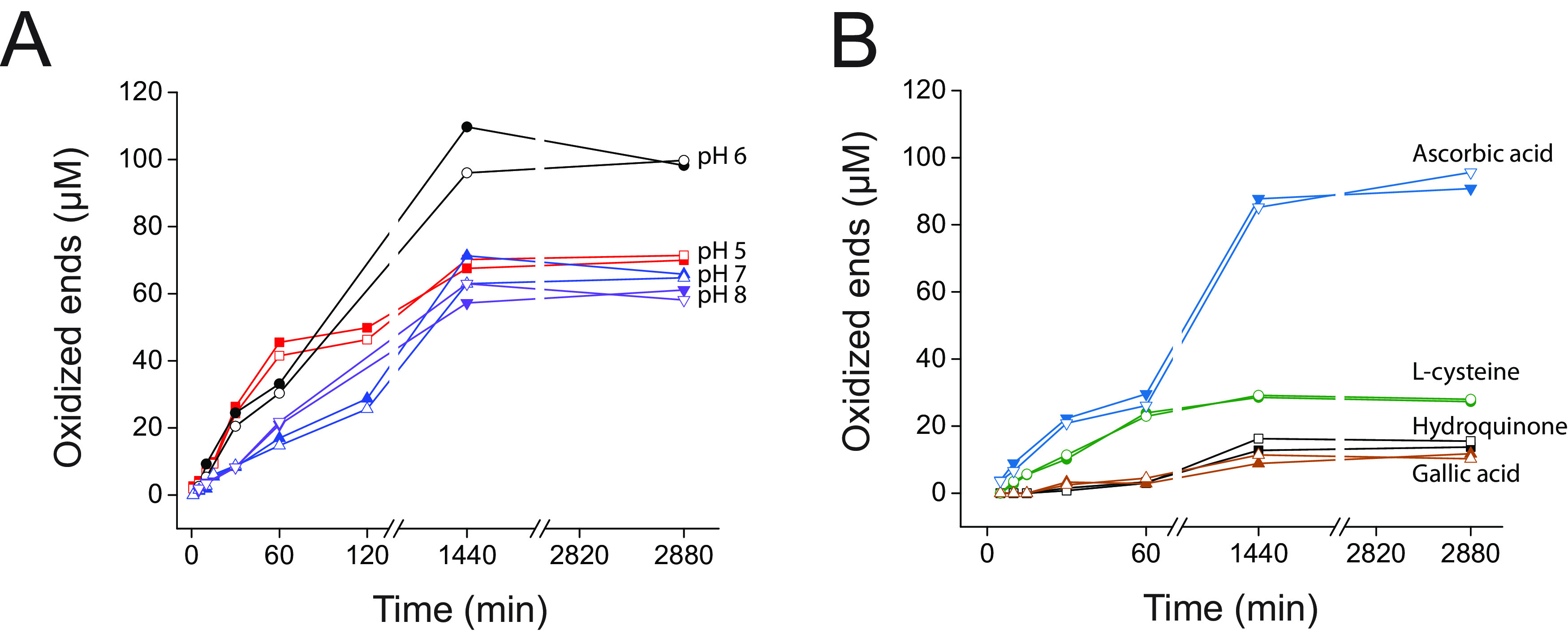
Cellulomonas flavigena is a saprotrophic bacterium that encodes, within its genome, four predicted lytic polysaccharide monooxygenases (LPMOs) from Auxiliary Activity family 10 (AA10). We showed previously that three of these cleave the plant polysaccharide cellulose by oxidation at carbon-1 (J. Li, L. Solhi, E.D. Goddard-Borger, Y. Mattieu et al., Biotechnol Biofuels 14:29, 2021, https://doi.org/10.1186/s13068-020-01860-3). Here, we present the biochemical characterization of the fourth C. flavigena AA10 member (CflaLPMO10D) as a chitin-active LPMO. Both the full-length CflaLPMO10D-Carbohydrate-Binding Module family 2 (CBM2) and catalytic module-only proteins were produced in Escherichia coli using the native general secretory (Sec) signal peptide. To quantify chitinolytic activity, we developed a high-performance anion-exchange chromatography-pulsed amperometric detection (HPAEC-PAD) method as an alternative to the established hydrophilic interaction liquid ion chromatography coupled with UV detection (HILIC-UV) method for separation and detection of released oxidized chito-oligosaccharides. Using this method, we demonstrated that CflaLPMO10D is strictly active on the β-allomorph of chitin, with optimal activity at pH 5 to 6 and a preference for ascorbic acid as the reducing agent. We also demonstrated the importance of the CBM2 member for both mediating enzyme localization to substrates and prolonging LPMO activity. Together with previous work, the present study defines the distinct substrate specificities of the suite of C. flavigena AA10 members. Notably, a cross-genome survey of AA10 members indicated that chitinolytic LPMOs are, in fact, rare among Cellulomonas bacteria. IMPORTANCE Species from the genus Cellulomonas have a long history of study due to their roles in biomass recycling in nature and corresponding potential as sources of enzymes for biotechnological applications. Although Cellulomonas species are more commonly associated with the cleavage and utilization of plant cell wall polysaccharides, here, we show that C. flavigena produces a unique lytic polysaccharide monooxygenase with activity on β-chitin, which is found, for example, in arthropods. The limited distribution of orthologous chitinolytic LPMOs suggests adaptation of individual cellulomonads to specific nutrient niches present in soil ecosystems. This research provides new insight into the biochemical specificity of LPMOs in Cellulomonas species and related bacteria, and it raises new questions about the physiological function of these enzymes.

Keywords: AA10; CBM2; Cellulomonas flavigena; HPAEC-PAD; LPMO; carbohydrate-binding module; chitin; lytic polysaccharide monooxygenase.


Conflict of interest statement

The authors declare no conflict of interest.

Figures

FIG 1
FIG 1
Primary sequence analysis and predicted tertiary structure of CflaLPMO10D. (A) Sequence alignment of CflaLPMO10D with chitin-active JdLPMO10A (30) in which it shares 50% sequence identity. All predicted secondary structure features are predicted and labeled above the alignment. The L2 loop region is indicated by a red rectangle under the alignment, the conserved chitin-binding motif (Y[W]EPQSVE) is indicated by a dotted green box, and the catalytic residues are indicated by a purple star. (B) Homology model of the full-length CflaLPMO10D showing the AA10 domain appended to a CBM2 domain. AA10 catalytic residues and CBM binding residues are labeled beside each respective residue. The L2 loop of the catalytic module, which contains the chitin-binding motif, is highlighted in red. The Phyre2 protein fold recognition server (104) was used to generate structural models. The catalytic and CBM2 domains utilized 5 AA10 templates with 100% confidence (PDB IDs 2BEM, 5L2V, 5AA7, 6T5Z, and 5WSZ [25, 30, 105–107]) and four CBM2 templates with 100% confidence (PDB IDs 3NDY, 1EXH, 5F7E, and 2RTT [73, 86, 108, 109]), respectively.
FIG 2
FIG 2
Substrate preference and product profile of CflaLPMO10D. (A) SDS-PAGE analysis of bound and unbound fractions following incubation of full-length and catalytic-only CflaLPMO10D with insoluble cellulose and chitin substrates (1:100 [wt/wt] protein-to-substrate loading; an equivalent amount of sample was loaded in each well). Bovine serum albumin (BSA) used as a negative control. The first lane of each experiment contains a reference control (100%) for pixel densitometry analysis (given below each protein band). (B) HPAEC-PAD chromatogram of soluble assay supernatant when 1 μM CflaLPMO10D-CBM2 (full-length, solid black trace) or CflaLPMO10D (catalytic module only, dotted black trace) was incubated with 0.1% β-chitin and 1 mM ascorbate reducing agent. C1-oxidized chito-oligosaccharide standards (blue trace) were produced using an AA7 member from Polyporus brumalis (PbChi7A) (117). The chromatogram for each protein variant shows the absolute PAD response (nC, unscaled) and is commensurate with the higher oxidizing activity of the full-length protein (Fig. 3). The red trace represents a no-enzyme control reaction. (C) MALDI-TOF MS spectrum of CflaLPMO10D assay supernatant indicating presence of oxidized chito-oligosaccharides. Masses in black are monosodiated aldonic acid forms ([M+Na]+), and masses in red are disodiated aldonic acid forms ([M−H+2Na]+).
FIG 3
FIG 3
Quantitation of CflaLPMO10D activity on β-chitin. (A) Release of oxidized ends following incubation on β-chitin, as determined by HPAEC-PAD. Duplicate assays were performed in parallel, and the individual results at each time point are shown. Red trace, CflaLPMO10D-CBM2 activity; black trace, CflaLPMO10D catalytic domain activity. (B) Inactivation check of CflaLPMO10D catalytic domain comparing activity at 24 h following addition of either fresh β-chitin or fresh enzyme.
FIG 4
FIG 4
pH dependence and reducing agent specificity of CflaLPMO10D on β-chitin. (A) Quantitative pH dependence time course assays over 48 h. (B) Quantitative reducing agent specificity assays over 48 h. Duplicate assays were performed in parallel, and the individual results at each time point are shown.
FIG 5
FIG 5
Maximum-likelihood phylogenetic analysis of the catalytic domains of all characterized and putative Cellulomonas AA10 members from complete genomes deposited in GenBank as of November 2021 (see http://www.cazy.org/bC.html). G. trabeum LPMO9 (GtLPMO9B) (111) was used as the outgroup. GenBank accession numbers for each Cellulomonas AA10 member are given in Table 1. The following biochemically characterized AA10 members were used: SliLPMO10E (32), SgLPMO10F (34), SamLPMO10B (29), JdLPMO10A (30), BtLPMO10A (28), EfLPMO10A (123), LmLPMO10 (33), BaLPMO10A (37), BlLPMO10 (26), PtLPMO10D (25), VcLPMO10B (36), CBP21 (SmLPMO10A) (10), CjLPMO10B (45), HcAA10-12 (39), TtAA10A (42), CjLPMO10A (31), CflaLPMO10A-C and CfiLPMO10 (43), TfLPMO10B (46), SamLPMO10C (29), ScLPMO10C (46), Tma12 (27), ACV034 (35), MaLPMO10B (44), ScLPMO10B (46) KpLPMO10 (41), and TfLPMO10B (46).

References

    1. Fernando LD, Dickwella Widanage MC, Penfield J, Lipton AS, Washton N, Latgé J-P, Wang P, Zhang L, Wang T. 2021. Structural polymorphism of chitin and chitosan in fungal cell walls from solid-state NMR and principal component analysis. Front Mol Biosci 8:727053. 10.3389/fmolb.2021.727053. - DOI - PMC - PubMed
    1. Peniche C, Argüelles-Monal W, Goycoolea FM. 2008. Chitin and chitosan: major sources, properties and applications, p 517–542. In Belgacem MN, Gandini A (ed), Monomers, polymers and composites from renewable resources. Elsevier, Oxford, UK.
    1. Merzendorfer H, Zimoch L. 2003. Chitin metabolism in insects: structure, function and regulation of chitin synthases and chitinases. J Exp Biol 206:4393–4412. 10.1242/jeb.00709. - DOI - PubMed
    1. Elieh-Ali-Komi D, Hamblin MR. 2016. Chitin and chitosan: production and application of versatile biomedical nanomaterials. Int J Advanced Res 4:411–427. - PMC - PubMed
    1. Klemm D, Heublein B, Fink H-P, Bohn A. 2005. Cellulose: fascinating biopolymer and sustainable raw material. Angew Chem Int Ed Engl 44:3358–3393. 10.1002/anie.200460587. - DOI - PubMed

Publication types

LinkOut - more resources