. 2022 Aug 9;88(15):e0088322.
doi: 10.1128/aem.00883-22. Epub 2022 Jul 14.

Regulation of l- and d-Aspartate Transport and Metabolism in Acinetobacter baylyi ADP1

Affiliations

Regulation of l- and d-Aspartate Transport and Metabolism in Acinetobacter baylyi ADP1

Stacy R Bedore et al. Appl Environ Microbiol. .

Abstract

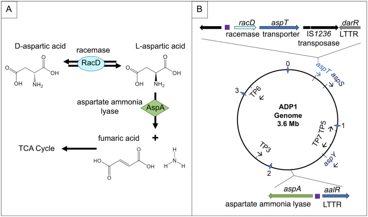
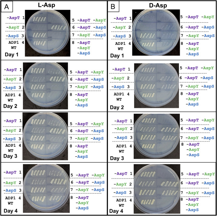
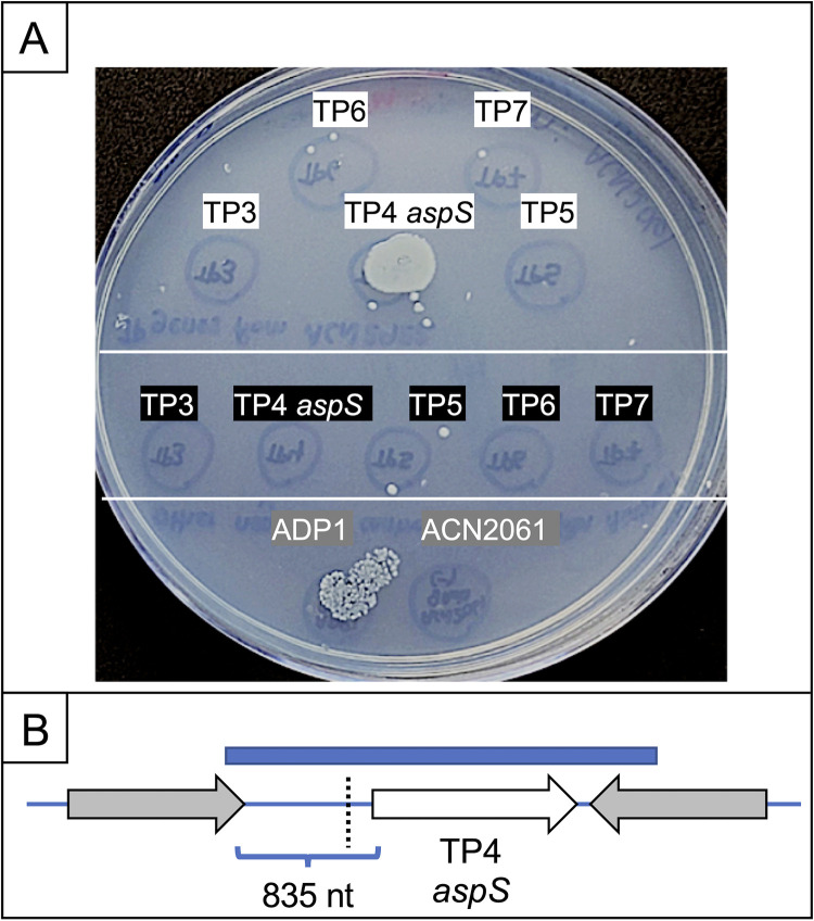
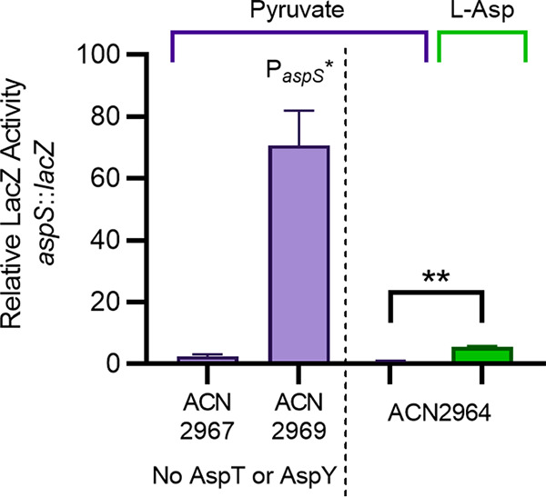
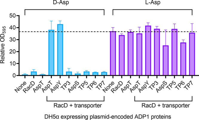
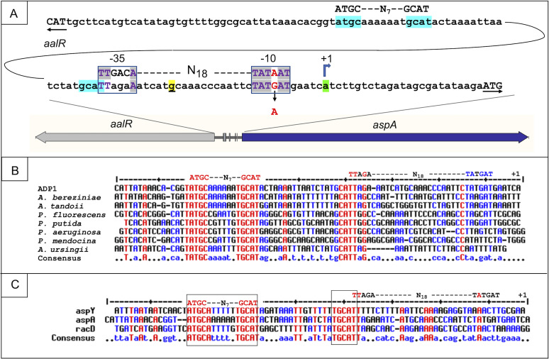
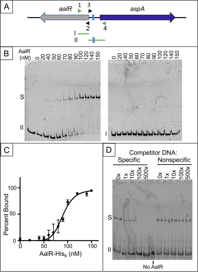
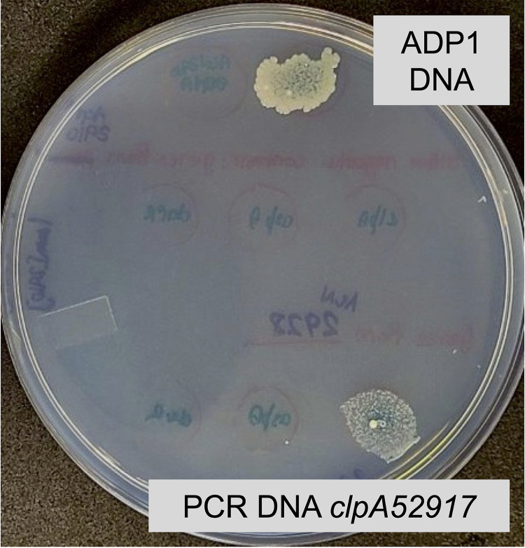
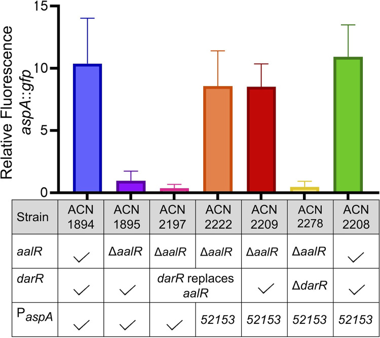
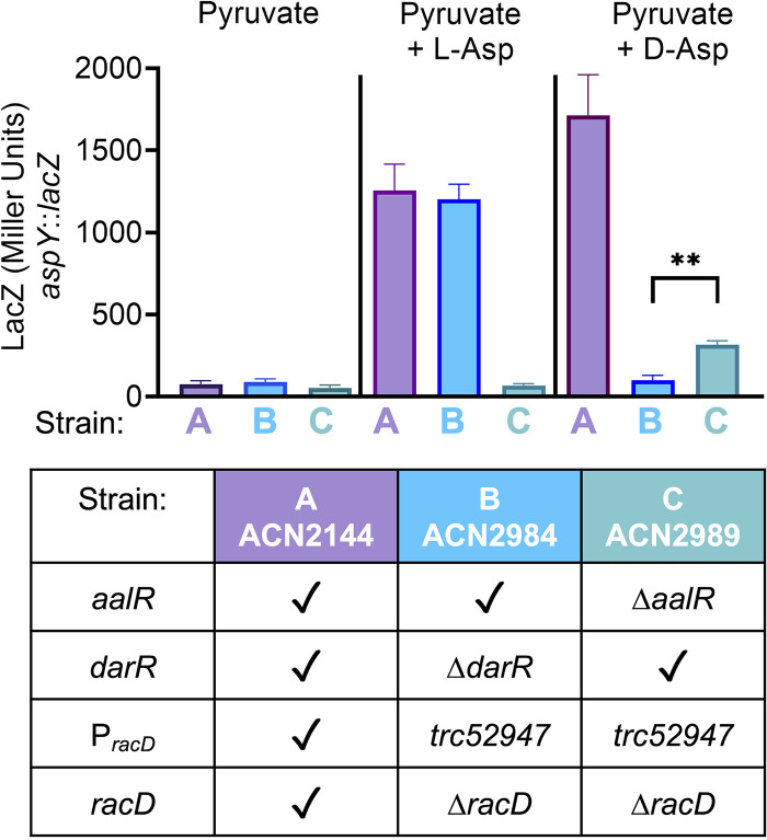
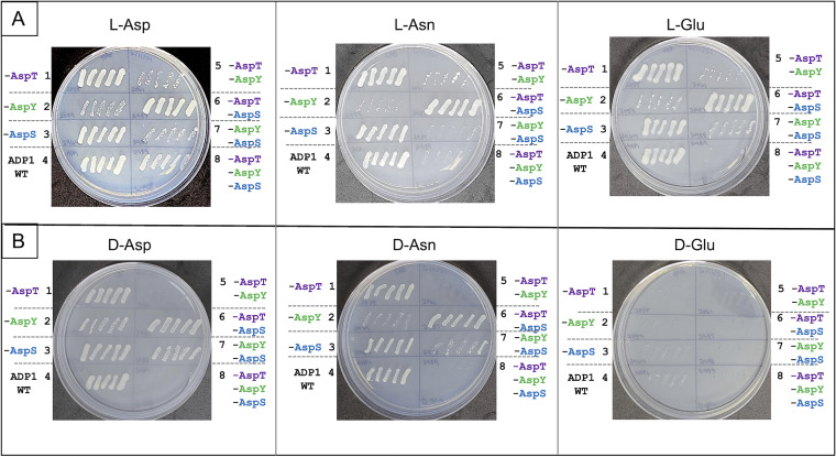
The regulated uptake and consumption of d-amino acids by bacteria remain largely unexplored, despite the physiological importance of these compounds. Unlike other characterized bacteria, such as Escherichia coli, which utilizes only l-Asp, Acinetobacter baylyi ADP1 can consume both d-Asp and l-Asp as the sole carbon or nitrogen source. As described here, two LysR-type transcriptional regulators (LTTRs), DarR and AalR, control d- and l-Asp metabolism in strain ADP1. Heterologous expression of A. baylyi proteins enabled E. coli to use d-Asp as the carbon source when either of two transporters (AspT or AspY) and a racemase (RacD) were coexpressed. A third transporter, designated AspS, was also discovered to transport Asp in ADP1. DarR and/or AalR controlled the transcription of aspT, aspY, racD, and aspA (which encodes aspartate ammonia lyase). Conserved residues in the N-terminal DNA-binding domains of both regulators likely enable them to recognize the same DNA consensus sequence (ATGC-N7-GCAT) in several operator-promoter regions. In strains lacking AalR, suppressor mutations revealed a role for the ClpAP protease in Asp metabolism. In the absence of the ClpA component of this protease, DarR can compensate for the loss of AalR. ADP1 consumed l- and d-Asn and l-Glu, but not d-Glu, as the sole carbon or nitrogen source using interrelated pathways. IMPORTANCE A regulatory scheme was revealed in which AalR responds to l-Asp and DarR responds to d-Asp, a molecule with critical signaling functions in many organisms. The RacD-mediated interconversion of these isomers causes overlap in transcriptional control in A. baylyi. Our studies improve understanding of transport and regulation and lay the foundation for determining how regulators distinguish l- and d-enantiomers. These studies are relevant for biotechnology applications, and they highlight the importance of d-amino acids as natural bacterial growth substrates.

Keywords: ADP1; Acinetobacter baylyi; DarR; LTTR; LysR; aspartate; racemase; regulation; transport.


Conflict of interest statement

The authors declare no conflict of interest.

Figures

FIG 1
FIG 1
d- and l-Asp catabolism pathway and genes in A. baylyi ADP1. (A) d-Asp is converted to l-Asp by a racemase, RacD. Next, l-Asp is cleaved by a lyase, AspA. Fumarate is further metabolized in the tricarboxylic acid (TCA) cycle. (B) The racD gene (ACIAD_RS01510) is adjacent to a gene (ACIAD_RS01515), designated aspT, predicted to encode a d-Asp transporter. The chromosomal positions and orientations of aspT and six additional genes encoding paralogous transport proteins (TP) (Fig. S2) are indicated. (The distance from the origin in megabases is indicated by numbered diamonds.) Two of the TP genes (TP2 and TP4) were renamed aspY and aspS. As described in the text, our data support a regulatory scheme in which two LTTRs, DarR and AalR, control a regulon that includes aspA, racD, aspT, and aspY. Purple boxes indicate operator-promoter regions upstream of aspA and the racD-aspT operon that are subject to LTTR-activated transcription.
FIG 2
FIG 2
Patched colonies of the wild type (WT) and aspT, aspY, and aspS mutants. Five comparably sized pyruvate-grown colonies were taken from a single plate for each strain and patched in the same position on a plate with l-Asp (A) or d-Asp (B) as the carbon source. Photographs were taken after 1, 2, 3, and 4 days of incubation (from top to bottom) at 37°C. Patches on other plates, including a final plate with LB medium as a positive control, are not shown here. The following strains are in these regions of each patched plate: 1, ACN2035, no AspT; 2, ACN2059, no AspY; 3, ACN2964, no AspS; 4, ADP1, WT; 5, ACN2061, no AspT or AspY; 6, ACN2987, no AspT or AspS; 7, ACN2988, no AspY or AspS; 8, ACN2967, no AspT, AspY, or AspS.
FIG 3
FIG 3
Transformation assay. (A) ACN2061 (lacking AspY and AspT) was spread on a plate with d-Asp as the carbon source. This recipient, which is d-Asp, grew only in 2 of 12 spots where specific cell-free donor DNA was dropped and could generate transformants in which the donor DNA replaced the corresponding chromosomal region of the recipient. PCR fragments were made with genomic template DNA from ACN2921 (d-Asp+). PCR products that each encompass the sequence of genes for TP3, TP4, TP5, TP6, or TP7 were dropped in spots (white labels with black text). Comparable fragments, made with genomic template DNA from ACN2061, were dropped in spots (black labels with white text). At the bottom, genomic DNA from ADP1 (able to reintroduce aspT and aspY) and DNA from ACN2061 served as positive and negative controls, respectively. (B) Schematic of the relative positions of the TP4 (aspS) coding sequence (white arrow), the PCR product (blue rectangle) used in the transformation assay, and a mutation (dotted line) identified in ACN2921. At this position, 200 nt upstream of the aspS coding sequence, the wild-type sequence (GTTTA) is altered by the insertion of one extra T (GTTTTA). This mutation (allele aspS52921) creates a variant aspS promoter (PaspS*). No mutations occurred in the aspS (TP4) coding sequence of ACN2921.
FIG 4
FIG 4
LacZ expression from the wild-type aspS promoter (PaspS) in ACN2967 and ACN2964 or the mutated promoter (in ACN2969), which was identified in the d-Asp+ mutant (allele aspS52921, PaspS* [Fig. 3]). Each strain has a chromosomal aspS::lacZ transcriptional reporter. LacZ activity is shown relative to that of ACN2964, which is otherwise wild type, grown on pyruvate (150 Miller units). Purple bars indicate that strains were grown on pyruvate as the sole carbon source. The green bar corresponds to LacZ activity when ACN2964 was grown on l-Asp as the sole carbon source. Error bars show the standard deviation from at least three biological replicates, each with two technical replicates. The significance in a t test of ACN2964 grown on pyruvate compared to the same strain grown in l-Asp is indicated by ** (P < 0.0001).
FIG 5
FIG 5
Heterologous expression of A. baylyi ADP1 proteins in E. coli DH5α. Cultures of DH5α carrying either a plasmid with no ADP1 DNA (pJPK13 [None]) or derivatives of this plasmid encoding the indicated proteins were grown in media with d-Asp (left side) or l-Asp (right side) as the carbon source. Growth measurements (OD595), taken 72 h after inoculation, are shown relative to that of DH5α(pJPK13) in d-Asp medium (OD595 = 0.03). The growth of this strain on l-Asp (OD595 = 1) is indicated by a dashed line. The following plasmids encode the ADP1 proteins indicated in parentheses: pBAC1680 (RacD), pBAC1683 (AspT), pBAC1673 (RacD and AspT), pBAC1941 (RacD and AspY), pBAC1942 (RacD and TP3), pBAC1943 (RacD and AspS), pBAC1944 (RacD and TP5), pBAC1945 (RacD and TP6), and pBAC1946 (RacD and TP7). Error bars represent the standard deviation from at least 5 biological replicates.
FIG 6
FIG 6
Operator-promoter sequences of aspA, aspY, and racD (A) Predicted regions for AalR binding/regulation are highlighted in turquoise. Part of this highlighted region abuts the −35 portion of the aspA promoter (boxed). The promoter mutation (PaspA52153 [red text]), shown near the aspA transcriptional start site (+1 [green]), increases the resemblance to a consensus σ70 E. coli promoter, shown above the ADP1 sequence. The transcriptional start site for aalR, on the opposite DNA strand, was localized to a 45-nt region between the underlined nucleotide (yellow) and the aspA coding sequence. (B) The ADP1 aspA promoter (+1 site at the right side) is aligned with predicted aspA DNA in Acinetobacter and Pseudomonas strains. Conserved sequences are marked in red or blue, depending on how many sequences are identical. Many red sequences correspond to the regions highlighted in turquoise in panel A. The putative AalR binding site (ATGC-N7-GCAT) and the ADP1 promoter sequence are shown above the alignment. (C) The aspA sequence from panel B is aligned with regions upstream of aspY and racD in ADP1. The right-most nucleotide (top line) is the experimentally determined transcriptional start site for aspY (+1). The aspS sequence is not included because there was insufficient similarity to allow alignment.
FIG 7
FIG 7
AalR-His6 binding to the operator-promoter region of aspA assessed by EMSA. (A) Two 6-FAM-labeled PCR products were used. At the start of the aalR coding sequence, a 202-bp fragment I (made with primers 6-FAM-ALS68 and ALS67 [arrowheads 1 and 2]), has no predicted AalR-binding site. Fragment II (241 bp, made with primers ALS66 and 6-FAM-ALS65 [arrowheads 3 and 4]) contains DNA between the divergent coding sequences, including the predicted AalR binding site (turquoise highlighting in Fig. 6). (B) Binding assays used purified AalR-His6 at the indicated tetramer concentrations (nM) with fragment I or II (2 nM) in the presence of 1 mM l-Asp. A representative EMSA is shown for each fragment. Unbound DNA is indicated with the fragment number; shifted AalR-DNA complex is labeled “S.” (C) A plot of the percent AalR-His6-bound fragment II versus nM concentration of tetrameric AalR-His6 from EMSAs performed in the presence of l-Asp is shown (results from two replicate experiments; vertical bars indicate standard error). Nonlinear regression analysis of the data for saturation binding of a single site (PRISM software) provided an estimate of the Kd value (93.3 ± 6.5 nM) for AalR-His6 binding of fragment II in the presence of the effector l-Asp. (D) A representative competition EMSA assessing the specificity of AalR-His6 binding to fragment II in the presence of 1 mM l-Asp is shown. (Four replicate assays were performed.) Binding reactions were performed with 280 nM AalR-His6; the molar excess of specific (unlabeled fragment II) and nonspecific (sonicated herring sperm DNA) competitor DNA relative to the 6-FAM-labeled fragment II (2 nM) is indicated above each lane of the gel.
FIG 8
FIG 8
A clpA mutation transforms a ΔaalR recipient, enabling d-Asp+ growth. A transformation assay with an ACN1280 culture spread on the surface of a d-Asp plate is shown. ADP1 DNA dropped on the bacterial lawn enabled d-Asp+ growth, presumably by allelic replacement that restores aalR. A PCR product carrying the mutated clpA allele of ACN2917 also enabled d-Asp+ growth. Additional DNA dropped on the plate in different spots failed to confer growth: PCR products carrying DNA of clpA, darR, and aspA using template DNA from the recipient, ACN1280. PCR products carrying DNA of darR and aspA from the d-Asp+ clpA mutant (ACN2917) were also dropped on as donor DNA and failed to confer d-Asp+ growth by allelic replacement.
FIG 9
FIG 9
Relative fluorescence of strains with an aspA::gfp transcriptional fusion. All strains have a chromosomal transcriptional fusion controlled by either the wild-type aspA promoter (PAspA) or the aspA promoter with one mutation, which was identified in ACN2153 (PaspA52153 promoter allele indicated as “52153”). Differences in genetic backgrounds are indicated for each strain in the table. A checkmark indicates the wild-type allele for aalR, darR, or PaspA. Fluorescence is shown per OD595 relative to that of ACN1895 (average florescence normalized to OD595 = 10). Error bars represent the standard deviation from at least 6 biological replicates. All strains were grown on pyruvate with d-Asp added as an inducer.
FIG 10
FIG 10
LacZ expression from the aspY promoter. All strains have a chromosomal transcriptional fusion (17) controlled by the aspY promoter. Differences in genetic backgrounds are indicated. A checkmark signifies the wild-type allele for aalR, darR, PracD (the promoter controlling racD and aspT), and racD. ACN2984 (strain B) and ACN2989 (strain C) carry a racD deletion (ΔracD52953) to prevent the interconversion of d-Asp and l-Asp. In these strains, an engineered constitutive promoter (PracD_trc52947) allows aspT transcription regardless of the presence of DarR. All strains were grown on pyruvate. d-Asp or l-Asp was added as an inducer where noted. The significance in the unpaired t test of ACN2989 compared to ACN2984 grown on pyruvate plus d-Asp is indicated by ** (P < 0.0001).
FIG 11
FIG 11
Growth of the wild type and transport mutants on l- and d-isomers of Asp, Glu, or Asn as the sole carbon source. Colonies were patched as described in the legend to Fig. 2. The images shown were taken after 4 days of incubation. The strains are ACN2035 (no AspT) in region 1, ACN2059 (no AspY) in region 2, ACN2964 (no AspS) in region 3, ADP1 (wild type) in region 4, ACN2061 (no AspT or AspY) in region 5, ACN2987 (no AspT or AspS) in region 6, ACN2988 (no AspY or AspS) in region 7, and ACN2967 (no AspT, AspY, or AspS) in region 8.

Similar articles

Cited by

References

    1. Ota N, Shi T, Sweedler JV. 2012. d-Aspartate acts as a signaling molecule in nervous and neuroendocrine systems. Amino Acids 43:1873–1886. 10.1007/s00726-012-1364-1. - DOI - PMC - PubMed
    1. Bastings J, van Eijk HM, Olde Damink SW, Rensen SS. 2019. d-Amino acids in health and disease: a focus on cancer. Nutrients 11:2205. 10.3390/nu11092205. - DOI - PMC - PubMed
    1. Cava F, Lam H, De Pedro MA, Waldor MK. 2011. Emerging knowledge of regulatory roles of d-amino acids in bacteria. Cell Mol Life Sci 68:817–831. 10.1007/s00018-010-0571-8. - DOI - PMC - PubMed
    1. Kobayashi J. 2019. d-Amino acids and lactic acid bacteria. Microorganisms 7:690. 10.3390/microorganisms7120690. - DOI - PMC - PubMed
    1. Wang R, Zhang Z, Sun J, Jiao N. 2020. Differences in bioavailability of canonical and non-canonical d-amino acids for marine microbes. Sci Total Environ 733:139216. 10.1016/j.scitotenv.2020.139216. - DOI - PubMed

Publication types

MeSH terms

Supplementary concepts