. 2022 Aug 18;90(8):e0012922.
doi: 10.1128/iai.00129-22. Epub 2022 Jul 11.

Mycoplasma pneumoniae Compared to Streptococcus pneumoniae Avoids Induction of Proinflammatory Epithelial Cell Responses despite Robustly Inducing TLR2 Signaling

Affiliations

Mycoplasma pneumoniae Compared to Streptococcus pneumoniae Avoids Induction of Proinflammatory Epithelial Cell Responses despite Robustly Inducing TLR2 Signaling

R C A de Groot et al. Infect Immun. .

Abstract

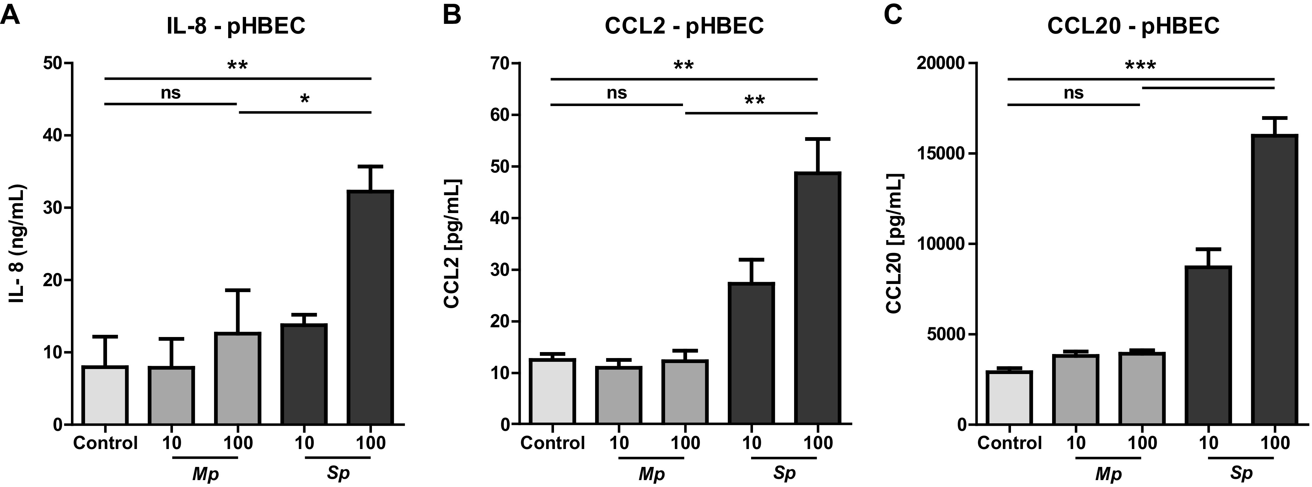
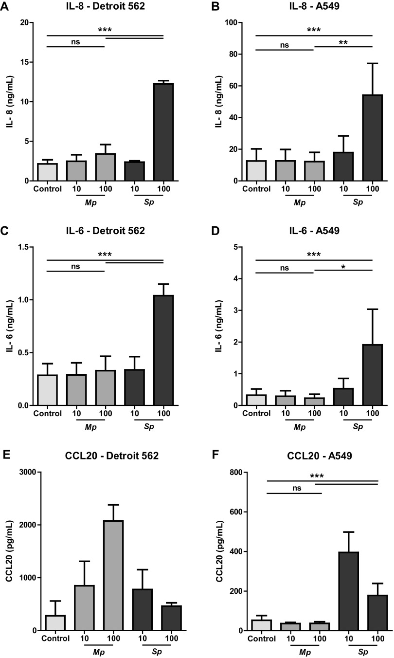
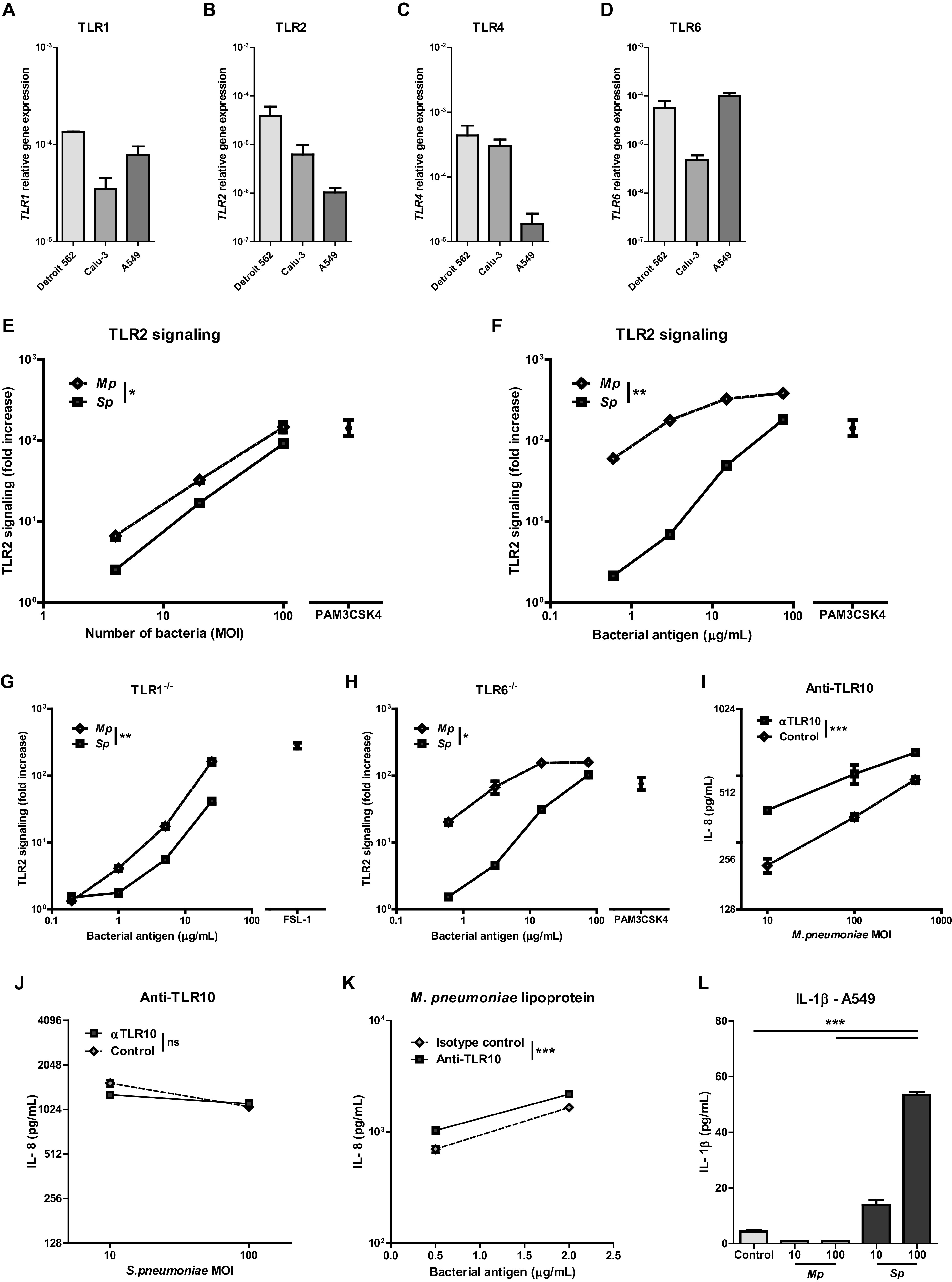
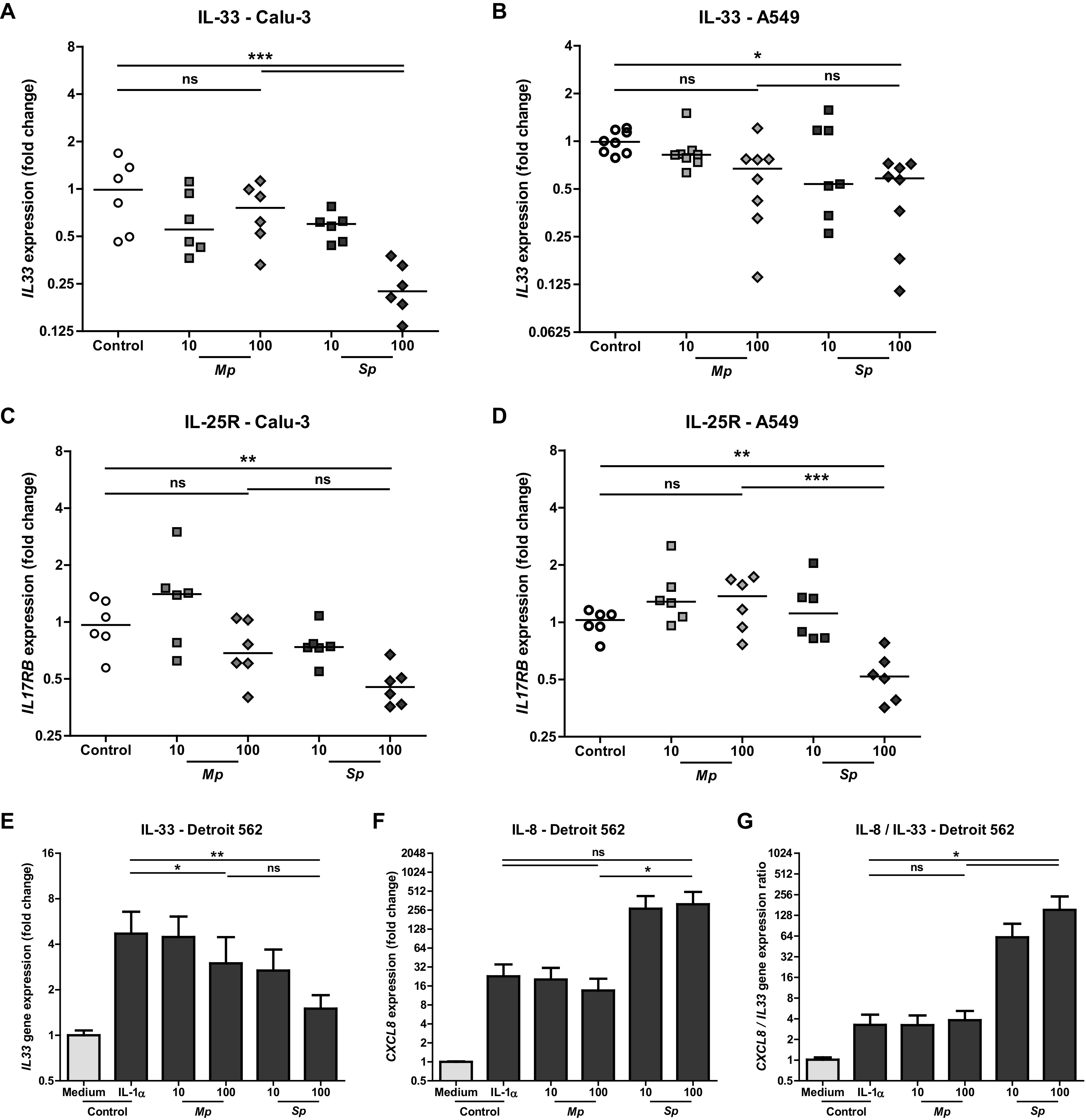
Mycoplasma pneumoniae and Streptococcus pneumoniae are the most common bacterial causes of pneumonia in children. The clinical characteristics of pneumonia differ significantly between the two bacteria. We aimed to elucidate the differences in pathogenesis between M. pneumoniae and S. pneumoniae by characterizing the respiratory epithelial cell immune response to both pathogens. Using primary human bronchial epithelial cells in air-liquid interface cultures, we observed lower production of the proinflammatory cytokines interleukin-6 (IL-6) and IL-8 in response to M. pneumoniae than to S. pneumoniae. In contrast to the differences in proinflammatory cytokine production, Toll-like receptor 2 (TLR2)-mediated signaling in response to M. pneumoniae was stronger than to S. pneumoniae. This difference largely depended on TLR1 and not TLR6. We found that M. pneumoniae, but not S. pneumoniae, also induced signaling of TLR10, a coreceptor of TLR2 that has inhibitory properties. M. pneumoniae-induced TLR10 signaling on airway epithelial cells was partially responsible for low IL-8 production, as blocking TLR10 by specific antibodies increased cytokine production. M. pneumoniae maintained Th2-associated cytokine production by epithelial cells, which concurs with the known association of M. pneumoniae infection with asthma. M. pneumoniae left IL-33 levels unchanged, whereas S. pneumoniae downregulated IL-33 production both under homeostatic and Th2-promoting conditions. By directly comparing M. pneumoniae and S. pneumoniae, we demonstrate that M. pneumoniae avoids induction of proinflammatory cytokine response despite its ability to induce robust TLR2 signaling. Our new findings suggest that this apparent paradox may be partially explained by M. pneumoniae-induced signaling of TLR2/TLR10.

Keywords: IL-33; IL-8; Mycoplasma pneumoniae; Streptococcus pneumoniae; TLR10; TLR2; host response; pneumonia; primary bronchial epithelial cells; respiratory pathogens.


Conflict of interest statement

The authors declare no conflict of interest.

Figures

FIG 1
FIG 1
Reduced activation of primary human bronchial epithelial cells by M. pneumoniae compared to S. pneumoniae. (A to C) Primary human bronchial epithelial cells cultured in air-liquid interface were stimulated with live M. pneumoniae or S. pneumoniae at a multiplicity of infection of 10 or 100. To reduce potential influence of patient-specific characteristics, primary cells of three different donors were used for all experiments. (A) Levels of IL-8 in culture medium after 24 h of stimulation. (B) Levels of CCL-2 in culture medium after 24 h of stimulation. (C) Levels of CCL20 in culture medium after 24 h of stimulation. Combined data of three individual donors from two independent experiments. For every donor, we performed two biological replicates of which we took two technical replicates. Bars represent means, and error bars show standard error of the mean (SEM). *, P < 0.05; **, P < 0.01 (repeated measures ANOVA with post hoc Bonferroni correction).
FIG 2
FIG 2
M. pneumoniae-induced cytokine production is reduced compared to S. pneumoniae-induced production in both upper and lower respiratory tract epithelial cells. Respiratory epithelial cell lines Detroit 562 and A549 were stimulated with live M. pneumoniae and S. pneumoniae at a multiplicity of infection of 10 or 100. (A to D) IL-8/IL-6 protein concentration in culture supernatant harvested after 24 h of stimulation (n = 5/condition). (E and F) CCL20 protein levels in culture supernatant harvested after 24 h of stimulation. (A to D) Data of at least two independent experiments. (E and F) Data of three experiments. All measurements consist of two technical replicates. (A to F) Bars represent means, and error bars show standard error of the mean. *, P < 0.05; **, P < 0.01; ***, P < 0.001 (ANOVA with post hoc Bonferroni correction).
FIG 3
FIG 3
In contrast to M. pneumoniae, S. pneumoniae lowered Th2-associated cytokine production in resting and IL-1 alpha stimulated respiratory epithelial cells. (A) TLR1, TLR2, TLR4, and TLR6 relative gene expression in unstimulated respiratory epithelial cells (n = 3 to 4/group). (E and F) TLR2 signaling as assessed by fold increase in bioluminescence of TLR2-luciferase reporter cells upon stimulation with different doses of heat-killed bacteria compared to vehicle controls (n = 3/dose). (G) TLR2 signaling as assessed by fold increase in bioluminescence of TLR1−/− TLR2-luciferase reporter cells stimulated with multiple doses of bacteria (n = 3/dose). (H) TLR2 signaling as assessed by fold increase in bioluminescence of TLR6−/− TLR2-luciferase reporter cells stimulated with multiple doses of bacteria (n = 3/dose). (I and J) IL-8 levels in culture medium after 24 h of stimulation of A549 cells with M. pneumoniae or S. pneumoniae in the presence of TLR10 blocking antibody or isotype control. (K) IL-8 levels in culture medium after 24 h of stimulation of M. pneumoniae lipoproteins with either a TLR10-blocking antibody or isotype control. (L) IL-1β levels in culture medium after 24 h of stimulation with live M. pneumoniae or S. pneumoniae at an MOI of 10 or 100. (A to D) Data of two independent experiments. (E to L) Data shown of one representative experiment. Bars and dots represent group means, and error bars show SEM. **, P ≤ 0.01; ***, P ≤ 0.001 (comparing 50% effective concentration [EC50] of fitted dose-response curves or 2-way ANOVA).
FIG 4
FIG 4
S. pneumoniae lowered Th2-associated cytokine production in resting and IL-1 alpha stimulated respiratory epithelial cells, whereas M. pneumoniae did not. Respiratory epithelial cell lines Detroit 562, Calu-3, and A549 were stimulated with live M. pneumoniae and S. pneumoniae with a multiplicity of infection of 10 or 100. (A and B) IL33 mRNA as fold increase to controls after 5 h of stimulation (n = 6 to 8/condition). (C and D) IL17RB mRNA as fold increase to controls after 5 h of stimulation (n = 6/condition). (E to G) Detroit 562 cells were incubated with IL-1 alpha and simultaneously stimulated with live M. pneumoniae or S. pneumoniae at a multiplicity of infection of 10 or 100. IL33 and CXCL8 gene expressions were assessed after 5 h. (n = 4/condition). Combined data of at least two independent experiments. All measurements consist of two technical replicates. (A to D) Data points represent biological replicates, and lines group medians. ANOVA with post hoc Bonferroni correction. (E to G) Bars represent group means, and error bars show SEM. Repeated measures ANOVA with post hoc Bonferroni correction. *, P < 0.05; **, P < 0.01; ***, P < 0.001.

References

    1. World Health Organization. 2016. Pneumonia fact sheet. World Health Organization, Geneva, Switzerland. Accessed 16 April 2018.
    1. Jain S, Williams DJ, Arnold SR, Ampofo K, Bramley AM, Reed C, Stockmann C, Anderson EJ, Grijalva CG, Self WH, Zhu Y, Patel A, Hymas W, Chappell JD, Kaufman RA, Kan JH, Dansie D, Lenny N, Hillyard DR, Haynes LM, Levine M, Lindstrom S, Winchell JM, Katz JM, Erdman D, Schneider E, Hicks LA, Wunderink RG, Edwards KM, Pavia AT, McCullers JA, Finelli L, Team CES. 2015. Community-acquired pneumonia requiring hospitalization among U.S. children. N Engl J Med 372:835–845. doi:10.1056/NEJMoa1405870. - DOI - PMC - PubMed
    1. de Groot RCA, Meyer Sauteur PM, Unger WWJ, van Rossum AMC. 2017. Things that could be Mycoplasma pneumoniae. J Infect 74(Suppl 1):S95–S100. doi:10.1016/S0163-4453(17)30198-6. - DOI - PubMed
    1. Meyer Sauteur PM, Krautter S, Ambroggio L, Seiler M, Paioni P, Relly C, Capaul R, Kellenberger C, Haas T, Gysin C, Bachmann LM, van Rossum AMC, Berger C. 2020. Improved diagnostics help to identify clinical features and biomarkers that predict Mycoplasma pneumoniae community-acquired pneumonia in children. Clin Infect Dis 71:1645–1654. doi:10.1093/cid/ciz1059. - DOI - PMC - PubMed
    1. Ishida T, Miyashita N, Nakahama C. 2007. Clinical differentiation of atypical pneumonia using Japanese guidelines. Respirology 12:104–110. doi:10.1111/j.1440-1843.2006.00927.x. - DOI - PubMed

LinkOut - more resources