. 2022 Aug 31;7(4):e0016922.
doi: 10.1128/msphere.00169-22. Epub 2022 Jul 5.

Impact of Prior Infection on SARS-CoV-2 Antibody Responses in Vaccinated Long-Term Care Facility Staff

Affiliations

Impact of Prior Infection on SARS-CoV-2 Antibody Responses in Vaccinated Long-Term Care Facility Staff

Emily N Gallichotte et al. mSphere. .

Abstract

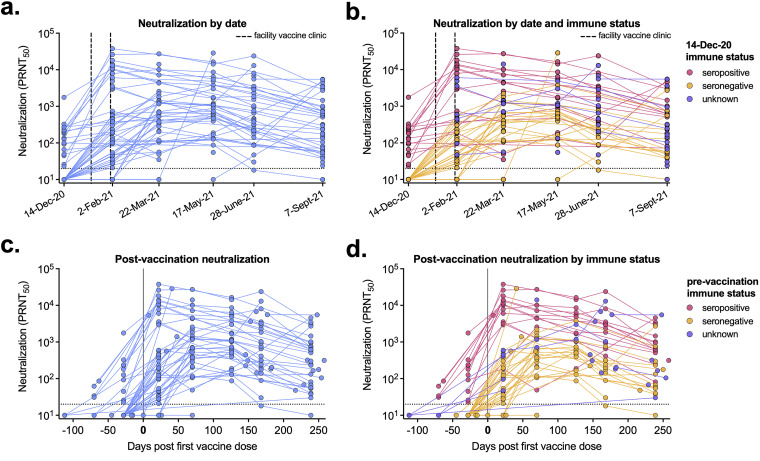
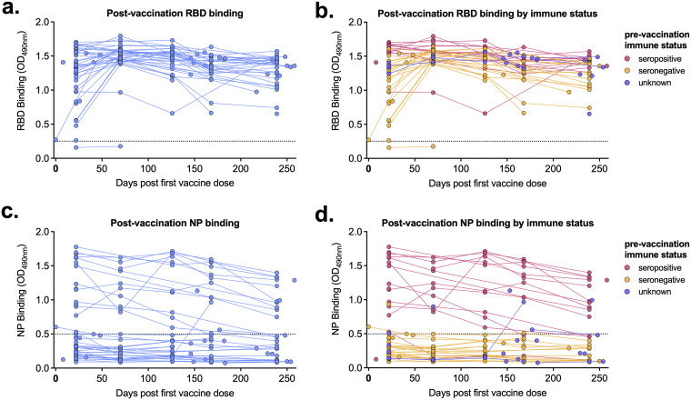
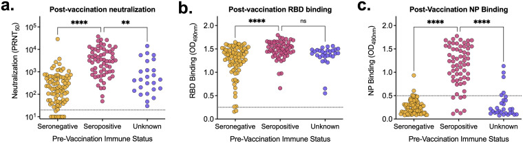
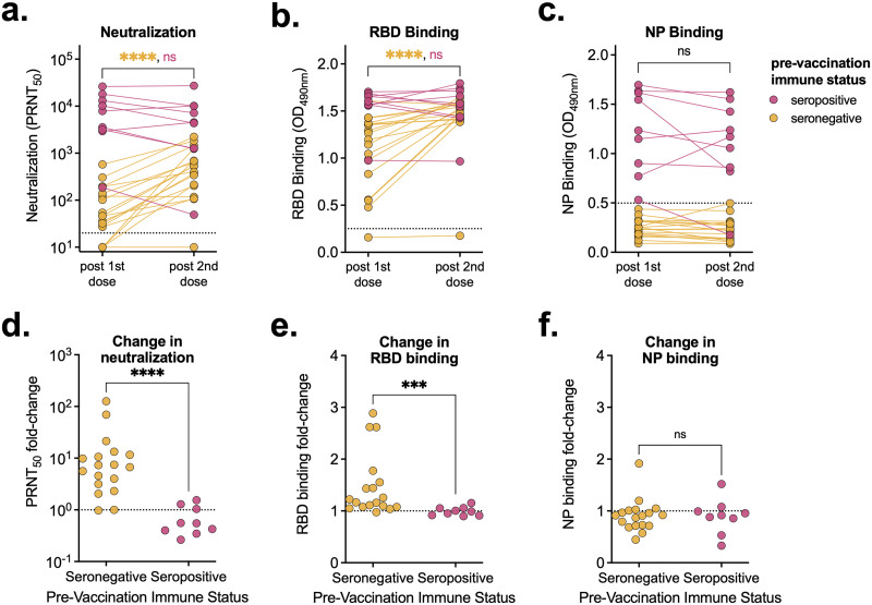
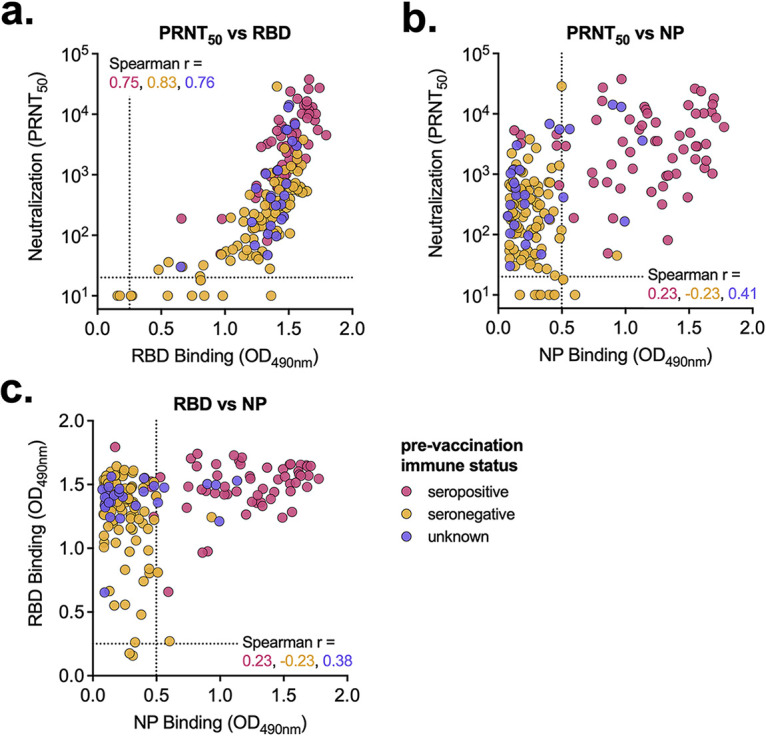
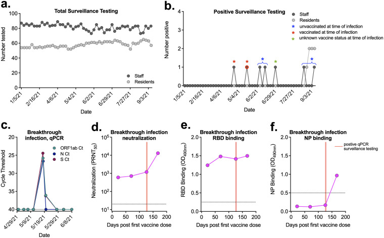
Severe acute respiratory syndrome coronavirus 2 (SARS-CoV-2) emerged in 2019 and has resulted in millions of deaths worldwide. Certain populations are at higher risk for infection, especially staff and residents at long-term care facilities (LTCF), due to the congregant living setting and high proportions of residents with many comorbidities. Prior to vaccine availability, these populations represented large fractions of total coronavirus disease 2019 (COVID-19) cases and deaths in the United States. Due to the high-risk setting and outbreak potential, staff and residents were among the first groups to be vaccinated. To define the impact of prior infection on the response to vaccination, we measured antibody responses in a cohort of staff members at an LTCF, many of whom were previously infected by SARS-CoV-2. We found that neutralizing, receptor-binding domain (RBD)-binding, and nucleoprotein (NP)-binding antibody levels were significantly higher after the full vaccination course in individuals that were previously infected and that NP antibody levels could discriminate individuals with prior infection from vaccinated individuals. While an anticipated antibody titer increase was observed after a vaccine booster dose in naive individuals, a boost response was not observed in individuals with previous COVID-19 infection. We observed a strong relationship between neutralizing antibodies and RBD-binding antibodies postvaccination across all groups, whereas no relationship was observed between NP-binding and neutralizing antibodies. One individual with high levels of neutralizing and binding antibodies experienced a breakthrough infection (prior to the introduction of Omicron), demonstrating that the presence of antibodies is not always sufficient for complete protection against infection. These results highlight that a history of COVID-19 exposure significantly increases SARS-CoV-2 antibody responses following vaccination. IMPORTANCE Long-term care facilities (LTCFs) have been disproportionately impacted by COVID-19, due to their communal nature, the high-risk profile of residents, and the vulnerability of residents to respiratory pathogens. In this study, we analyzed the role of prior natural immunity to SARS-CoV-2 in postvaccination antibody responses. The LTCF in our cohort experienced a large outbreak, with almost 40% of staff members becoming infected. We found that individuals that were infected prior to vaccination had higher levels of neutralizing and binding antibodies postvaccination. Importantly, the second vaccine dose significantly boosted antibody levels in those that were immunologically naive prior to vaccination, but not in those that had prior immunity. Regardless of the prevaccination immune status, the levels of binding and neutralizing antibodies were highly correlated. The presence of NP-binding antibodies could be used to identify individuals that were previously infected when prevaccination immune status was not known. Our results reveal that vaccination antibody responses differ depending on prior natural immunity.

Keywords: COVID-19; SARS-CoV-2; correlate of protection; neutralizing antibodies; vaccines.


Conflict of interest statement

The authors declare no conflict of interest.

Figures

FIG 1
FIG 1
Postvaccination serum neutralizing levels vary by prior infection. (A to D) Neutralization titers (PRNT50) for each serum sample are shown by blood sample collection date (92 participants, 260 samples total) (A, B) or by days post-first vaccine dose (68 participants, 226 samples total) (C, D). (B, D) Serum sample data are labeled based on participants’ prevaccination immune status. (B) Pink, seropositive, 23 participants, 86 samples; orange, seronegative, 38 participants, 127 samples; purple, unknown immune status, 31 participants, 47 samples. (D) Pink, seropositive, 16 participants, 70 samples; orange, seronegative, 32 participants, 121 samples; purple, unknown immune status, 20 participants, 35 samples. Dashed lines represent the limit of detection (PRNT50 = 20). Samples without neutralization detected are plotted at half the limit of detection (PRNT50 = 10).
FIG 2
FIG 2
Postvaccination receptor-binding domain (RBD)- and nucleoprotein (NP)-binding levels are higher in previously infected individuals. (A to D) RBD-binding (A, B) and NP-binding (C, D) levels for each serum sample are shown by days post-first vaccine dose (68 participants, 178 samples total). (B, D) Serum sample data are labeled based on participants’ prevaccination immune status. Pink, seropositive, 17 participants, 61 samples; orange, seronegative, 31 participants, 87 samples; purple, unknown immune status, 20 participants, 30 samples. Dashed line represents background level for each assay. OD490, optical density at 490 nm.
FIG 3
FIG 3
Prevaccination immune status impacts postvaccination antibody levels. (A to C) All postvaccination neutralization titers (A) and RBD-binding (B) and NP-binding (C) values were aggregated and stratified based on prevaccination immune status. Pink, seropositive, n = 90; orange, seronegative, n = 63; purple, unknown immune status, n = 25. (A) Dashed line represents the limit of detection. Samples without neutralization detected are plotted at half the limit of detection (PRNT50 = 10). (B, C) Dashed line represents the background level for each assay. Tukey’s multiple-comparison one-way analysis of variance (ANOVA) was used to determine statistical significance. **, P < 0.01; ****, P < 0.0001; ns, not significant.
FIG 4
FIG 4
A second vaccine dose only increases antibody levels in seronegative individuals. (A to C) Neutralization titers (A) and RBD-binding (B) and NP-binding (C) values of serum samples from individuals following their first and second vaccine doses (3 weeks after the first dose and 7 weeks after the second dose), stratified by prevaccination immune status (seropositive, n = 9; seronegative, n = 18). (D to F) Fold changes between neutralization titers (D) and RBD-binding (E) and NP-binding (F) values relative to levels following participants’ first vaccine doses. (A) Dashed line represents the limits of detection. Samples without neutralization detected are plotted at half the limit of detection (PRNT50 = 10). (B, C) Dashed line represents the background level for each assay. Mann-Whitney test was used to determine statistical significance. ***, P < 0.001; ****, P < 0.0001; ns, not significant.
FIG 5
FIG 5
Vaccine-elicited antibody levels are similarly correlated regardless of immune status. (A to C) Postvaccination serum samples were compared by neutralization versus RBD binding (A), neutralization versus NP binding (B), and RBD binding versus NP binding (C). Serum sample data are labeled based on prevaccination immune status. Pink, seropositive, 17 participants, 61 samples; orange, seronegative, 33 participants, 91 samples; purple, unknown immune status, 18 participants, 26 samples. (A) Dashed line represents the limit of detection. Samples without neutralization detected are plotted at half the limit of detection. (B, C) Dashed line represents the background level for each assay. Spearman r values for each group (seropositive, seronegative, and unknown) are noted.
FIG 6
FIG 6
Breakthrough infection in a vaccinated individual. (A) Total surveillance testing on staff and residents at the LTCF each week. (B) Number of staff and residents that tested positive each week. Blue asterisks indicate staff members that were unvaccinated at time of infection (brackets show positive tests from the same individual). Red asterisks indicate staff members that were vaccinated prior to infection. Green asterisk represents a staff member with unknown vaccine status at time of infection. Detailed data for individual represented by symbol with red outline are given in panels C to F. (C) qRT-PCR surveillance testing for three viral targets from the individual with a breakthrough infection that was seronegative prior to vaccination. (D to F) Neutralization titers (D) and RBD-binding (E) and NP-binding (F) values postvaccination and pre- and post-breakthrough infection. Red lines correspond to date of positive qRT-PCR surveillance testing. (C, D) Dashed lines represent limits of detection. (E, F) Dashed line represents the background level for each assay.

References

    1. The New York Times. 16 February 2022. Coronavirus in the U.S.: latest map and case count. https://www.nytimes.com/interactive/2021/us/covid-cases.html.
    1. Centers for Medicare & Medicaid Services. 16 February 2022. COVID-19 nursing home data. https://data.cms.gov/covid-19/covid-19-nursing-home-data. - PubMed
    1. Ochieng N, Chidambaram P, Garfield R, Neuman T. 14 January 2021. Factors associated with COVID-19 cases and deaths in long-term care facilities: findings from a literature review. Kaiser Family Foundation, San Francisco, CA.
    1. Tang O, Bigelow BF, Sheikh F, Peters M, Zenilman JM, Bennett R, Katz MJ. 2020. Outcomes of nursing home COVID-19 patients by initial symptoms and comorbidity: results of universal testing of 1970 residents. J Am Med Dir Assoc 21:1767–1773.e1. doi: 10.1016/j.jamda.2020.10.011. - DOI - PMC - PubMed
    1. Ouslander JG, Grabowski DC. 2020. COVID-19 in nursing homes: calming the perfect storm. J Am Geriatr Soc 68:2153–2162. doi: 10.1111/jgs.16784. - DOI - PubMed

Publication types