. 2022 Aug 31;10(4):e0072822.
doi: 10.1128/spectrum.00728-22. Epub 2022 Jul 14.

Evaluation of 3-Deoxy-D-Arabino-Heptulosonate 7-Phosphate Synthase (DAHPS) as a Vulnerable Target in Mycobacterium tuberculosis

Affiliations

Evaluation of 3-Deoxy-D-Arabino-Heptulosonate 7-Phosphate Synthase (DAHPS) as a Vulnerable Target in Mycobacterium tuberculosis

Luiza Galina et al. Microbiol Spectr. .

Abstract

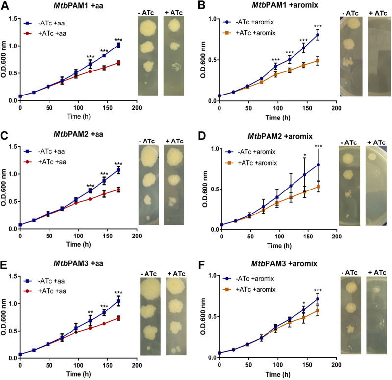
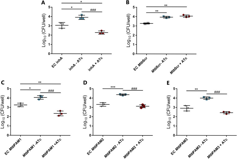
Tuberculosis (TB) remains one of the leading causes of death due to a single pathogen. The emergence and proliferation of multidrug-resistant (MDR-TB) and extensively drug-resistant strains (XDR-TB) represent compelling reasons to invest in the pursuit of new anti-TB agents. The shikimate pathway, responsible for chorismate biosynthesis, which is a precursor of important aromatic compounds, is required for Mycobacterium tuberculosis growth. The enzyme 3-deoxy-d-arabino-heptulosonate 7-phosphate synthase (MtbDAHPS) catalyzes the first step in the shikimate pathway and it is an attractive target for anti-tubercular agents. Here, we used a CRISPRi system to evaluate the DAHPS as a vulnerable target in M. tuberculosis. The silencing of aroG significantly reduces the M. tuberculosis growth in both rich medium and, especially, in infected murine macrophages. The supplementation with amino acids was only able to partially rescue the growth of bacilli, whereas the Aro supplement (aromix) was enough to sustain the bacterial growth at lower rates. This study shows that MtbDAHPS protein is vulnerable and, therefore, an attractive target to develop new anti-TB agents. In addition, the study contributes to a better understanding of the biosynthesis of aromatic compounds and the bacillus physiology. IMPORTANCE Determining the vulnerability of a potential target allows us to assess whether its partial inhibition will impact bacterial growth. Here, we evaluated the vulnerability of the enzyme 3-deoxy-d-arabino-heptulosonate 7-phosphate synthase (DAHPS) from M. tuberculosis by silencing the DAHPS-coding aroG gene in different contexts. These results could lead to the development of novel and potent anti-tubercular agents in the near future.

Keywords: CRISPRi; aroG; macrophages; shikimate pathway; vulnerability.


Conflict of interest statement

The authors declare no conflict of interest.

Figures

FIG 1
FIG 1
Location of PAM sequences inside MSMEG_4244 and aroG locus used in this study. (A) From left to right: 5′-NNAGGAC-3′, 5′-NNGGAAC-3′, 5′-NNAGCAG-3′. (B) From left to right: 5′-NNGGAAG-3′, 5′-NNAGCAG-3′, and 5′-NNGGCAG-3′. The repression strength of each PAM sequence, according to Rock et al. (2017) (16) is also described.
FIG 2
FIG 2
aroG silencing in M. smegmatis does not affect growth in vitro. (A to E). M. smegmatis growth curves and dilution spots in the presence or absence of Atc 100 ng/mL. mmpL3 (A) and scrambled sgRNAs were used as positive and negative controls, respectively (B). Three different aroG sequences were used MsgPAM1, MsgPAM2, and MsgPAM3 (C to E). Growth curves of M. smegmatis at 37°C in 7H9-OADC medium containing Atc 100 ng/mL. Cultures were inoculated to a starting A600 of 0.07. Data are the mean ± standard deviation from biological and technical duplicates. Statistical analysis was performed using one-way ANOVA analysis followed by Bonferroni’s posttest. ***, P < 0.001.
FIG 3
FIG 3
aroG silencing in M. tuberculosis results in growth perturbation in vitro. (A to E). M. tuberculosis growth curves and dilution spots in the presence or absence of Atc 100 ng/mL. inhA (A) and scrambled sgRNAs were used as positive and negative control, respectively (B). (C to E) aroG was silenced using three different PAM sequences (MtbPAM1, MtbPAM2, and MtbPAM3). Growth curves of M. tuberculosis at 37°C in 7H9-OADC medium containing Atc 100 ng/mL. Cultures were inoculated to a starting A600 of 0.07. Data are the mean ± standard deviation from biological and technical duplicates. Statistical analysis was performed using one-way ANOVA analysis followed by Bonferroni’s posttest. *, P < 0.03; **, P < 0.002; ***, P < 0.001.
FIG 4
FIG 4
mRNA levels in cultures expressing inhA, (control) or aroG-targeting sgRNAs induced with different levels of ATc. mRNA levels are expressed relative to a scrambled sgRNA control with 100 ng/mL ATc in M. tuberculosis. Results are the mean ± standard deviation of two experimental duplicates from two biological replicates. §§§, P < 0.001; ***, P < 0.001; ###, P < 0.001; $$$, P < 0.001.
FIG 5
FIG 5
Effect of aroG silencing on M. tuberculosis in supplemented media. (A, C, E). M. tuberculosis growth curves and dilution spots in the presence or absence of Atc 100 ng/mL with aa supplement. (B, D, F) M. tuberculosis growth curves and dilution spots in the presence or absence of Atc 100 ng/mL with the aromix supplement. Cultures were inoculated to a starting A600 of 0.07. Data are the mean ± standard deviation from biological and technical duplicates. Statistical analysis was performed using two-way ANOVA analysis followed by Bonferroni’s posttest. *, P < 0.03; **, P < 0.002; ***, P < 0.001.
FIG 6
FIG 6
Evaluation M. tuberculosis aroG silencing in macrophages. Comparative analysis of intracellular growth profile of controls (A, B) and aroG knockdown strains of M. tuberculosis (C, E). Intracellular bacterial load was determined by counting viable bacteria (CFU). EC, early control; MtbScr, scrambled. Error bars represent the SD from at least three measurements. Statistical significance is determined by using one-way ANOVA analysis followed by Bonferroni’s posttest. *, P < 0.03; **, P < 0.002; ###, P < 0.001.

References

    1. WHO. 2021. Global tuberculosis report 2021. World Health Organization.
    1. Lechartier B, Rybniker J, Zumla A, Cole ST. 2014. Tuberculosis drug discovery in the post-post-genomic era. EMBO Mol Med 6:158–168. doi:10.1002/emmm.201201772. - DOI - PMC - PubMed
    1. Bentley R. 1990. The shikimate pathway—a metabolic tree with many branches. Crit Rev Biochem Mol Biol 25:307–383. doi:10.3109/10409239009090615. - DOI - PubMed
    1. Parish T, Stoker NG. 2002. The common aromatic amino acid biosynthesis pathway is essential in Mycobacterium tuberculosis. Microbiology (Reading) 148:3069. doi:10.1099/00221287-148-10-3069. - DOI - PubMed
    1. Reichau S, Jiao W, Walker SR, Hutton RD, Baker EN, Parker EJ. 2011. Potent inhibitors of a shikimate pathway enzyme from Mycobacterium tuberculosis: combining mechanism- and modeling-based design. J Biol Chem 286:16197–16207. doi:10.1074/jbc.M110.211649. - DOI - PMC - PubMed

Publication types

MeSH terms

Substances

LinkOut - more resources