. 2022 Aug 31;10(4):e0169322.
doi: 10.1128/spectrum.01693-22. Epub 2022 Jul 14.

Generating Heterokaryotic Cells via Bacterial Cell-Cell Fusion

Affiliations

Generating Heterokaryotic Cells via Bacterial Cell-Cell Fusion

Shraddha Shitut et al. Microbiol Spectr. .

Abstract

Fusion of cells is an important and common biological process that leads to the mixing of cellular contents and the formation of multinuclear cells. Cell fusion occurs when distinct membranes are brought into proximity of one another and merge to become one. Fusion holds promise for biotechnological innovations, for instance, for the discovery of urgently needed new antibiotics. Here, we used antibiotic-producing bacteria that can proliferate without their cell wall as a model to investigate cell-cell fusion. We found that fusion between genetically distinct cells yields heterokaryons that are viable, contain multiple selection markers, and show increased antimicrobial activity. The rate of fusion induced using physical and chemical methods was dependent on membrane fluidity, which is related to lipid composition as a function of cellular age. Finally, by using an innovative system of synthetic membrane-associated lipopeptides, we achieved targeted fusion between distinctly marked cells to further enhance fusion efficiency. These results provide a molecular handle to understand and control cell-cell fusion, which can be used in the future for the discovery of new drugs. IMPORTANCE Cell-cell fusion is instrumental in introducing different sets of genes in the same environment, which subsequently leads to diversity. There is need for new protocols to fuse cells of different types together for biotechnological applications like drug discovery. We present here wall-deficient cells as a platform for the same. We identify the fluidity of the membrane as an important characteristic for the process of fusion. We demonstrate a cell-specific approach for fusion using synthetically designed peptides yielding cells with modified antibiotic production profiles. Overall, wall-deficient cells can be a chassis for innovative metabolite production by providing an alternative method for cell-cell fusion.

Keywords: cell fusion; cell membranes; cell wall deficient; coiled-coil peptides; heterokaryon; lipopeptides; membrane fluidity; protoplast fusion; wall deficiency.


Conflict of interest statement

The authors declare no conflict of interest.

Figures

FIG 1
FIG 1
L-form system and cell-cell fusion. (A) The wild-type Kitasatospora viridifaciens delta L-form strain was genetically modified to either express apramycin resistance and green fluorescence (AG) or hygromycin resistance and red fluorescence (HR). Each reporter pair (antibiotic resistance plus fluorescence gene) was introduced via a plasmid by using the ɸC31 integration system. (B) Strains AG and HR plated on a single antibiotic (either apramycin or hygromycin) to confirm resistance. AG showed growth on apramycin (top) and HR showed growth on hygromycin (bottom), with no cross-resistance observed. (C) Visual confirmation of fluorescence reporters by microscopy indicated a positive signal in the green channel for AG and in the red channel for HR. No bleed-through was observed between channels. Bar, 5 μm. (D) Fusion can be obtained by two approaches, namely, nonspecific (via centrifugation or PEG) and cell specific (coiled-coil lipopeptides). The process of fusion and the outcome differs in both cases. For nonspecific fusion, the membranes come together by dehydration induced by PEG or by physical centrifugal force. In case of coiled-coil lipopeptides (CPE4 and CPK4), they dock in the membrane using the cholesterol anchor and pull together opposing membranes upon complementary coiling. This complementarity results in fusion of only oppositely labeled cells, unlike that in the nonspecific methods. (E) Confirmation of phenotype after fusion. Monocultures of AG and HR plated on medium with both antibiotics showed no growth. Only cells that have undergone fusion grow in the presence of both apramycin and hygromycin.
FIG 2
FIG 2
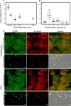
Cell-cell fusion of L-forms. (A and B) Nonspecific cell fusion was carried out using either a physical method (centrifugation) (A) or chemical method (PEG) (B). The fusion efficiency was calculated by dividing the total cell count obtained on double selection media with the cell count of the individual parent strain (AG or HR). Increasing centrifugal force leads to a decrease in efficiency (one-way analysis or variance [ANOVA], F = 15, P = 9.77 × 10−9, group-wise comparison with Tukey’s honestly significant difference [HSD]). PEG concentrations also affected fusion efficiency (one-way ANOVA, F = 22, P = 0.033, group-wise comparison with Tukey’s HSD) with 10 w% resulting in the highest efficiency of fusion. (C) Fluorescence microscopy of colony biomass on double antibiotic media after fusion via centrifugation (top two rows) or via PEG 10 w% (bottom two rows). Fluorescence expression (EGFP and mCherry) is indicated as a percentage in the top right corner of each image and was calculated using ImageJ/Fiji. The overlay image (third column) shows the percentage or area occupied by both green and red pixels and was slightly higher for PEG-induced fusion. A cell suspension from colony biomass that was cultured in LPB medium with both antibiotics and imaged at a higher magnification also shown. Scale bar for colony biomass = 100 μm; scale bar for cell suspension = 5 μm.
FIG 3
FIG 3
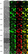
Viability of fused cells. Growth and division of fused cell were tracked over time with bright-field (BF) and fluorescence (GFP and mCherry) microscopy. Images were taken every 10 min for a total of 16 h. The panels (column-wise, left to right: bright-field, EGFP, mCherry, overlay) consist of a select few images over this time period (labeled on the left, in minutes). White triangles indicate growing cells and membrane extensions, while arrows indicate a lysed cell. Cell growth was characterized by deviation from the circular shape, membrane extension, and formation of smaller circular daughter cells. Fluorescence was maintained during this process of cell growth, as seen in the EGFP and mCherry channels. Lysed cells, on the other hand, immediately lost fluorescence and shape (t = 800 and t = 900). Images were taken from Video S1, available in the supplemental material.
FIG 4
FIG 4
Membrane fluidity affects L-form fusion. (A) Fluidity of L-form membranes was quantified as a generalized polarization (GP) value, using the Laurdan dye assay. A strong positive correlation was obtained between GP value and the period of growth, indicating a decrease in membrane fluidity with increasing culture age (Spearman’s rank correlation test). Age of the culture also had an effect on fusion efficiency (inset, 2-sample t test, P = 2.22 × 10−6, n = 3) with young 2-day-old cultures fusing more efficiently than older 7-day-old cultures. (B) Analysis of membrane lipids of cultures from different periods of growth (1, 3, 5, and 7 days) indicated a change in the percentage of saturated and unsaturated fatty acids over time. Specifically, triglyceraldehyde (TG) and phosphatidylethanolamine (PE) showed a strong decrease between days 1 and 3. Both lipids are required for fluidity of the membrane. (C) Positive correlation obtained between GP value and the percentage of PEG used, indicating a decrease in membrane fluidity with increasing concentration of PEG (Spearman’s rank correlation test). (D) The GP value showed a strong negative correlation with fusion efficiency. A low percentage of PEG (10%) leads to slightly more fluid membranes compared to those exposed to a high PEG percentage (50%), which resulted in higher fusion (Spearman’s rank correlation test). The grayscale (bottom left corner) indicates the PEG percentage, which ranged from 10 to 50%.
FIG 5
FIG 5
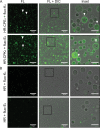
Coiled-coil lipopeptides integrate in L-form membranes. (A) Confocal microscopy images (fluorescence [FL] and overlay [FL +differential interference contrast, or DIC]), indicating peptide CPE4 or CPK4 insertion into the L-form membranes and coiled-coil formation with complementary peptides (fluo-K4 or fluo-E4). White arrows indicate clear membrane insertions. (B) In the absence of CPE4 or CPK4, no binding of the complementary fluorescent peptide (fluo-K4 or fluo-E4) was observed. Experiments were performed at 30°C, and L-forms in P-buffer were incubated with 10 μM CPE4 or CPK4 for 30 min. Subsequently, the unbound peptide was washed via centrifugation and the complementary fluorescent peptides were added. Scale bar = 5 μm.
FIG 6
FIG 6
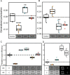
Coiled-coil lipopeptides increase membrane fluidity and cell-specific fusion. (A) Strain AR showed an increased fluidity on treatment with PEG (P = 3.06 × 10−6), a decrease in fluidity on treatment with CPE4 (P = 2.13 × 10−3), and no change in fluidity with CPK4 (one-way ANOVA, F = 36, P = 4.59 × 10−18, followed by Tukey’s pairwise comparison) compared to the control (dotted line). (B) Strain HR showed increased fluidity (low GP value) when treated with CPE4 (P = 3.11 × 10−3) and CPK4 (P = 1.4 × 10−2) compared to the control (dotted line), whereas no significant change was found when treated with 10% PEG (one-way ANOVA, F = 36, P = 2.83 × 10−18, followed by Tukey’s pairwise comparison). Dotted line is for comparison of GP values to the control where no peptide or PEG was added. (C) The AG and HR strains were individually treated with either PEG, CPE4, or CPK4 at different peptide concentrations to assess the effects on fusion efficiency. Interestingly, PEG resulted in a low level of fusion despite increasing fluidity, because of its nonspecific nature. The combination of AG-CPK4 and HR-CPE4 resulted in the highest fusion efficiency relative to the basal level. The increase in relative fusion efficiency was concentration dependent as well as peptide dependent (one-way ANOVA, F = 30, P = 3.47 × 10−14, followed by Tukey’s pairwise comparison). (D) The AG and HR strains were first treated with either PEG, CPE4, or CPK4. These strains were then directly plated on double selection media in the absence (gray boxes) or presence (black boxes) of 10 w% PEG to assess the effect on fusion efficiency. Interestingly, PEG resulted in low fusion, despite increasing fluidity because of its nonspecific nature when washed away prior to plating, but it produced a high efficiency when present during the plating. The treatment with peptides also showed a higher efficiency when in the presence of PEG (Kruskall-Wallis chi-square = 24.84, P = 5.4 × 10−5, followed by Dunnett’s pairwise comparison) compared to the control, where no peptide or PEG was added (dotted line).

References

    1. Chen EH, Grote E, Mohler W, Vignery A. 2007. Cell–cell fusion. FEBS Lett 581:2181–2193. doi:10.1016/j.febslet.2007.03.033. - DOI - PubMed
    1. Ogle BM, Cascalho M, Platt JL. 2005. Biological implications of cell fusion. Nat Rev Mol Cell Biol 6:567–575. doi:10.1038/nrm1678. - DOI - PubMed
    1. Zito F, Lampiasi N, Kireev I, Russo R. 2016. United we stand: adhesion and molecular mechanisms driving cell fusion across species. Eur J Cell Biol 95:552–562. doi:10.1016/j.ejcb.2016.09.002. - DOI - PubMed
    1. Chernomordik L, Kozlov MM, Zimmerberg J. 1995. Lipids in biological membrane fusion. J Membr Biol 146:1–14. doi:10.1007/BF00232676. - DOI - PubMed
    1. Smeijers AF, Markvoort AJ, Pieterse K, Hilbers PAJ. 2006. A detailed look at vesicle fusion. J Phys Chem B 110:13212–13219. doi:10.1021/jp060824o. - DOI - PubMed

Publication types

LinkOut - more resources