. 2022 Aug 31;10(4):e0163922.
doi: 10.1128/spectrum.01639-22. Epub 2022 Jul 12.

Human Air-Liquid-Interface Organotypic Airway Cultures Express Significantly More ACE2 Receptor Protein and Are More Susceptible to HCoV-NL63 Infection than Monolayer Cultures of Primary Respiratory Epithelial Cells

Affiliations

Human Air-Liquid-Interface Organotypic Airway Cultures Express Significantly More ACE2 Receptor Protein and Are More Susceptible to HCoV-NL63 Infection than Monolayer Cultures of Primary Respiratory Epithelial Cells

Gino Castillo et al. Microbiol Spectr. .

Abstract

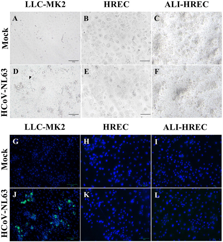
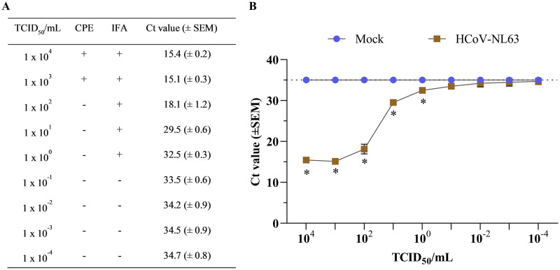
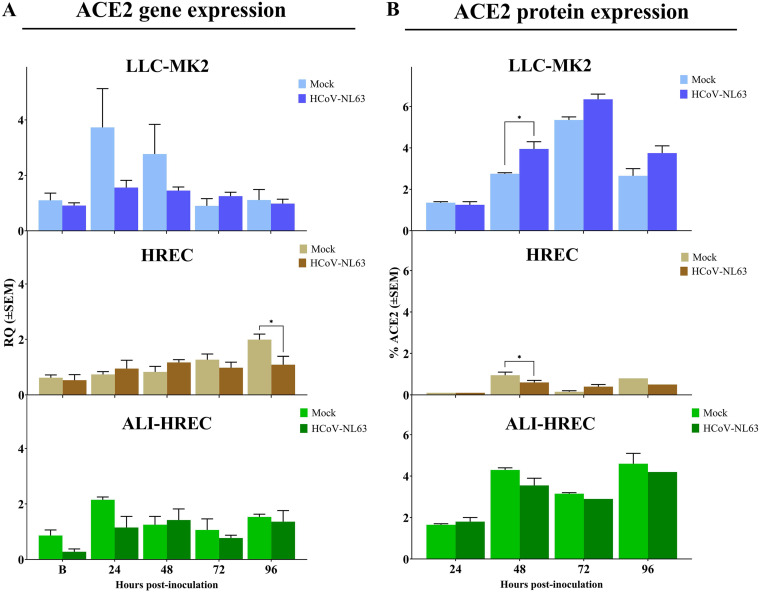
Human coronavirus NL63 (HCoV-NL63) is commonly associated with mild respiratory tract infections in infants, being that the respiratory epithelial cells are the main target for infection and initial replication of this virus. Standard immortalized cells are highly permissive to HCoV-NL63, and they are routinely used for isolation and propagation of the virus from clinical specimens. However, these cell lines are not the natural cell target of the virus and lack sufficient complexity to mimic the natural infection process in vivo. This study comparatively evaluated the differences on the susceptibility to HCoV-NL63 infection and virus replication efficiency of submerged monolayer cultures of LLC-MK2 and primary human respiratory epithelial cells (HRECs) and organotypic airway cultures of respiratory cells (ALI-HRECs). Productive viral infection and growth kinetics were assessed by morphologic examination of cytopathic effects, immunofluorescence, reverse transcription quantitative real-time PCR, and flow cytometry. Results from this study showed higher susceptibility to HCoV-NL63 infection and replication in LLC-MK2 cells followed by ALI-HRECs, with very low susceptibility and no significant virus replication in HRECs. This susceptibility was associated with the expression levels of angiontensin-converting enzyme 2 (ACE2) receptor protein in LLC-MK2, ALI-HRECs, and HRECs, respectively. Remarkably, organotypic ALI-HREC cultures expressed significantly more ACE2 receptor protein and were more susceptible to HCoV-NL63 infection than monolayer cultures of HREC. The ACE2 receptor is, therefore, a critical factor for susceptibility to HCoV-NL63 infection and replication, as is the type of culture used during infection studies. IMPORTANCE HCoV-NL63 is widespread globally, accounting for a significant number of respiratory infections in children and adults. HCoV-NL63 gains entrance into respiratory epithelial cells via the ACE2 receptor, the same cell receptor used by severe acute respiratory syndrome coronavirus (SARS-CoV) and SARS-CoV-2. Thus, HCoV-NL63 has been suggested as safe surrogate for studying disease mechanisms and therapeutic interventions against SARS-like CoVs, while working under BSL-2 conditions. The present study not only showed the critical role of ACE2 for effective HCoV-NL63 infection and replication, but also shed light on the need of more refined and complex in vitro organotypic models that recapitulate the proxy of air-liquid respiratory epithelia cell composition, structure, and functionality. These cultures have broaden virological studies toward improving our understanding of how coronaviruses cause disease and transmission not just within humans but also in animal populations.

Keywords: ACE2; Alphacoronavirus; HCoV-NL63; LLC-MK2; coronavirus; infection; organotypic airway cultures; primary human respiratory epithelial cells; upper respiratory tract.


Conflict of interest statement

The authors declare no conflict of interest.

Figures

FIG 1
FIG 1
Microscopic evaluation of the susceptibility of LLC-MK2 cells and human respiratory epithelial cells (HRECs) to HCoV-NL63 infection. Confluent monolayer (submerged) cultures of LLC-MK2 cells and HRECs were inoculated with HCoV-NL63 at 1 × 105 TCID50/mL or mock inoculated (infection medium) and incubated for 96 h (n = 3). (A to F) Brightfield images of LLC-MK2 and HREC cultures at 96 h postinoculation (hpi). Mock-inoculated with LLC-MK2 cells (A), HRECs (B), and ALI-HRECs (C). (D) Cytopathic effects (CPE) in HCoV-NL63-infected LLC-MK2 cells, including clumping of cells and cytoplasmic stranding with cell detachment (see arrows). (E to F) Absence of CPE in HREC and ALI-HREC cultures. (G to L) Immunofluorescence staining for HCoV-NL63 N protein (green) and DAPI nuclear counterstain (blue). The indirect immunofluorescence assay (IFA) was performed using an IgG1 mouse monoclonal anti-HCoV-NL63 N protein (2D4; Ingenasa-Eurofins) at a final concentration of 0.25 μg/mL, followed by incubation with 20 μg/mL of FITC-conjugated goat anti-mouse IgG antibody (KPL; SeraCare Life Sciences Inc.), while cell nuclei were stained using NucBlue Fixed cell ReadyProbes reagent (DAPI; Invitrogen, Thermo Fisher Scientific). (G to I) Absence of unspecific fluorescence in mock-inoculated IFA controls for LLC-MK2 cells (G), HRECs (H), and ALI-HRECs (I). (J to L) HCoV-NL63 N protein (green) was detected in LLC-MK2 cells (J) but not in HRECs (K) nor in ALI-HRECs (L). Scale bar = 100 μm.
FIG 2
FIG 2
Titration HCoV-NL63 cultured in LLC-MK2 cells. (A) Comparing HCoV-NL63 detection at different viral dilutions after 120 h postinoculation (hpi) using cytopathic effects (CPE), indirect immunofluorescence assay (IFA), and real-time quantitative PCR (RT-qPCR). The presence of CPE and immunofluorescence against the HCoV-NL63 N protein is represented with a (+) sign, while the absence of infection is represented with a (−) sign. Viral RNA detection was determined using RT-qPCR and expressed as mean threshold cycle (CT) values (±standard error of the mean [SEM]) (B) Graph representing mean CT values plotted from nine 10-fold serial dilutions of HCoV-NL63 (n = 7)- or mock-inoculated (infection medium) LLC-MK2 cells (n = 1) after 120 hpi. The mean CT values were determined from collected supernatants using rRT-qPCR targeting the HCoV-NL63 nucleocapsid (N) gene. Samples with a (CT) value >35 were considered negatives. * Statistically significant difference denoted with P < 0.05.
FIG 3
FIG 3
HCoV-NL63 growth kinetics in monolayer cultures of LLC-MK2 cells and primary human respiratory epithelial cells (HRECs), and organotypic airway cultures of human respiratory epithelial cells (ALI-HRECs) inoculated with a viral dose of 1 × 105 TCID50/mL. The first time point (denoted as B) corresponded to the collection immediately after inoculum removal and subsequent washing step, i.e., 2 h postinoculation (hpi) for LLC-MK2 or HRECs, and 6 hpi for ALI-HRECs. Viral RNA detection was determined using real time quantitative PCR (RT-qPCR) and expressed as mean threshold cycle (CT) value (±standard error of the mean [SEM]). A CT value of 35 was used as cut-off. (A) Viral kinetic curve generated using HCoV-NL63 N gene-based RT-qPCR data from HCoV-NL63- (1 × 105 TCID50/mL) or mock-inoculated culture supernatants (n = 3) of LLC-MK2 cells and HRECs, and basolateral media from ALI-HRECs. Additionally, a 100 μL LHC medium (Thermo Fisher Scientific) was added onto ALI-HRECs to also collect an apical wash for each time point. (B) Viral kinetic curve generated using cell pellets (n = 3) from LLC-MK2 cells, HRECs, and ALI-HRECs. Significant statistical differences (P < 0.01) across virus-inoculated cultures are denoted with a [LLC-MK2 versus HREC], b [LLC-MK2 versus ALI-HREC], and c [HREC versus ALI-HREC] for each time point. No viral RNA amplification was detected at any time point in any of the mock-inoculated cultures (LLC-MK2, HRECs, and ALI-HRECs), being the CT values significantly different (P < 0.0001) compared to the obtained for the virus-inoculated cultures throughout the study. However, for clarity, the significant differences are not denoted in the graph.
FIG 4
FIG 4
Flow cytometric analysis of subpopulations of LLC-MK2 cells, HRECs, and ALI-HRECs inoculated with HCoV-NL63 (1 × 105 TCID50/mL) based on the relative expression of the HCoV-NL63 N protein (A) and the ACE2 receptor protein (B). Cell percentages are expressed as mean ± standard error of the mean (SEM). Statistically significant differences (P < 0.01) are denoted with a [LLC-MK2 versus HREC], b [LLC-MK2 versus ALI-HREC], and c [HREC versus ALI-HREC] for each time point.
FIG 5
FIG 5
Effect of HCoV-NL63 infection on the expression of ACE2 receptor within each culture type or cell population, i.e., monolayer cultures of LLC-MK2 cells and HRECs, and organotypic ALI-HRECs. (A) Gene expression levels of angiotensin-converting enzyme 2 (ACE2) were determined by quantitative PCR (qPCR) and expressed as relative quantification (RQ) values (±SEM) for HCoV-NL63- and mock-inoculated cells. (B) Protein expression levels of ACE2 were determined by flow cytometry analysis and expressed as percentages (±SEM) for HCoV-NL63- and mock-inoculated cells. Expression levels were compared at 24, 48, 72, and 96 h postinoculation (hpi) for each culture type. * Statistically significant difference denoted with P < 0.05.

Similar articles

Cited by

References

    1. International Committee on Taxonomy of Viruses (ICTV). 2020. ICTV taxonomy history: human coronavirus NL63. https://talk.ictvonline.org/taxonomy/p/taxonomy-history?taxnode_id=20200.... Accessed May 3, 2022.
    1. van der Hoek L, Pyrc K, Jebbink MF, Vermeulen-Oost W, Berkhout RJ, Wolthers KC, Wertheim-van Dillen PM, Kaandorp J, Spaargaren J, Berkhout B. 2004. Identification of a new human coronavirus. Nat Med 10:368–373. doi:10.1038/nm1024. - DOI - PMC - PubMed
    1. Fielding BC. 2011. Human coronavirus NL63: a clinically important virus? Future Microbiol 6:153–159. doi:10.2217/fmb.10.166. - DOI - PMC - PubMed
    1. Bastien N, Anderson K, Hart L, Van Caeseele P, Brandt K, Milley D, Hatchette T, Weiss EC, Li Y. 2005. Human coronavirus NL63 infection in Canada. J Infect Dis 191:503–506. doi:10.1086/426869. - DOI - PMC - PubMed
    1. Bastien N, Robinson JL, Tse A, Lee BE, Hart L, Li Y. 2005. Human coronavirus NL-63 infections in children: a 1-year study. J Clin Microbiol 43:4567–4573. doi:10.1128/JCM.43.9.4567-4573.2005. - DOI - PMC - PubMed

Substances

LinkOut - more resources