

Interaction of Liberibacter Solanacearum with Host Psyllid Vitellogenin and Its Association with Autophagy
- PMID: 35863005
- PMCID: PMC9430699
- DOI: 10.1128/spectrum.01577-22
Interaction of Liberibacter Solanacearum with Host Psyllid Vitellogenin and Its Association with Autophagy
Abstract
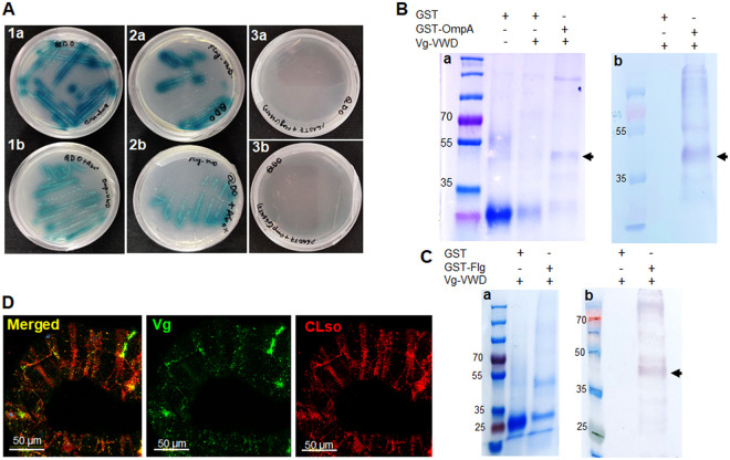
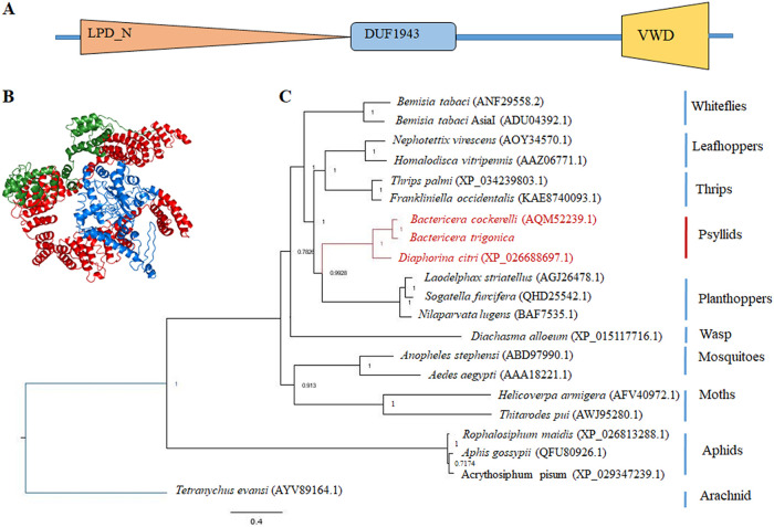
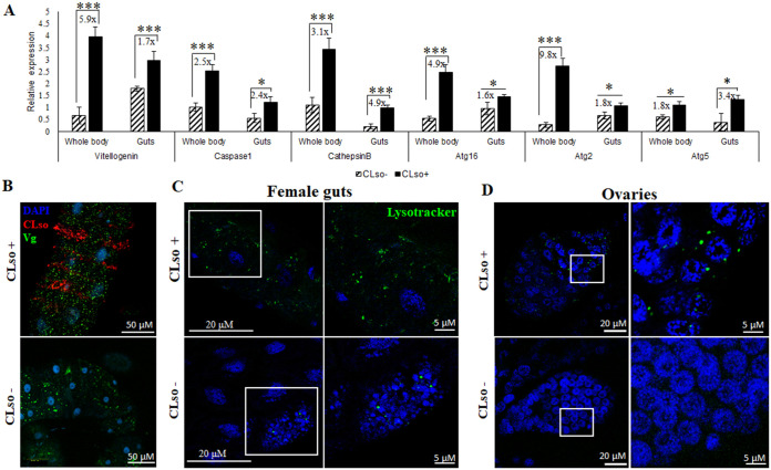
Candidatus Liberibacter solanacearum (CLso) haplotype D, transmitted by the carrot psyllid Bactericera trigonica, is a major constraint for carrot production in Israel. Unveiling the molecular interactions between the psyllid vector and CLso can facilitate the development of nonchemical approaches for controlling the disease caused by CLso. Bacterial surface proteins are often known to be involved in adhesion and virulence; however, interactions of CLso with carrot psyllid proteins that have a role in the transmission process has remained unexplored. In this study, we used CLso outer membrane protein (OmpA) and flagellin as baits to screen for psyllid interacting proteins in a yeast two-hybrid system assay. We identified psyllid vitellogenin (Vg) to interact with both OmpA and flagellin of CLso. As Vg and autophagy are often tightly linked, we also studied the expression of autophagy-related genes to further elucidate this interaction. We used the juvenile hormone (JH-III) to induce the expression of Vg, thapsigargin for suppressing autophagy, and rapamycin for inducing autophagy. The results revealed that Vg negatively regulates autophagy. Induced Vg expression significantly suppressed autophagy-related gene expression and the levels of CLso significantly increased, resulting in a significant mortality of the insect. Although the specific role of Vg remains obscure, the findings presented here identify Vg as an important component in the insect immune responses against CLso and may help in understanding the initial molecular response in the vector against Liberibacter. IMPORTANCE Pathogen transmission by vectors involves multiple levels of interactions, and for the transmission of liberibacter species by psyllid vectors, much of these interactions are yet to be explored. Candidatus Liberibacter solanacearum (CLso) haplotype D inflicts severe economic losses to the carrot industry. Understanding the specific interactions at different stages of infection is hence fundamental and could lead to the development of better management strategies to disrupt the transmission of the bacteria to new host plants. Here, we show that two liberibacter membrane proteins interact with psyllid vitellogenin and also induce autophagy. Altering vitellogenin expression directly influences autophagy and CLso abundance in the psyllid vector. Although the exact mechanism underlying this interaction remains unclear, this study highlights the importance of immune responses in the transmission of this disease agent.
Keywords: Liberibacter; autophagy; outer membrane proteins; psyllid; vitellogenin.
Conflict of interest statement
The authors declare no conflict of interest.
Figures
Similar articles
-
The plant pathogenic bacterium Candidatus Liberibacter solanacearum induces calcium-regulated autophagy in midgut cells of its insect vector Bactericera trigonica.Microbiol Spectr. 2023 Sep 28;11(5):e0130123. doi: 10.1128/spectrum.01301-23. Online ahead of print. Microbiol Spectr. 2023. PMID: 37768086 Free PMC article.
-
Association of endoplasmic reticulum associated degradation (ERAD) with the transmission of Liberibacter solanacearum by its psyllid vector.Insect Mol Biol. 2023 Aug;32(4):436-449. doi: 10.1111/imb.12842. Epub 2023 Apr 15. Insect Mol Biol. 2023. PMID: 37060303
-
Differential expression of "Candidatus Liberibacter solanacearum" genes and prophage loci in different life stages of potato psyllid.Sci Rep. 2024 Jul 15;14(1):16248. doi: 10.1038/s41598-024-65156-4. Sci Rep. 2024. PMID: 39009624 Free PMC article.
-
Interactions of Liberibacter Species with Their Psyllid Vectors: Molecular, Biological and Behavioural Mechanisms.Int J Mol Sci. 2022 Apr 5;23(7):4029. doi: 10.3390/ijms23074029. Int J Mol Sci. 2022. PMID: 35409386 Free PMC article. Review.
-
A comprehensive review of zebra chip disease in potato and its management through breeding for resistance/tolerance to 'Candidatus Liberibacter solanacearum' and its insect vector.Pest Manag Sci. 2022 Sep;78(9):3731-3745. doi: 10.1002/ps.6913. Epub 2022 May 6. Pest Manag Sci. 2022. PMID: 35415948 Review.
Cited by
-
The plant pathogenic bacterium Candidatus Liberibacter solanacearum induces calcium-regulated autophagy in midgut cells of its insect vector Bactericera trigonica.Microbiol Spectr. 2023 Sep 28;11(5):e0130123. doi: 10.1128/spectrum.01301-23. Online ahead of print. Microbiol Spectr. 2023. PMID: 37768086 Free PMC article.
-
Treatment of Rapamycin and Evaluation of an Autophagic Response in the Gut of Bactericera cockerelli (Sulč).Insects. 2023 Jan 31;14(2):142. doi: 10.3390/insects14020142. Insects. 2023. PMID: 36835711 Free PMC article.
-
Accumulation and Transmission of 'Candidatus Liberibacter solanacearum' Haplotypes by the Nymphs of Two Psyllid Vectors.Insects. 2023 Dec 16;14(12):956. doi: 10.3390/insects14120956. Insects. 2023. PMID: 38132629 Free PMC article.
-
Interaction between the flagellum of Candidatus Liberibacter asiaticus and the vitellogenin-like protein of Diaphorina citri significantly influences CLas titer.Front Microbiol. 2023 Apr 18;14:1119619. doi: 10.3389/fmicb.2023.1119619. eCollection 2023. Front Microbiol. 2023. PMID: 37143541 Free PMC article.
-
An insect virus differentially alters gene expression among life stages of an insect vector and enhances bacterial phytopathogen transmission.J Virol. 2025 Jan 31;99(1):e0163024. doi: 10.1128/jvi.01630-24. Epub 2024 Dec 23. J Virol. 2025. PMID: 39714167 Free PMC article.
References
-
- Mawassi M, Dror O, Bar-Joseph M, Piasezky A, Sjölund JM, Levitzky N, Shoshana N, Meslenin L, Haviv S, Porat C, Katsir L, Kontsedalov S, Ghanim M, Zelinger-Reichert E, Arnsdorf YM, Gera A, Bahar O. 2018. ‘Candidatus Liberibacter solanacearum’ is tightly associated with carrot yellows symptoms in Israel and transmitted by the prevalent psyllid vector Bactericera trigonica. Phytopathology 108:1056–1066. doi:10.1094/PHYTO-10-17-0348-R. - DOI - PubMed
-
- Ghosh S, Jassar O, Kontsedalov S, Lebedev G, Wang C, Turner D, Levy A, Ghanim M. 2019. A transcriptomics approach reveals putative interaction of Candidatus Liberibacter solanacearum with the endoplasmic reticulum of its psyllid vector. Insects 10:279. doi:10.3390/insects10090279. - DOI - PMC - PubMed
-
- Capoor SP, Rao DG, Viswanath SM. 1967. Diaphorina citri Kuway., a vector of the greening disease of citrus in India. Indian J Agric Sci 37:572–576.
-
- Ammar ED. 1994. Propagative transmission of plant and animal viruses by insects: factors affecting vector specificity and competence. Adv Dis Vector Res:289–331.
Publication types
MeSH terms
Substances
LinkOut - more resources
Full Text Sources
Research Materials
