Review
. 2022 Sep 14;122(17):14066-14084.
doi: 10.1021/acs.chemrev.1c01062. Epub 2022 Jul 21.

Three-Dimensional Visualization of Viral Structure, Entry, and Replication Underlying the Spread of SARS-CoV-2

Affiliations
Review

Three-Dimensional Visualization of Viral Structure, Entry, and Replication Underlying the Spread of SARS-CoV-2

James W Saville et al. Chem Rev. .

Abstract

The global spread of SARS-CoV-2 has proceeded at an unprecedented rate. Remarkably, characterization of the virus using modern tools in structural biology has also progressed at exceptional speed. Advances in electron-based imaging techniques, combined with decades of foundational studies on related viruses, have enabled the research community to rapidly investigate structural aspects of the novel coronavirus from the level of individual viral proteins to imaging the whole virus in a native context. Here, we provide a detailed review of the structural biology and pathobiology of SARS-CoV-2 as it relates to all facets of the viral life cycle, including cell entry, replication, and three-dimensional (3D) packaging based on insights obtained from X-ray crystallography, cryo-electron tomography, and single-particle cryo-electron microscopy. The structural comparison between SARS-CoV-2 and the related earlier viruses SARS-CoV and MERS-CoV is a common thread throughout this review. We conclude by highlighting some of the outstanding unanswered structural questions and underscore areas that are under rapid current development such as the design of effective therapeutics that block viral infection.


Conflict of interest statement

The authors declare the following competing financial interest(s): S.S. is the founder and CEO of Gandeeva Therapeutics Inc.

Figures

Figure 1
Figure 1
Timeline of the COVID-19 pandemic. Events are divided into general milestones (blue), variants of concern (red), and vaccine and therapeutic developments (green). The number of global COVID-19-associated deaths (gray) and vaccine doses administered (green) are graphed per month over the course of the COVID-19 pandemic (ref (10)).
Figure 2
Figure 2
Summary of SARS-CoV-2 vaccines in development. The 10 vaccines in phase 4 are the following: nucleic acids mRNA-1273 (Moderna), BNT162 (Pfizer/BioNTech), and mRNA-1273.351 (Moderna); viral vectors ChAdOx1 (AstraZeneca/Oxford), Ad5-nCoV (CanSino Biologics), and JNJ-78436735 (Johnson & Johnson); protein-based MVC-COV1901 (Medigen); whole virus CoronaVac (Sinovac), BBIBP-CorV (Beijing Institute of Biological Products), and BIBP (Sinopharm) (refs ( and 20)). Adapted with permission from ref (20). Copyright 2022 Gavi, the Vaccine Alliance.
Figure 3
Figure 3
Emergence and global prevalence of the D614G and variant of concern lineages of SARS-CoV-2. Sequence data was downloaded from the Global Initiative on Sharing All Influenza Data (GISAID) and graphed as weekly totals (refs ( and 8)). D614 and G614 genotype prevalence is shown from January to September 2020, and variant of concern lineage prevalence is shown from September 2020 to April 2022.
Figure 4
Figure 4
Summary of progress toward SARS-CoV-2 structural characterization. (A) Schematic of the SARS-CoV-2 genome with structurally characterized proteins indicated in full color. Proteins that have not yet been characterized are displayed through homology modeling and are shown as semitransparent images (NSP4/6/12, E protein, M protein). Protein illustrations were generated using Illustrate (ref (34)). (B) The number of X-ray crystallography, cryo-EM, and all SARS-CoV-2 protein structures deposited into the RCSB protein data bank (PDB) over the first 18 months of the COVID-19 pandemic (ref (35)).
Figure 5
Figure 5
Three-dimensional model of a coronavirus particle. Membrane (M), spike (S), envelope (E), and nucleocapsid (N) structural proteins are shown. Models for E and M proteins were obtained from https://sars3d.com/ and were manually (not experimentally) arranged on the surface of a 3D model of the virion rendered using EMD-30430. Adapted with permission from ref (47). Copyright 2020 Elsevier.
Figure 6
Figure 6
Spike protein distribution, conformations, and tilt angles in authentic SARS-CoV-2 virions. (A) Tomographic slices of four representative SARS-CoV-2 virions and side projections of three individual S proteins. (B) Three-dimensional model of a single SARS-CoV-2 virion derived from subtomogram averaging. Prefusion S proteins are colored in blue with up RBDs colored pink. Postfusion S protein densities are colored in orange. (C) Prefusion and postfusion S protein trimer densities obtained by subtomogram averaging and fitted with PDBs 6VXX and 6XRA, respectively. (D) Prefusion trimer conformations as observed on intact virions. The densities corresponding to three closed, one open, and two open RBDs are fitted with PDBs 6VXX, 6VYB, and 6X2B, respectively, with protomers containing up RBDs colored in blue. (E) Averaging of trimer subsets is shown for pools centered at 0°, 30°, and 60° from the normal, as well as for two rotations of the S protein relative to the tilt direction. Adapted with permission from ref (53). Copyright 2020 Ke et al. http://creativecommons.org/licenses/by/4.0/.
Figure 7
Figure 7
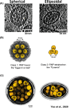
Packing of the ribonucleoprotein (RNP) complex within spherical and ellipsoidal SARS-CoV-2 particles. (A) Representative tomogram slices (5 Å thick) of spherical and ellipsoid viral particles. RNPs are visible as granular densities within the viral lumen. (B) Hexon and pyramid in situ ultrastructure reconstructions of the RNP. There was an approximately 2-fold increase in pyramid RNP reconstructions in ellipsoid viruses compared to spherical viruses. (C) Representative RNP packing arrangements in spherical and ellipsoid SARS-CoV-2 virions. Adapted with permission from ref (47). Copyright 2020 Elsevier.
Figure 8
Figure 8
Overview of coronavirus cell-entry mechanisms. (A) Members of the α- and β-coronavirus genera and their major associated cellular receptors. (B) Model of coronavirus receptor-mediated membrane-fusion mechanism between viral and cellular membranes. Adapted from ref (72). Copyright 2018 Xia et al. http://creativecommons.org/licenses/by/4.0/. (C) Endocytosis (a) and membrane fusion (b) pathways of coronavirus cell entry. Created with BioRender.
Figure 9
Figure 9
Structural insights into the SARS-CoV-2 S protein–ACE2 interaction. (A) Cryo-EM structure of the SARS-CoV-2 RBD–ACE2–B0AT1 protein complex reported by Yan et al. (6M17). The complex is shown as a colorized ribbon model and molecular surface with the RBD, ACE2, and B0AT1 shown in red, blue, and green, respectively. (B) Superposition of ACE2-complexed SARS-CoV (2AJF, brown) and SARS-CoV-2 (6M0J, purple) RBDs aligned by the RBD (refs ( and 93)). The ACE2 structure for the SARS-CoV-2 complex is shown alone to simplify the RBD–ACE2 interface. The major structural discrepancy between the SARS-CoV and SARS-CoV-2 RBDs is circled with a black dotted line. The side chains of residues mutated in variants of concern (prior to the Omicron variant) are shown and labeled in red. (C) The same as in panel B, but mutated residues are shown for the Omicron BA.2 variant.
Figure 10
Figure 10
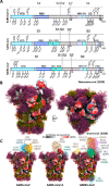
Glycosylation of the SARS-CoV-2, SARS-CoV, and MERS-CoV spike proteins. (A) Schematic representation of the SARS-CoV-2, SARS-CoV, and MERS-CoV protein open reading frames with glycosylation sites indicated (refs ( and 95)). Adapted with permission from ref (95). Copyright 2020 Watanabe et al. http://creativecommons.org/licenses/by/4.0/. (B) Moss surface representation of SARS-CoV-2 S protein glycosylation from molecular dynamic simulations performed by Grant et al. (ref (101)). Glycans are shown in ball-and-stick representations and colorized accordingly: M9, green; M5, dark yellow; hybrid, orange; complex, pink. The S protein surface (6VSB) is colored according to antibody accessibility from black (least accessible) to red (most accessible). The RBD in the up conformation is circled in blue. Adapted with permission from ref (101). Copyright 2020 Grant et al. http://creativecommons.org/licenses/by/4.0/. (C) The same as in panel B, but for SARS-CoV, SARS-CoV-2, and MERS-CoV S proteins and with available S protein–antibody structures overlapped.
Figure 11
Figure 11
Overview of RNA translation and replication, viral packaging, and release of the SARS-CoV-2 virion. Schematic representations (top) and experimental data (bottom) of the cellular machinery and viral proteins involved in (A) genome translation and initial polypeptide processing, (B) replication of genomic and subgenomic RNA, (C) assembly of the virion at the ER–Golgi intermediate compartment (ERGIC), and (D) final egress of the viral particle into the extracellular environment. (A, bottom) Mpro dimer surface model (6LU7) (ref (31)) colored by chain and the substrate binding pocket (inset) depicting the bound Mpro inhibitor N3 (sticks). Experimental data from panels B–D show tomographic slices from cryo-ET studies of (B) murine hepatitis virus (MHV) or (C and D) SARS-CoV-2-infected cells highlighting the transport of RNA through a molecular DMV pore, budding of a SARS-CoV-2 virion, and a viral exit tunnel, respectively. Panel B was adapted with permission from ref (138). Copyright 2020 Wolff et al. http://creativecommons.org/licenses/by/4.0/. Panel C was adapted with permission from ref (125). Copyright 2020 Klein et al. http://creativecommons.org/licenses/by/4.0/. Panel D was adapted with permission from ref (139). Copyright 2021 Mendonça et al. http://creativecommons.org/licenses/by/4.0/. Created with BioRender.
Figure 12
Figure 12
Structure of the SARS-CoV-2 multiprotein replication–transcription complex (RTC). Surface representation (PDB 7KRN) (ref (143)) of the RTC highlighting the relative positions of the RNA-dependent RNA polymerase (RdRp, NSP12), processivity cofactors (NSP7 and NSP8), and the viral helicase (NSP13) as determined by single-particle cryo-EM. The NTP entry tunnel (inset) plays a critical role in the backtracking/proofreading function of the RTC, as erroneously incorporated ribonucleotides are frayed into the entry tunnel where they can then be removed by the 3′-5′ exonuclease, NSP14, to ensure high-fidelity replication of the viral genome. Created with BioRender.
Figure 13
Figure 13
Therapeutic discovery during the COVID-19 pandemic. These data were compiled by the Biotechnology Innovation Organization and are presented here as the cumulative monthly number of antiviral, treatment, and vaccine therapies in development (ref (160)). Antivirals are defined here as drugs that interact directly with the virus or disrupt its ability to replicate. Treatments are defined here as drugs that treat various COVID-19-associated illnesses resulting from SARS-CoV-2 viral infection. Vaccines are defined here as prophylactic therapeutics that stimulate immunity against SARS-CoV-2.

Similar articles

Cited by

References

    1. Xu R.-H.; He J.-F.; Evans M. R.; Peng G.-W.; Field H. E.; Yu D.-W.; Lee C.-K.; Luo H.-M.; Lin W.-S.; Lin P.; et al. Epidemiologic Clues to SARS Origin in China. Emerging Infectious Diseases 2004, 10, 1030–1037. 10.3201/eid1006.030852. - DOI - PMC - PubMed
    1. Zhong N.; Zheng B.; Li Y.; Poon L.; Xie Z.; Chan K.; Li P.; Tan S.; Chang Q.; Xie J.; et al. Epidemiology and cause of severe acute respiratory syndrome (SARS) in Guangdong, People’s Republic of China, in February, 2003. Lancet 2003, 362, 1353–1358. 10.1016/S0140-6736(03)14630-2. - DOI - PMC - PubMed
    1. Heymann D. L.; Mackenzie J. S.; Peiris M. SARS legacy: outbreak reporting is expected and respected. Lancet 2013, 381, 779–781. 10.1016/S0140-6736(13)60185-3. - DOI - PMC - PubMed
    1. Listings of WHO’s Response to COVID-19. World Health Organization, June 29, 2020. https://www.who.int/news/item/29-06-2020-covidtimeline (accessed 2021-09-07).
    1. Boni M. F.; Lemey P.; Jiang X.; Lam T. T.-Y.; Perry B. W.; Castoe T. A.; Rambaut A.; Robertson D. L. Evolutionary origins of the SARS-CoV-2 sarbecovirus lineage responsible for the COVID-19 pandemic. Nature Microbiology 2020, 5, 1408–1417. 10.1038/s41564-020-0771-4. - DOI - PubMed

Publication types

Grants and funding