Case Reports
. 2022 Jul 22;7(1):248.
doi: 10.1038/s41392-022-01034-7.

Inhibition of pancreatic EZH2 restores progenitor insulin in T1D donor

Affiliations
Case Reports

Inhibition of pancreatic EZH2 restores progenitor insulin in T1D donor

Keith Al-Hasani et al. Signal Transduct Target Ther. .

Abstract

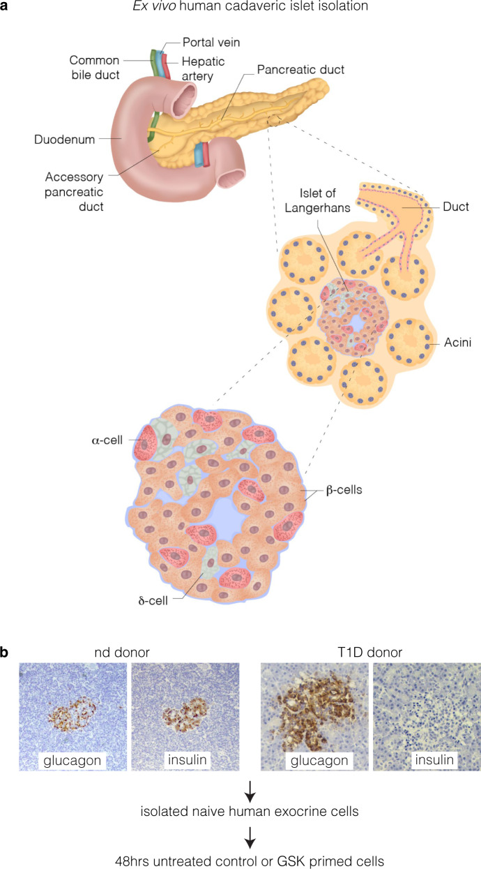
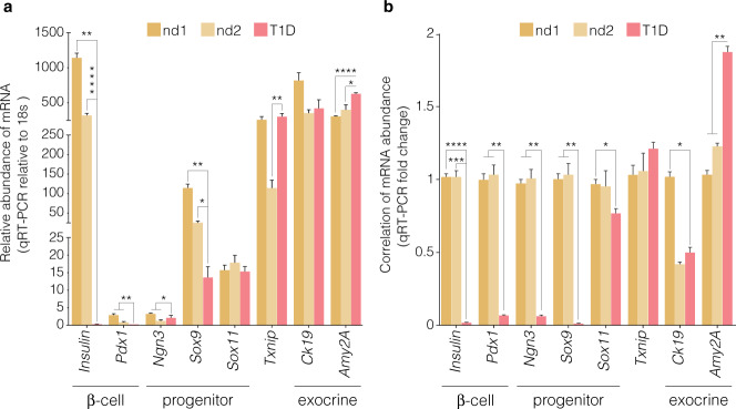
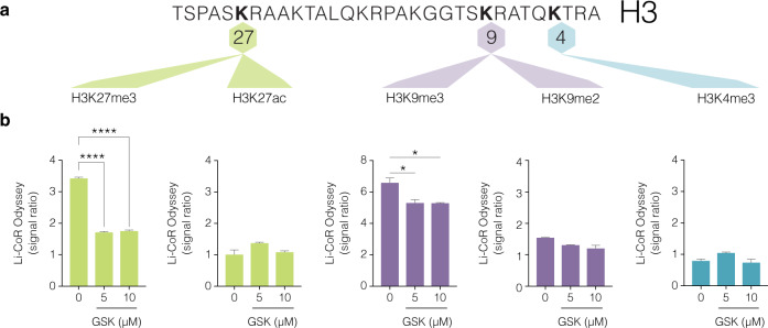
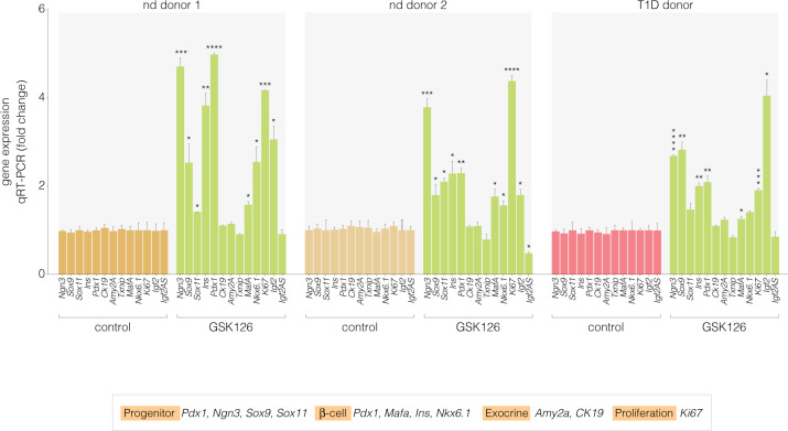
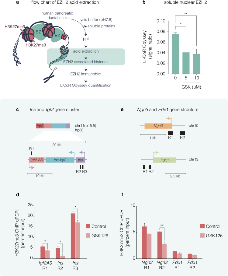
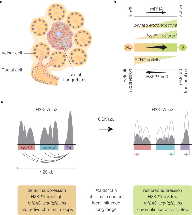
Type 1 diabetes (T1D) is an autoimmune disease that selectively destroys insulin-producing β-cells in the pancreas. An unmet need in diabetes management, current therapy is focussed on transplantation. While the reprogramming of progenitor cells into functional insulin-producing β-cells has also been proposed this remains controversial and poorly understood. The challenge is determining why default transcriptional suppression is refractory to exocrine reactivation. After the death of a 13-year-old girl with established insulin-dependent T1D, pancreatic cells were harvested in an effort to restore and understand exocrine competence. The pancreas showed classic silencing of β-cell progenitor genes with barely detectable insulin (Ins) transcript. GSK126, a highly selective inhibitor of EZH2 methyltransferase activity influenced H3K27me3 chromatin content and transcriptional control resulting in the expression of core β-cell markers and ductal progenitor genes. GSK126 also reinstated Ins gene expression despite absolute β-cell destruction. These studies show the refractory nature of chromatin characterises exocrine suppression influencing β-cell plasticity. Additional regeneration studies are warranted to determine if the approach of this n-of-1 study generalises to a broader T1D population.


Conflict of interest statement

The authors declare no competing interests.

Figures

Fig. 1
Fig. 1
Human cadaveric ex vivo exocrine isolation. a Schematic of the human pancreas emphasising the ductal endocrine and exocrine organisation. Pancreatic islet illustrated showing the major cell types. Pancreatic exocrine cells were isolated from cadaveric tissue derived from two non-diabetic and a T1D donor. b Representative immunohistochemical insulin and glucagon staining in the non-diabetic donor and T1D donor. Insulin and glucagon expression are indicated by the brown staining in human islets. Note the complete absence of insulin in type 1 diabetic donor
Fig. 2
Fig. 2
Distinguishable expression of embryonic and β-cell mRNA indices from naïve pancreatic exocrine cells isolated from donors. Abundance of Ins, Pdx1, Ngn3, Sox9 and Txnip mRNA. a Expression analyses in naïve exocrine cells showing the comparative abundance of mRNA relative to 18s assessed by qRT-PCR. Relative expression of mRNA isolated from non-diabetic (nd) and type 1 diabetic (T1D) donors. b Correlation of mRNA expression of Ins, Pdx1, Ngn3, Sox9 and Txnip displayed as a function of fold change. Expression analyses of mRNA isolated from non-diabetic (nd) and type 1 diabetic (T1D) donors. Significance is calculated by comparing nd1 v T1D and nd2 v T1D. Student t test, *P < 0.05, **P < 0.01, ***P < 0.001, ****P < 0.0001. Error bars represent SEM, n = 2
Fig. 3
Fig. 3
Pharmacological inhibition of EZH2 activity by GSK126 diminishes H3K27me3 and not H3K27ac protein content in human pancreatic ductal cells. a Partial histone H3 lysine map of sites for methylation and acetylation. The transcriptionally suppressive marks, H3K27me3, H3K9me3 and H3K9me2 including permissive histone marks, H3K27ac and H3K4me3 are shown. b Dose-dependent increase of GSK126 (5 or 10 µM) for 48 h attenuates H3K27me3 in human pancreatic ductal cells. Histones and their associated proteins were prepared by acid extraction. Quantification levels of H3K27me3, H3K27ac, H3K9me2, H3K9me3 and H3K4me3 were calculated and adjusted to overall histone H3 using Li-COR Odyssey. The signal ratio calculated was as follows; H3K27me3/total H3, H3K27ac/total H3, H3K9me3/total H3, H3K9me2/total H3, and H3K4me3/total H3. Vehicle control is DMSO. Ordinary one-way ANOVA was performed on Control vs GSK-126 (*P < 0.05, ****P < 0.0001 error bars are SEM, n = 3)
Fig. 4
Fig. 4
GSK-126 restores the expression of islet indices in non-diabetic and type 1 diabetic pancreatic exocrine cells. Correlation of mRNA abundance of Ngn3, Sox9, Sox11, Ins, Pdx1, Ck19, Amy2A, Txnip, Mafa, Nkx6.1, Igf2 and Igf2AS displayed as fold change by qRT-PCR. Significant changes in mRNA abundance were not detected for the ductal marker (Ck19), acinar marker (Amy2A) and the glucose-sensor (Txnip). Student’s t-test was performed on Control vs GSK-126 (*P < 0.05, **P < 0.01, ***P < 0.001, ****P < 0.0001 error bars are SEM, n = 3)
Fig. 5
Fig. 5
GSK126 influences the refractory H3K27me3 content on the insulin chromatin domain in human non-diabetic exocrine cells. a Flow chart of EZH2 acid extraction from human pancreatic ductal cells. Purification method of acid histone-associated protein fraction isolated from heterochromatin isolates derived from pancreatic ductal nuclei. Extraction of histone-binding EZH2 protein fraction from heterochromatin involve acid homogenisation and precipitation (ppt) with 5 M H2SO4. Isolated histone-binding proteins with EZH2 were fractionated and quantified using Li-COR Odyssey. b Dose-dependent increase of GSK126 (5 or 10 µM) for 48 h attenuates EZH2 in human pancreatic ductal cells. Quantification levels of EZH2 were calculated and adjusted to unmodified histone H3 using Li-COR Odyssey. The signal ratio was calculated as EZH2/overall H3. Vehicle control was DMSO. Ordinary one-way ANOVA was performed on Control vs GSK-126 (*P < 0.05, **P < 0.01, error bars are SEM, n = 3). c Insulin domain was assessed using amplimers that were specifically designed to distinguish promoter regions (R) of the Ins and Igf2AS genes. d Quantitative PCR analysis of DNA in chromatin immunoprecipitated (ChIP) with anti-H3K27me3 antibody. Vehicle control was DMSO. e DNA was assessed using amplimers that specifically recognise the promoter regions (R) of the Ngn3 and Pdx1 genes. f Quantitative PCR analysis of DNA in ChIP with anti-H3K27me3 antibody. Vehicle control was DMSO. Data represented as the mean Input signal against specific H3K27me3 abundance. Student’s t-test was performed on Control vs GSK-126 (*P < 0.05, **P < 0.01 error bars are SEM, n = 2)
Fig. 6
Fig. 6
Schematic representation of pancreatic progenitor differentiation and the refractory influence of H3K27me3 content on the insulin chromatin domain in human exocrine cells. a Organisation of the human endocrine and exocrine pancreas showing the main pancreatic ductal tree connecting the acinar bundle. b The inability to influence transcriptional expression in the exocrine and endocrine pancreas is in accordance with default transcriptional suppression mediated by EZH2 dependent H3K27me3. Conversion of default repression state in the acinar and ductal (AD) cells is influenced by pharmacological inhibition of EZH2 by GSK126 to prime β-cell lineage regeneration and restore insulin expression. c Proposed model of the Ins chromatin domain in pancreatic cells. Default transcriptional suppression is characterised with H3K27me3 rich regions can influence chromatin conformation, and suppress the expression of Ins, Igf2AS and Ins-Igf2 genes postulated by long-distance chromatinised-looping. We propose GSK126 attenuates EZH2 activity to influence local H3K27me3 chromatin content and long-distance interactions that function on the Ins chromatin domain and gene expression

Similar articles

Cited by

References

    1. Gribben C, et al. Ductal Ngn3-expressing progenitors contribute to adult beta-cell neogenesis in the pancreas. Cell Stem Cell. 2021;28:2000–2008 e2004. doi: 10.1016/j.stem.2021.08.003. - DOI - PMC - PubMed
    1. Gu G, Dubauskaite J, Melton DA. Direct evidence for the pancreatic lineage: NGN3+ cells are islet progenitors and are distinct from duct progenitors. Development. 2002;129:2447–2457. doi: 10.1242/dev.129.10.2447. - DOI - PubMed
    1. Schwitzgebel VM, et al. Expression of neurogenin3 reveals an islet cell precursor population in the pancreas. Development. 2000;127:3533–3542. doi: 10.1242/dev.127.16.3533. - DOI - PubMed
    1. Gouzi M, Kim YH, Katsumoto K, Johansson K, Grapin-Botton A. Neurogenin3 initiates stepwise delamination of differentiating endocrine cells during pancreas development. Dev. Dyn. 2011;240:589–604. doi: 10.1002/dvdy.22544. - DOI - PubMed
    1. Gu G, Brown JR, Melton DA. Direct lineage tracing reveals the ontogeny of pancreatic cell fates during mouse embryogenesis. Mech. Dev. 2003;120:35–43. doi: 10.1016/S0925-4773(02)00330-1. - DOI - PubMed

Publication types