Clinical Trial
. 2022 Jul 21;13(1):4119.
doi: 10.1038/s41467-022-31262-y.

A phase I/II study of triple-mutated oncolytic herpes virus G47∆ in patients with progressive glioblastoma

Affiliations
Clinical Trial

A phase I/II study of triple-mutated oncolytic herpes virus G47∆ in patients with progressive glioblastoma

Tomoki Todo et al. Nat Commun. .

Abstract

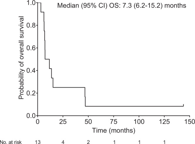
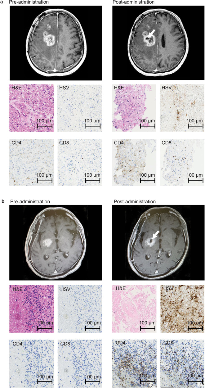
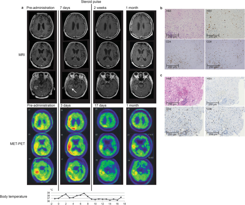
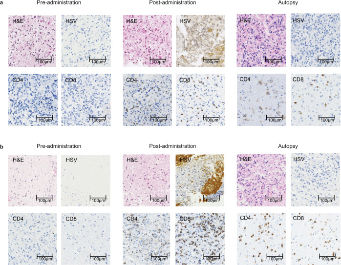
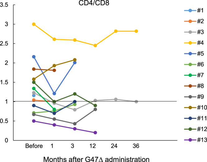
Here, we report the results of a phase I/II, single-arm study (UMIN-CTR Clinical Trial Registry UMIN000002661) assessing the safety (primary endpoint) of G47∆, a triple-mutated oncolytic herpes simplex virus type 1, in Japanese adults with recurrent/progressive glioblastoma despite radiation and temozolomide therapies. G47Δ was administered intratumorally at 3 × 10⁸ pfu (low dose) or 1 × 10⁹ pfu (set dose), twice to identical coordinates within 5-14 days. Thirteen patients completed treatment (low dose, n = 3; set dose, n = 10). Adverse events occurred in 12/13 patients. The most common G47Δ-related adverse events were fever, headache and vomiting. Secondary endpoint was the efficacy. Median overall survival was 7.3 (95%CI 6.2-15.2) months and the 1-year survival rate was 38.5%, both from the last G47∆ administration. Median progression-free survival was 8 (95%CI 7-34) days from the last G47∆ administration, mainly due to immediate enlargement of the contrast-enhanced area of the target lesion on MRI. Three patients survived >46 months. One complete response (low dose) and one partial response (set dose) were seen at 2 years. Based on biopsies, post-administration MRI features (injection site contrast-enhancement clearing and entire tumor enlargement) likely reflected tumor cell destruction via viral replication and lymphocyte infiltration towards tumor cells, the latter suggesting the mechanism for "immunoprogression" characteristic to this therapy. This study shows that G47Δ is safe for treating recurrent/progressive glioblastoma and warrants further clinical development.


Conflict of interest statement

T.T. owns the patent right for G47∆ in Japan (“Viral vectors and their use in therapeutic methods.” 4212897). All other authors have no conflict of interest to declare.

Figures

Fig. 1
Fig. 1. Kaplan–Meier curve for overall survival from the last G47∆ administration (FAS population; N = 13).
Source data are provided as a Source Data file.
Fig. 2
Fig. 2. MRI and histology studies in two patients (a: Patient #6 and b: Patient #10).
MRI showed enlargement of the entire enhanced lesion with clearing of contrast enhancement at the injection site (arrows) post-G47Δ administration. All specimens from pre-administration were negative for HSV-1 immunostaining. Second biopsy specimens obtained before the second injection from the same coordinates of the first G47∆ injection 7 days (a) and 6 days (b) post-administration showed decreased number of tumor cells, possibly due to tumor cell destruction associated with viral replication (H&E) and positive HSV-1 immunostaining. An increase in infiltration of CD4+ and CD8+ T lymphocytes towards remaining tumor cells was observed. Pathology images are representative of four biopsy specimens.
Fig. 3
Fig. 3. MET-PET and histology findings of Patient #5.
a MRI, MET-PET images and the chart of body temperature shown in parallel. MRI showed enlargement of the enhanced legion with clearing of contrast enhancement at the injection site (arrow) 7 days after the second dose of G47∆. MET-PET performed 3 days after the second dose showed decreased MET uptake at the site of G47∆ injection but a substantially increased uptake in a vast area of the right cerebral hemisphere. Immediately after initiation of 7-day steroid pulse therapy, the enlarged enhanced lesion on MRI returned to pre-G47∆ treatment size, the increased MET uptake markedly reduced, and the fever subsided. The reduced MET uptake event remained so at 1 month. b and c Biopsy specimen obtained before the second injection from the same coordinates of the first G47∆ injection after 12 days. G47∆ replication was observed by positive HSV-1 immunostaining (b). Infiltration of CD4+ and CD8+ T lymphocytes was observed in tumor areas around G47∆ (b) as well as in tumor areas without G47∆ (c). Representative of four biopsy specimens. Source data for body temperature are provided as a Source Data file.
Fig. 4
Fig. 4. Serial MRI findings of two patients who showed responses.
a Patient #3. After receiving two doses of G47∆ (3 × 108 pfu/dose), a drastic decrease in size of the target lesion was observed at 6 months and a complete response was observed at 24 months, leaving areas of porencephaly. b Patient #4. After receiving two doses of G47∆ (1 × 109 pfu/dose), the entire target lesion increased in contrast enhancement size at 1 month, but then gradually decreased at 5 months, and ended with a marked decrease at 2 years. A portion of the lesion continued to show contrast enhancement, so this patient was considered PR. The lesion remains stable and this patient survives for >11 years.
Fig. 5
Fig. 5. Histological findings in autopsy cases.
a Patient #11: 6.2 months after receiving G47Δ and b Patient #13: 13.7 months after receiving G47Δ. Both brain tumor specimens from pre-administration showed negative immunostaining for HSV-1, and very scarce CD4+ and CD8+ T lymphocytes. Infiltration of CD4+ and CD8+ T lymphocytes and positive HSV-1 immunostaining were observed after G47∆ administration at the injection site in both cases. Representative of four biopsy specimens. At autopsy, both brain tumor specimens showed viable glioblastoma cells (H&E) together with intratumoral infiltration of CD4+ and CD8+ T lymphocytes. The infiltration of CD4+ and CD8+ T lymphocytes was shown to persist for >13 months. Autopsy brain tumor specimens were negative for HSV-1 immunostaining. Representative of three tissue samples.
Fig. 6
Fig. 6. Blood CD4/CD8 ratio.
Blood CD4/CD8 ratio was measured before G47∆ and 1, 3, 12, 24, and 36 months after the last G47∆ administration. Patient #4, a long-term survivor, maintained a high CD4/CD8 ratio. Source data are provided as a Source Data file.

References

    1. Stupp R, et al. Effects of radiotherapy with concomitant and adjuvant temozolomide versus radiotherapy alone on survival in glioblastoma in a randomised phase III study: 5-year analysis of the EORTC-NCIC trial. Lancet Oncol. 2009;10:459–466. doi: 10.1016/S1470-2045(09)70025-7. - DOI - PubMed
    1. Stupp R, et al. Effect of tumor-treating fields plus maintenance temozolomide vs maintenance temozolomide alone on survival in patients with glioblastoma: A randomized clinical trial. JAMA. 2017;318:2306–2316. doi: 10.1001/jama.2017.18718. - DOI - PMC - PubMed
    1. Ballman KV, et al. The relationship between six-month progression-free survival and 12-month overall survival end points for phase II trials in patients with glioblastoma multiforme. Neuro Oncol. 2007;9:29–38. doi: 10.1215/15228517-2006-025. - DOI - PMC - PubMed
    1. Taguchi S, Fukuhara H, Todo T. Oncolytic virus therapy in Japan: progress in clinical trials and future perspectives. Jpn J. Clin. Oncol. 2019;49:201–209. doi: 10.1093/jjco/hyy170. - DOI - PubMed
    1. Fukuhara H, Ino Y, Todo T. Oncolytic virus therapy: A new era of cancer treatment at dawn. Cancer Sci. 2016;107:1373–1379. doi: 10.1111/cas.13027. - DOI - PMC - PubMed

Publication types

Associated data