

Pigmentary lesions in eyes with rhegmatogenous retinal detachment with flap tears: a retrospective observational study
- PMID: 35864144
- PMCID: PMC9304380
- DOI: 10.1038/s41598-022-16508-5
Pigmentary lesions in eyes with rhegmatogenous retinal detachment with flap tears: a retrospective observational study
Abstract
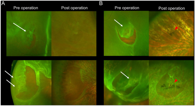
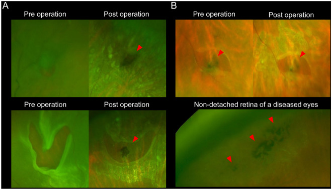
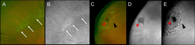
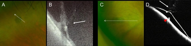
We included 97 patients with unilateral rhegmatogenous retinal detachment (RRD) with posterior vitreous detachment who underwent vitrectomy, and examined pigmentary lesion (PL) characteristics around the sites of original tears using pre- and postoperative ultra-widefield scanning light ophthalmoscopy, green light fundus autofluorescence (FAF) imaging, and intraoperative digital video. If PL did not involve RRD, we used OCT to preoperatively assess any pathologic changes to the lesion. A total of 116 retinal tears (mean count, 1.2 ± 0.5; range, 1-4 per eye) were observed in the detached retina. Overall, 102 (88%), 63 (54%), 14 (12%), and 25 (22%) tears were accompanied by lattice degeneration (LD) or PL, both LD and PL, only LD, and only PL, respectively. In green FAF images, LD showed normal to mild-hyper fluorescence, whereas all PL showed hypofluorescence. On OCT, PL were located at the RPE level, while choroid abnormalities were unclear. In the retinal areas of 22 eyes, which were not affected by RRD, we observed PL without retinal tears; some were accompanied by vitreous traction and tractional retinal detachment. Pre-, intra-, and post-operative assessments of original flap tears suggested that PL might be a risk factor for RRD, developing alongside or separately from LD.
© 2022. The Author(s).
Conflict of interest statement
The authors declare no competing interests.
Figures
Similar articles
-
Spotlight on Lattice Degeneration Imaging Techniques.Clin Ophthalmol. 2023 Aug 16;17:2383-2395. doi: 10.2147/OPTH.S405200. eCollection 2023. Clin Ophthalmol. 2023. PMID: 37605766 Free PMC article. Review.
-
Retinal detachment after branch retinal vein occlusion: influence of the type of break on the outcome of vitreous surgery.Ophthalmology. 1997 Jan;104(1):27-32. doi: 10.1016/s0161-6420(97)30366-2. Ophthalmology. 1997. PMID: 9022100
-
[Multimodal Approaches for the Analysis of Retinal Functional Disorders―Focusing on Retinal Detachment].Nippon Ganka Gakkai Zasshi. 2017 Mar;121(3):185-231. Nippon Ganka Gakkai Zasshi. 2017. PMID: 30088405 Japanese.
-
Ultra-widefield fundus imaging in gas-filled eyes after vitrectomy.BMC Ophthalmol. 2017 Jul 3;17(1):114. doi: 10.1186/s12886-017-0510-7. BMC Ophthalmol. 2017. PMID: 28673266 Free PMC article.
-
Pars plana vitrectomy versus scleral buckling for repairing simple rhegmatogenous retinal detachments.Cochrane Database Syst Rev. 2019 Mar 8;3(3):CD009562. doi: 10.1002/14651858.CD009562.pub2. Cochrane Database Syst Rev. 2019. PMID: 30848830 Free PMC article.
Cited by
-
Black posterior vitreous detachment: A 10-year historical cohort of pigmented uveal tumors.Heliyon. 2024 Nov 21;11(1):e40533. doi: 10.1016/j.heliyon.2024.e40533. eCollection 2025 Jan 15. Heliyon. 2024. PMID: 39790883 Free PMC article.
-
Spotlight on Lattice Degeneration Imaging Techniques.Clin Ophthalmol. 2023 Aug 16;17:2383-2395. doi: 10.2147/OPTH.S405200. eCollection 2023. Clin Ophthalmol. 2023. PMID: 37605766 Free PMC article. Review.
References
Publication types
MeSH terms
LinkOut - more resources
Full Text Sources
Medical
Research Materials
