. 2022 Jul 21;12(1):12471.
doi: 10.1038/s41598-022-16691-5.

Neural specialization to human faces at the age of 7 months

Affiliations

Neural specialization to human faces at the age of 7 months

Santeri Yrttiaho et al. Sci Rep. .

Abstract

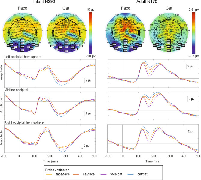
Sensitivity to human faces has been suggested to be an early emerging capacity that promotes social interaction. However, the developmental processes that lead to cortical specialization to faces has remained unclear. The current study investigated both cortical sensitivity and categorical specificity through event-related potentials (ERPs) previously implicated in face processing in 7-month-old infants (N290) and adults (N170). Using a category-specific repetition/adaptation paradigm, cortical specificity to human faces, or control stimuli (cat faces), was operationalized as changes in ERP amplitude between conditions where a face probe was alternated with categorically similar or dissimilar adaptors. In adults, increased N170 for human vs. cat faces and category-specific release from adaptation for face probes alternated with cat adaptors was found. In infants, a larger N290 was found for cat vs. human probes. Category-specific repetition effects were also found in infant N290 and the P1-N290 peak-to-peak response where latter indicated category-specific release from adaptation for human face probes resembling that found in adults. The results suggest cortical specificity to human faces during the first year of life. Encoding of unfamiliar cat stimuli might explain N290 amplification found in infants.


Conflict of interest statement

The authors declare no competing interests.

Figures

Figure 1
Figure 1
Human and cat face stimuli. Human face stimuli consisted of photographs of seven female individuals and were modified from images included in the Utrecht ECVP face database. The cat stimuli consisted of seven exemplars of short-haired cats including and resembling the depicted stimulus example. Low-level stimulus features were matched across stimuli using manipulations of the luminance histograms and spatial frequency spectra of the images. Top panel: example stimuli from human (left) and cat (right) categories. Middle panel: Luminance histograms from human (left) and cat (right) stimuli indicate brightness values (0–255) on the x-axis and count of pixels at each value on the y-axis. Bottom panel: Spatial frequency spectra of human (left) and cat (right) faces. The x-axis indicates frequency as cycles/image. The y-axis shows energy at frequency. Line-plots indicate the mean value across stimuli and error-bars indicate standard deviation across exemplars (for both histograms and spectra alike). The human face photograph depicted above, resembling those used in the current study, is included here with permission from the model. The cat photograph is a derivative of “European_shorthair_TUROK_cat_show_Turku_2010-03-27.JPG” by Heikki Siltala licensed under CC BY 3.0.
Figure 2
Figure 2
Schematic illustration of the stimulus-specific adaptation paradigm with two different stimulus categories of human (black squares) and cat (white squares) faces. ERPs were recorded in response to two different probe categories. Across separate stimulus conditions, the adaptor stimulus was chosen either from the same stimulus category as the probe or from the other stimulus category. In blocks with two different stimulus categories, human and cat faces alternated between consecutive trials.
Figure 3
Figure 3
ERP responses elicited by human faces and cat faces in 7-month-old infants (left) and in adult (right) participants. Topoplots (top) represent scalp topography of infant N290 and adult N170 response as difference from the preceding P1 response. Lower panels show response waveforms that are averaged from electrode arrays located at left, midline, and right occipital regions (see main text for channel selection). Data from 49 infant, and 21 adult participants, respectively.

References

    1. Sugita Y. Face perception in monkeys reared with no exposure to faces. Proc. Natl. Acad. Sci. 2008;105(1):394–398. doi: 10.1073/pnas.0706079105. - DOI - PMC - PubMed
    1. De Haan M, Johnson MH, Halit H. Development of face-sensitive event-related potentials during infancy: A review. Int. J. Psychophysiol. 2003;51(1):45–58. doi: 10.1016/S0167-8760(03)00152-1. - DOI - PubMed
    1. Jones W, Klin A. Attention to eyes is present but in decline in 2–6-month-old infants later diagnosed with autism. Nature. 2013;504(7480):427–431. doi: 10.1038/nature12715. - DOI - PMC - PubMed
    1. Peltola MJ, Yrttiaho S, Leppänen JM. Infants’ attention bias to faces as an early marker of social development. Dev. Sci. 2018;21(6):e12687. doi: 10.1111/desc.12687. - DOI - PubMed
    1. Le Grand R, Mondloch CJ, Maurer D, Brent HP. Expert face processing requires visual input to the right hemisphere during infancy. Nat. Neurosci. 2003;6(10):1108–1112. doi: 10.1038/nn1121. - DOI - PubMed

Publication types