. 2022 Jul 21;12(1):12491.
doi: 10.1038/s41598-022-16776-1.

Distinct antibody clones detect PD-1 checkpoint expression and block PD-L1 interactions on live murine melanoma cells

Affiliations

Distinct antibody clones detect PD-1 checkpoint expression and block PD-L1 interactions on live murine melanoma cells

Christina Martins et al. Sci Rep. .

Abstract

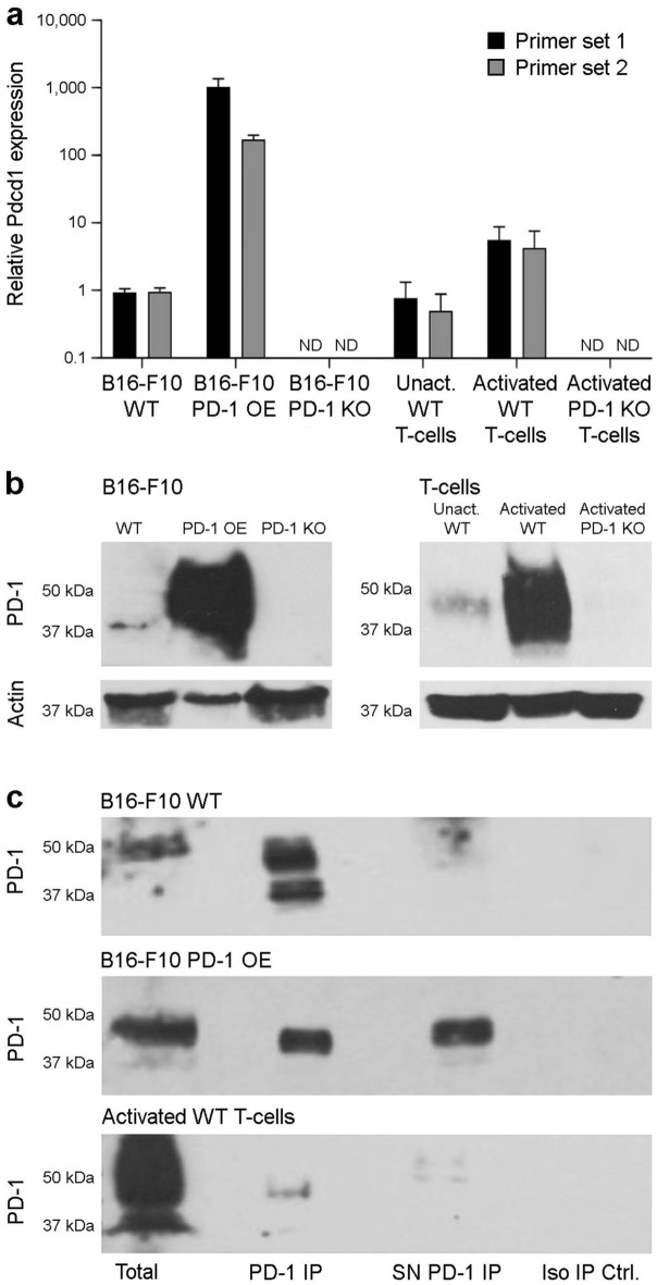
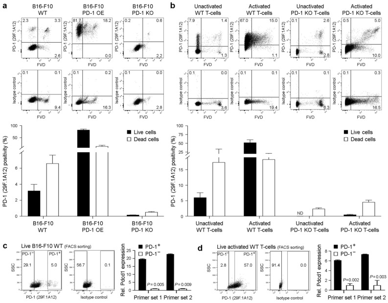
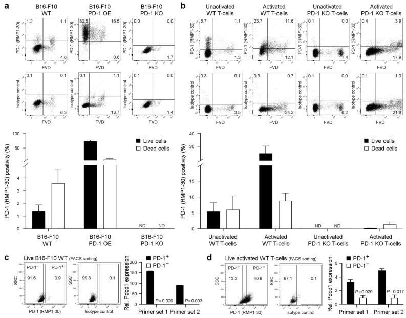
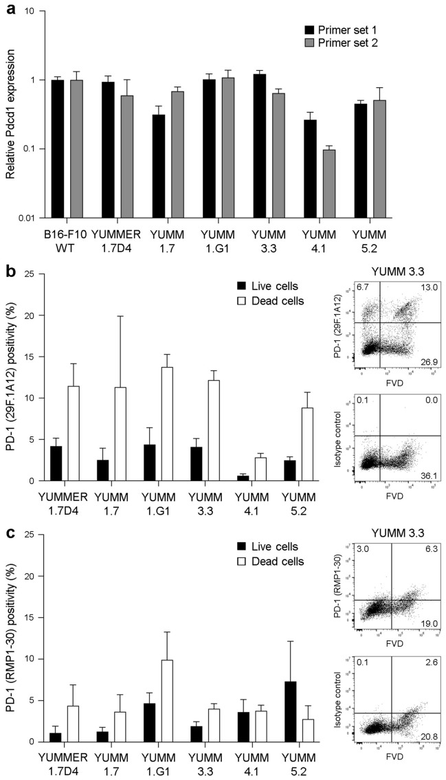
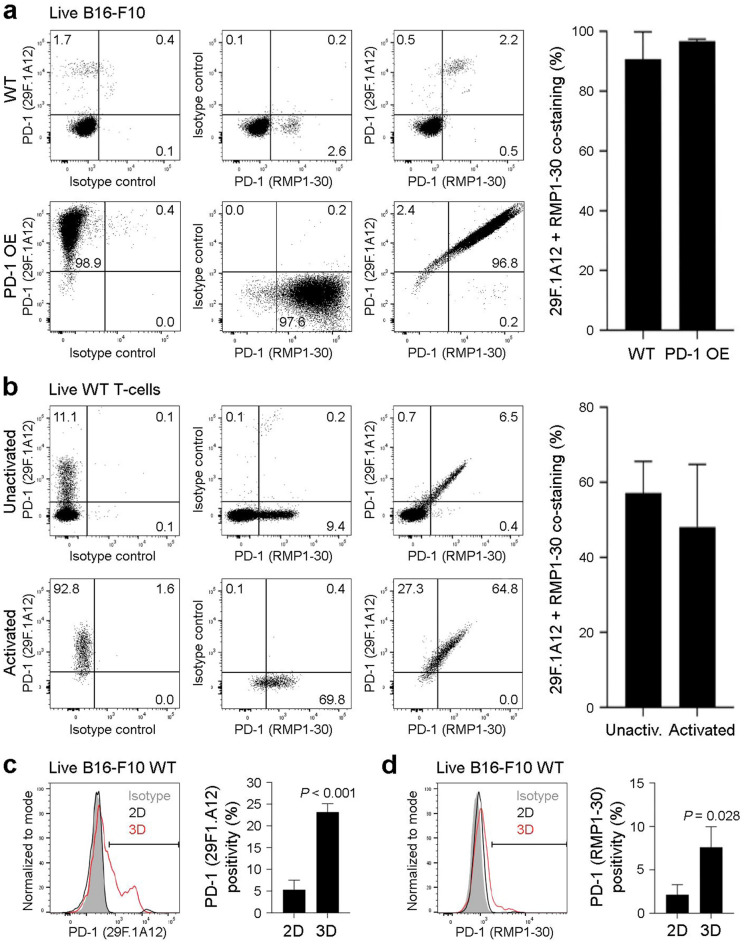
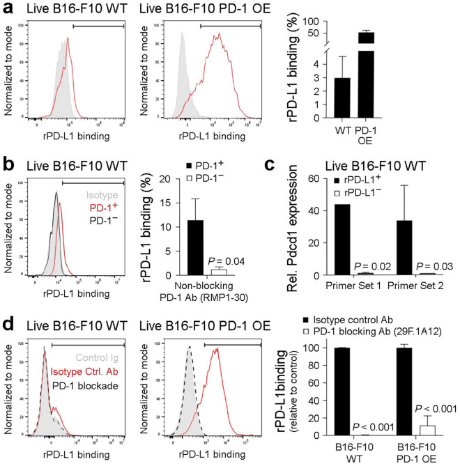
Monoclonal antibodies (abs) targeting the programmed cell death 1 (PD-1) immune checkpoint pathway have revolutionized tumor therapy. Because T-cell-directed PD-1 blockade boosts tumor immunity, anti-PD-1 abs have been developed for examining T-cell-PD-1 functions. More recently, PD-1 expression has also been reported directly on cancer cells of various etiology, including in melanoma. Nevertheless, there is a paucity of studies validating anti-PD-1 ab clone utility in specific assay types for characterizing tumor cell-intrinsic PD-1. Here, we demonstrate reactivity of several anti-murine PD-1 ab clones and recombinant PD-L1 with live B16-F10 melanoma cells and YUMM lines using multiple independent methodologies, positive and negative PD-1-specific controls, including PD-1-overexpressing and PD-1 knockout cells. Flow cytometric analyses with two separate anti-PD-1 ab clones, 29F.1A12 and RMP1-30, revealed PD-1 surface protein expression on live murine melanoma cells, which was corroborated by marked enrichment in PD-1 gene (Pdcd1) expression. Immunoblotting, immunoprecipitation, and mass spectrometric sequencing confirmed PD-1 protein expression by B16-F10 cells. Recombinant PD-L1 also recognized melanoma cell-expressed PD-1, the blockade of which by 29F.1A12 fully abrogated PD-1:PD-L1 binding. Together, our data provides multiple lines of evidence establishing PD-1 expression by live murine melanoma cells and validates ab clones and assay systems for tumor cell-directed PD-1 pathway investigations.


Conflict of interest statement

The authors declare no competing interests.

Figures

Figure 1
Figure 1
PD-1 is expressed by murine B16-F10 melanoma cells. (a) Relative PD-1 gene (Pdcd1) expression (mean ± s.d., two distinct primer sets) in wild-type (WT), PD-1-overexpressing (OE), and PD-1 knockout (KO) murine B16-F10 melanoma cells, and in unactivated and activated WT, and PD-1 KO T-cells from C57BL/6 mice, as determined by real-time quantitative RT-PCR. (b) Immunoblot of PD-1 protein expression and respective β-actin loading controls in cells as above. (c) Immunoprecipitation (IP) of PD-1 (lane 2) from B16-F10 WT (top), B16-F10 PD-1-OE melanoma (center), and activated WT T-cell (bottom) lysates and respective supernatants (SN, lane 3). PD-1 immunoblots of total protein (lane 1) and isotype IP negative controls (lane 4) are also depicted. PD-1 protein identity for immunoprecipitated bands was confirmed by mass spectrometry (MS)-based sequencing. All results are representative of at least n = 3 independent experiments. ND, not detected. Please see Supplementary Information for full-length Western blot images.
Figure 2
Figure 2
The anti-PD-1 29F.1A12 monoclonal antibody recognizes PD-1 surface protein on live B16-F10 melanoma cells. (a,b) Representative FACS plots of surface PD-1 protein expression (clone 29F.1A12) or isotype control staining and % positivity (mean ± s.e.m.) on live versus dead (fixable viability dye, FVD-negative or -positive, respectively) (a) B16-F10 wild-type (WT), PD-1-overexpressing (OE), or PD-1 knockout (KO) melanoma cells, or on (b) unactivated versus activated WT or PD-1 KO T-cells (C57BL/6), respectively. (c,d) Relative Pdcd1 expression (mean ± s.d., two distinct primer sets), as determined by real-time qPCR (right), in FACS-sorted (29F.1A12, left) live (FVD) (c) PD-1+ versus PD-1 B16-F10 WT melanoma cells or (d) respective activated WT (C57BL/6) T-cell subsets. All results are representative of at least n = 3 independent experiments. **p < 0.01; ND, not detected.
Figure 3
Figure 3
The anti-PD-1 RMP1-30 monoclonal antibody recognizes PD-1 surface protein on live B16-F10 melanoma cells. (a,b) Representative FACS plots of surface PD-1 protein expression (clone RMP1-30) or isotype control staining and % positivity (mean ± s.e.m.) on live versus dead (fixable viability dye, FVD-negative or -positive, respectively) (a) B16-F10 wild-type (WT), PD-1-overexpressing (OE), or PD-1 knockout (KO) melanoma cells, or on (b) unactivated versus activated WT or PD-1 KO T-cells (C57BL/6), respectively. (c,d) Relative Pdcd1 expression (mean ± s.d., two distinct primer sets), as determined by real-time qPCR (right), in FACS-sorted (RMP1-30, left) live (FVD) (c) PD-1+ versus PD-1 B16-F10 WT melanoma cells or (d) respective activated WT (C57BL/6) T-cell subsets. All results are representative of at least n = 3 independent experiments. *p < 0.01; **p < 0.01; ND, not detected.
Figure 4
Figure 4
The anti-PD-1 29F.1A12 and RMP1-30 antibody clones recognize PD-1 surface protein on live YUMM melanoma cells. (a) Relative PD-1 gene (Pdcd1) expression (mean ± s.d., two distinct primer sets) in wild-type B16-F10, YUMMER1.7D4, YUMM1.7, YUMM1.G1, YUMM3.3, YUMM4.1, and YUMM5.2 cells. (b,c) Representative FACS plots (YUMM3.3) of surface PD-1 protein expression (clones 29F.1A12 and RMP1-30, respectively) or isotype control staining and % positivity (mean ± s.e.m.) on live versus dead (fixable viability dye, FVD-negative or -positive, respectively) YUMM melanoma cell lines, as in (a).
Figure 5
Figure 5
The 29F.1A12 and RMP1-30 anti-PD-1 antibody clones recognize overlapping B16-F10 melanoma subpopulations. (a,b) Representative FACS plots of 29F.1A12 and RMP1-30 co-staining (left) and % dual positivity (mean ± s.e,m., calculated as a fraction of RMP1-30-reactive cells) of PD-1 surface protein expression (right) by live (FVD) (a) B16-F10 wild-type (WT) versus PD-1-overexpressing (OE) melanoma cells or (b) unactivated versus activated WT T-cells (C57BL/6) reveals co-localization of PD-1 antibody binding. (c,d) Representative histograms (left) and % positivity (mean ± s.e.m.) of PD-1 surface protein expression (right) by live (FVD) B16-F10 WT cells grown in standard (2D, black line) versus tumor spheroid (3D, red line) culture conditions, as determined by the (c) 29F.1A12 or (d) RMP1-30 anti-mouse PD-1 antibody clones. Results are representative of at least n = 3 independent experiments. *p < 0.01; ***p < 0.001.
Figure 6
Figure 6
The 29F.1A12 anti-PD-1 monoclonal antibody blocks PD-1 interactions with recombinant PD-L1 on live B16-F10 melanoma cells. (a,b) Representative histograms (left) and % recombinant (r)PD-L1 binding (mean ± s.e.m., right), to live (FVD) (a) B16-F10 wild-type (WT) or PD-1-overexpressing (OE) melanoma cells or (b) PD-1+ (red line) versus PD-1 (black line), RMP1-30-gated B16-F10 WT melanoma cell subset. (c) Relative Pdcd1 expression (mean ± s.d., two distinct primer sets), as determined by real-time qPCR, in live (FVD) FACS-sorted, rPD-L1+/− B16-F10 WT subpopulations. (d) Effect of antibody-mediated PD-1 blockade (29F.1A12, black dashed line) versus isotype control (red line) on rPD-L1 binding to B16-F10 WT or PD-1 OE melanoma cells. Shown are representative histogram plots (left) and % rPD-L1 relative to isotype control (mean ± s.e.m., right). Results are representative of at least n = 3 independent experiments. *p < 0.01; ***p < 0.001.

References

    1. Sharpe AH, Pauken KE. The diverse functions of the PD1 inhibitory pathway. Nat. Rev. Immunol. 2018;18:153–167. doi: 10.1038/nri.2017.108. - DOI - PubMed
    1. Robert C. A decade of immune-checkpoint inhibitors in cancer therapy. Nat. Commun. 2020;11:3801. doi: 10.1038/s41467-020-17670-y. - DOI - PMC - PubMed
    1. Sharma P, et al. The next decade of immune checkpoint therapy. Cancer Discov. 2021;11:838–857. doi: 10.1158/2159-8290.CD-20-1680. - DOI - PubMed
    1. Juneja VR, et al. PD-L1 on tumor cells is sufficient for immune evasion in immunogenic tumors and inhibits CD8 T cell cytotoxicity. J. Exp. Med. 2017;214:895–904. doi: 10.1084/jem.20160801. - DOI - PMC - PubMed
    1. Kleffel S, et al. Melanoma cell-intrinsic PD-1 receptor functions promote tumor growth. Cell. 2015;162:1242–1256. doi: 10.1016/j.cell.2015.08.052. - DOI - PMC - PubMed

Publication types

Substances