Clinical Trial
. 2022 Aug;28(8):1630-1639.
doi: 10.1038/s41591-022-01897-x. Epub 2022 Jul 21.

Intratumoral oncolytic herpes virus G47∆ for residual or recurrent glioblastoma: a phase 2 trial

Affiliations
Clinical Trial

Intratumoral oncolytic herpes virus G47∆ for residual or recurrent glioblastoma: a phase 2 trial

Tomoki Todo et al. Nat Med. 2022 Aug.

Erratum in

Abstract

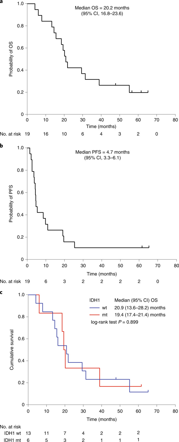
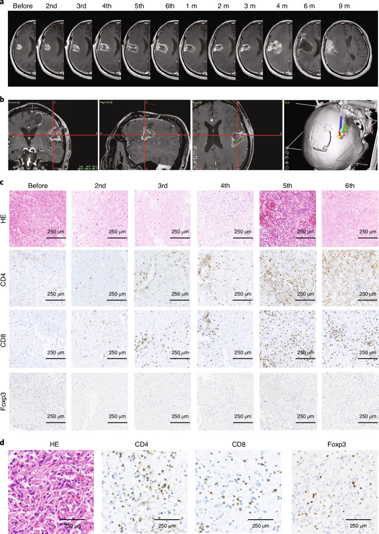
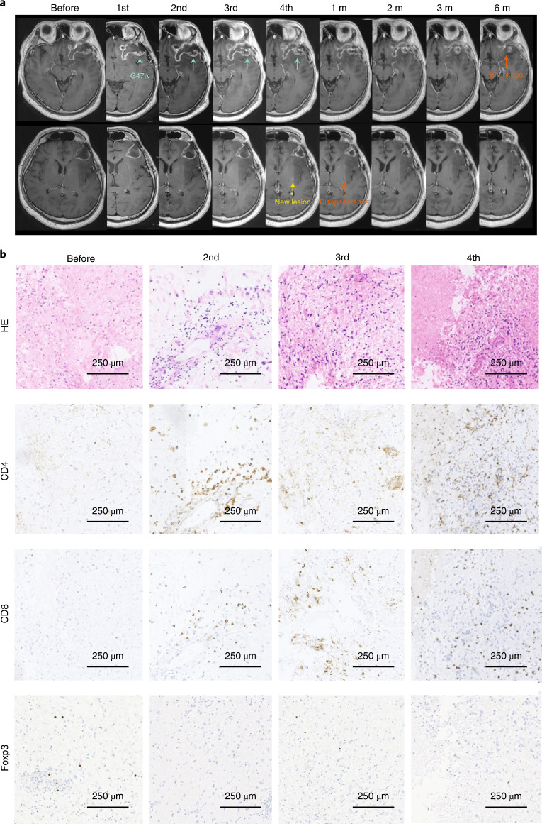
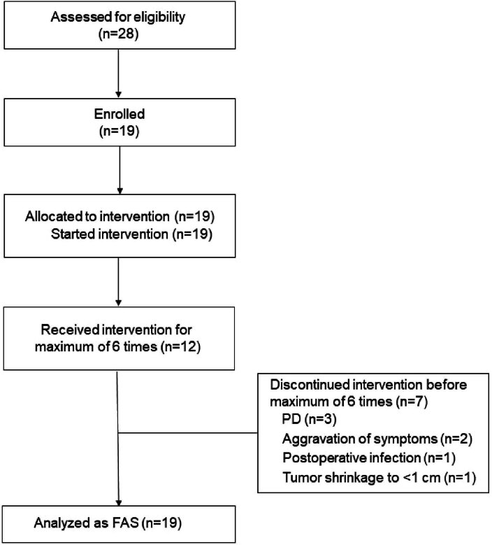
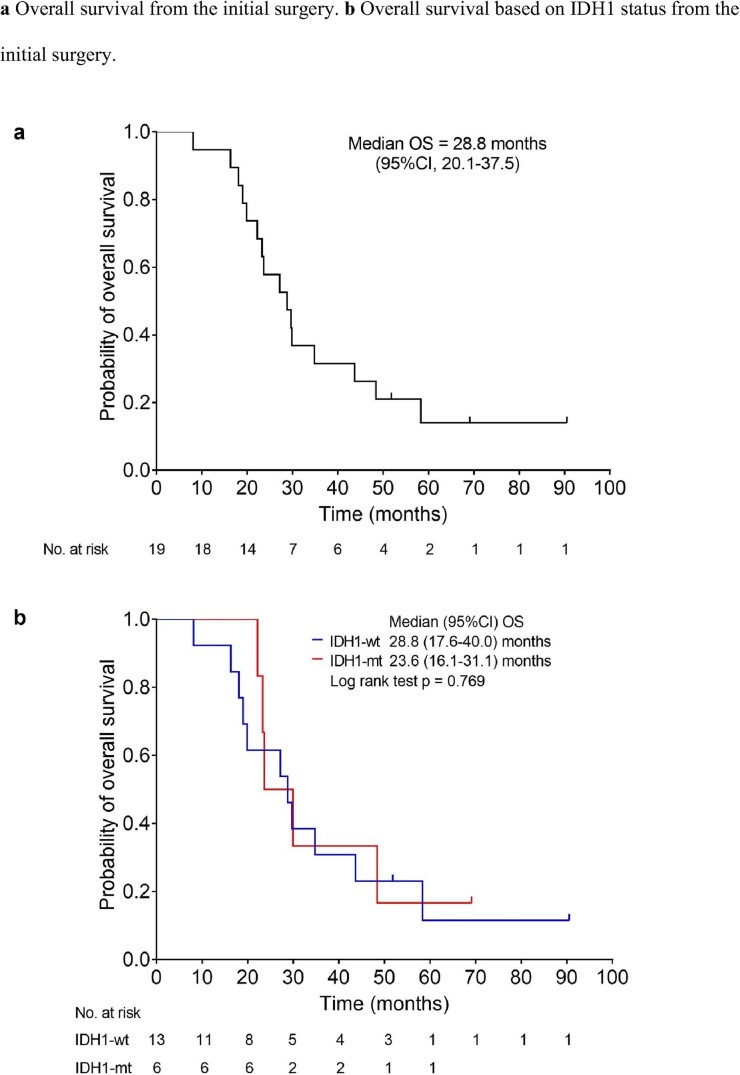
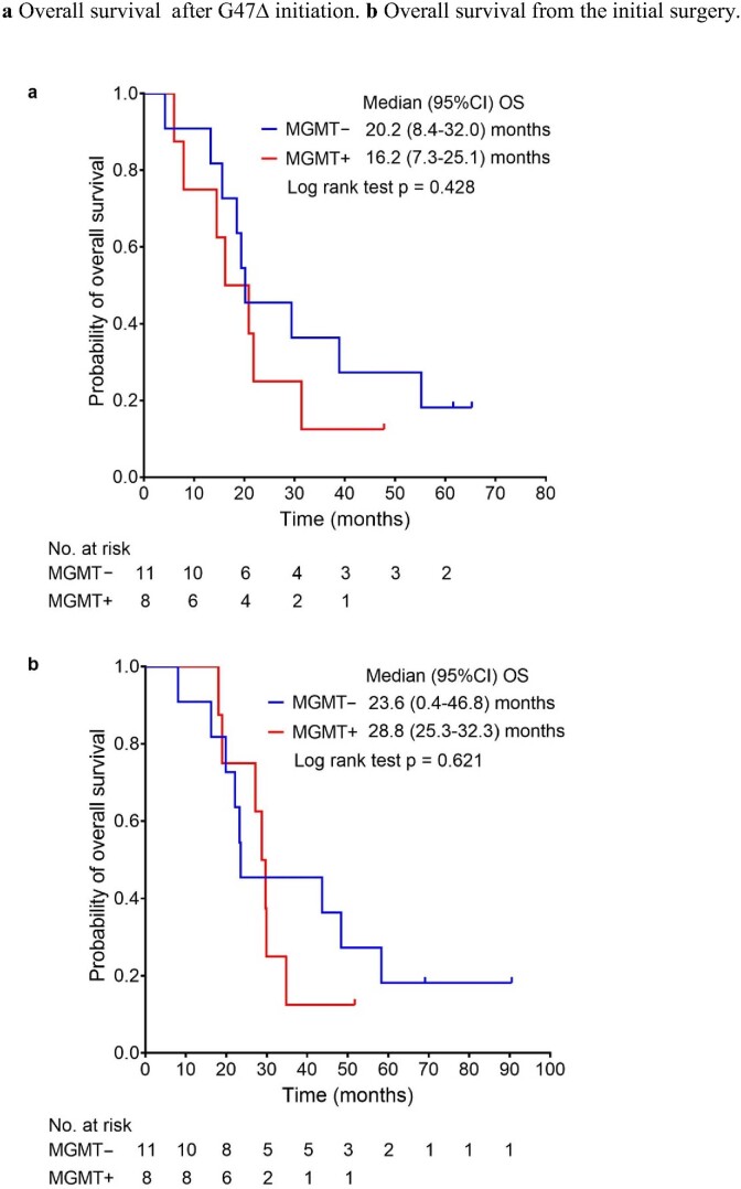
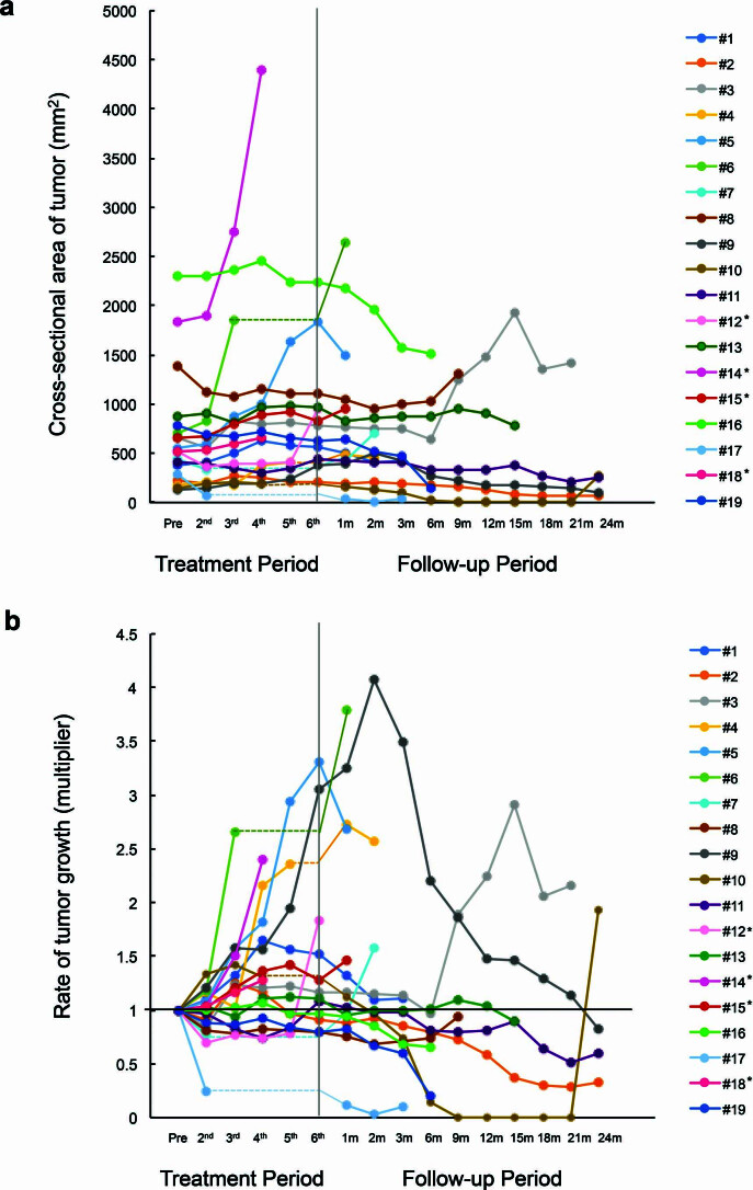
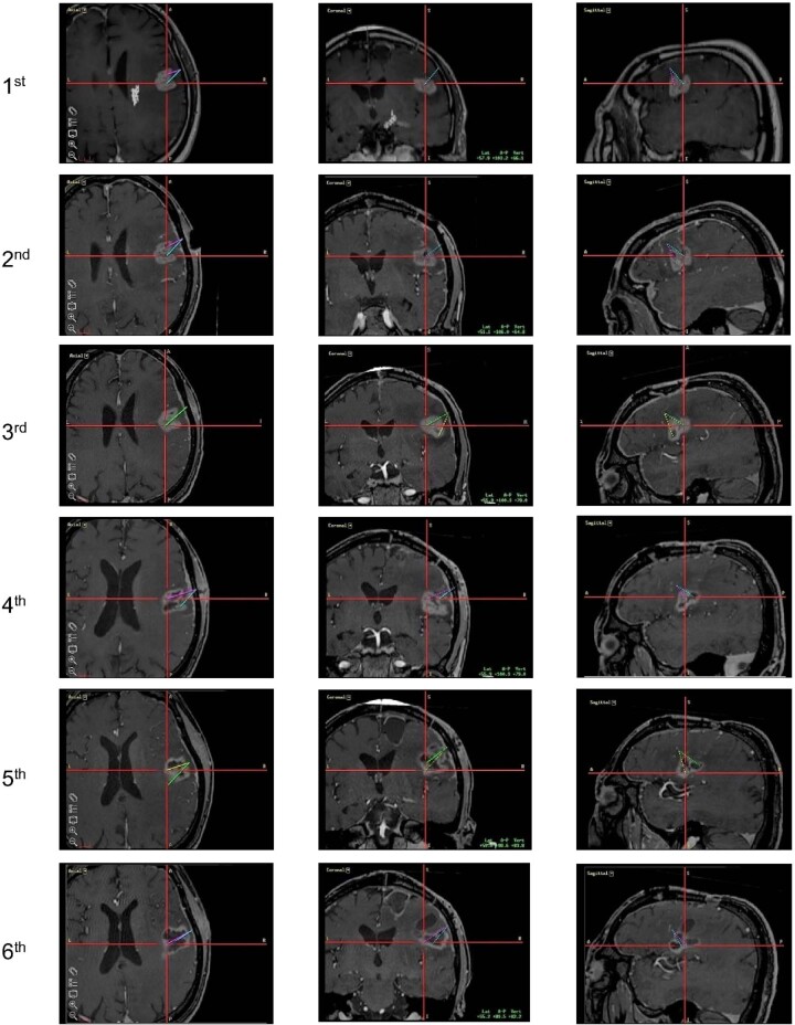
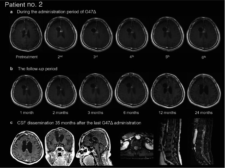
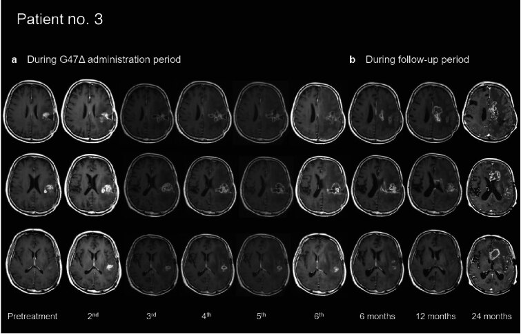
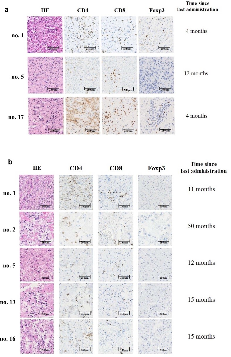
This investigator-initiated, phase 2, single-arm trial primarily assessed the efficacy of G47∆, a triple-mutated, third-generation oncolytic herpes simplex virus type 1, in 19 adult patients with residual or recurrent, supratentorial glioblastoma after radiation therapy and temozolomide (UMIN-CTR Clinical Trial Registry UMIN000015995). G47Δ was administered intratumorally and repeatedly for up to six doses. The primary endpoint of 1-yr survival rate after G47∆ initiation was 84.2% (95% confidence interval, 60.4-96.6; 16 of 19). The prespecified endpoint was met and the trial was terminated early. Regarding secondary endpoints, the median overall survival was 20.2 (16.8-23.6) months after G47∆ initiation and 28.8 (20.1-37.5) months from the initial surgery. The most common G47∆-related adverse event was fever (17 of 19) followed by vomiting, nausea, lymphocytopenia and leukopenia. On magnetic resonance imaging, enlargement of and contrast-enhancement clearing within the target lesion repeatedly occurred after each G47∆ administration, which was characteristic to this therapy. Thus, the best overall response in 2 yr was partial response in one patient and stable disease in 18 patients. Biopsies revealed increasing numbers of tumor-infiltrating CD4+/CD8+ lymphocytes and persistent low numbers of Foxp3+ cells. This study showed a survival benefit and good safety profile, which led to the approval of G47∆ as the first oncolytic virus product in Japan.


Conflict of interest statement

T.T. owns the patent right for G47∆ in Japan. All other authors have no conflict of interest to declare.

Figures

Fig. 1
Fig. 1. Kaplan-Meier curves after G47Δ initiation.
ac, Kaplan–Meier curves for OS after G47∆ initiation (a), PFS after G47∆ initiation (b) and OS based on IDH1 status after G47Δ initiation (c). The data were analyzed and Kaplan–Meier curves created on 1 March 2022. Source data
Fig. 2
Fig. 2. Representative case (patient no. 1) treated with G47Δ.
a, MRI images at indicated observation time points. Characteristic MRI changes were observed at every G47Δ administration, that is, a clearing of contrast-enhancement at the injection site and an enlargement of the entire target lesion. G47∆ was injected to different coordinates from previous injections, so the area of contrast-enhancement clearing increased as the doses increased, leading to a large hollow within the target lesion with an increase in diameter after six G47∆ doses. The MRI changes ceased after the last G47∆ injection and the target lesion stayed stable until 4 months after G47∆ therapy, when the tumor showed a regrowth. The regrown tumor was resected at 6 months but further regrew at 9 months, and the patient died of exacerbation of the disease at 16.2 months after G47∆ initiation. b, Planning MRI using StealthStation Surgical Navigation System at the 6th dose, displaying administration routes from the 2nd to 6th dose (10 injections) overlaid in the same image. c, Histology of biopsy specimens. Biopsies were performed before indicated injections and were obtained from coordinates different from previous G47∆ injections. CD4+ and CD8+ lymphocytes infiltrating within the tumor increased abundantly in number as G47∆ injections were repeated. In contrast, the number of tumor-infiltrating Foxp3+ cells remained low throughout repeated G47∆ injections. Representative of four biopsy specimens. d, Histology of resected tumor at regrowth. The numbers of tumor-infiltrating CD4+ and CD8+ lymphocytes remained high in the regrown tumor resected 6 months after the last G47∆ administration. A higher number of Foxp3+ cells are observed in the tumor at regrowth than in tumors during G47∆ treatment. Representative of three tissue samples. HE, haematoxylin and eosin; m, month(s).
Fig. 3
Fig. 3. Representative case (patient no. 10) showing a long-term efficacy via antitumor immunity.
a, MRI images at indicated observation time points. G47∆ was injected into the target lesion (green arrows), causing the characteristic appearance of contrast-enhancement clearing at the injection site and an enlargement of the entire target lesion. After four doses, a new lesion (yellow arrow) appeared in the left basal ganglia remote from the target lesion, so G47∆ therapy was terminated. However, at the first follow-up 1 month after the last G47∆ administration, the new lesion disappeared (orange arrow at 1 month). Eventually, in the observations that followed, the target lesion decreased in size (orange arrow at 6 months). Remote new lesions further appeared at 24 months and the patient died of exacerbation of the disease at 38.9 months after G47∆ initiation. b, Histology of biopsy specimens. Biopsies were performed before indicated injections and were obtained from coordinates different from previous G47∆ injections. Similar to patient no. 1, CD4+ and CD8+ lymphocytes infiltrating within the tumor increased abundantly in number as G47∆ injections were repeated, whereas the number of tumor-infiltrating Foxp3+ cells remained low throughout repeated G47∆ injections.
Extended Data Fig. 1
Extended Data Fig. 1. Patient disposition.
Of 28 patients who gave informed consent, 19 patients who matched the eligibility criteria were enrolled and composed the full analysis set (FAS).
Extended Data Fig. 2
Extended Data Fig. 2. Kaplan-Meier curves from the initial surgery.
a Overall survival from the initial surgery. b Overall survival based on IDH1 status from the initial surgery. Source data
Extended Data Fig. 3
Extended Data Fig. 3. Kaplan-Meier curves based on MGMT expression.
a Overall survival after G47Δ initiation. b Overall survival from the initial surgery. Source data
Extended Data Fig. 4
Extended Data Fig. 4. Time course of cross-sectional area of the target lesion of individual patients.
a Cross-sectional area (CSA) of the target lesion expressed as actual area at every assessment point. The median CSA of the tumor at baseline was 519.9 mm2 (range, 122.4-2,306.7 mm2). b CSA of the target lesion expressed as a ratio relative to the pre-treatment area. The target lesions tended to show a temporary increase in area during G47∆ treatment. Twelve patients (63.2%) showed a decrease in target lesion area for a certain time compared to pre-treatment. In patients who did not receive G47∆ for the maximum of 6 times, the period during which they were not administered is expressed as a horizontal dotted line. * indicates patients who received corticosteroids. Source data
Extended Data Fig. 5
Extended Data Fig. 5. Planning MRI of a representative case (patient no. 1).
Individual planning MRI of patient no. 1 using StealthStation Surgical Navigation System from the 1st to the 6th dose displaying 2 administration routes for each dose. Axial (left), coronal (middle) and sagittal (right) sections are shown for each dose. G47∆ was injected into viable tumor sites, depicted as contrast-enhanced portions on MRI, remaining from previous injections.
Extended Data Fig. 6
Extended Data Fig. 6. Representative case (patient no. 2) showing cerebrospinal fluid dissemination at progression.
a Characteristic MRI changes are observed with an overall lesion size increased during the administration period. b Shrinkage of the target lesion is observed at 12 months after the last G47Δ administration. c Cerebrospinal fluid (CSF) dissemination is observed in the brain and in the spinal cord at 35 months after the last G47Δ administration. Note that the target lesion is well controlled. The disease course since the initiation of G47Δ therapy was 55.2 months.
Extended Data Fig. 7
Extended Data Fig. 7. Representative case (patient no. 3) showing target lesion shrinkage and subsequent progression adjacent to but away from G47∆ injected sites.
a Characteristic MRI changes are observed, resulting in a large clearing area within, and an enlargement of, the target lesion. b The target lesion is reduced in size at 6 months after the last G47Δ administration. At 12 months, while the target lesion keeps shrinking, a progression is seen in the brain adjacent to the target lesion anterior-medially. The progression continues at 24 months. The disease course since the initiation of G47Δ therapy was 31.4 months.
Extended Data Fig. 8
Extended Data Fig. 8. Histology of regrown tumors at reoperation and brain lesions at autopsy.
a After G47∆ therapy, 3 patients underwent craniotomy for tumor resection of regrown tumors (no. 1 and no. 17 at 4 months and no. 5 at 12 months after the last G47∆ administration). Infiltrating numbers of CD4 + and CD8 + lymphocytes remained abundantly increased in the two cases at 4 months and to some extent in the case at 12 months. Increased numbers of Foxp3+ cells were found in the two cases at 4 months and both underwent reoperation, but not found in the case at 12 months. Each is representative of 3 tissue samples. b Among 5 patients who underwent autopsy, 4 patients died of tumor progression (no. 1, no. 2, no. 5 and no. 16) and an infiltration of CD4+ and CD8+ lymphocytes and a low number of Foxp3+ cells persisted at autopsy 11 to 50 months after the last G47Δ administration. In patient no. 13 whose target lesion was well controlled at the time of death, necrosis and calcification, but no viable tumor cells, were found in the brain at autopsy 15 months after the last G47∆ administration. Each is representative of 3 tissue samples.

Comment in

References

    1. Stupp, R. et al. Effect of tumor-treating fields plus maintenance temozolomide vs maintenance temozolomide alone on survival in patients with glioblastoma: a randomized clinical trial. JAMA318, 2306–2316 (2017). - PMC - PubMed
    1. Todo, T., Martuza, R. L., Rabkin, S. D. & Johnson, P. A. Oncolytic herpes simplex virus vector with enhanced MHC class I presentation and tumor cell killing. Proc. Natl Acad. Sci. USA98, 6396–6401 (2001). - PMC - PubMed
    1. Ma, W., He, H. & Wang, H. Oncolytic herpes simplex virus and immunotherapy. BMC Immunol.19, 40 (2018). - PMC - PubMed
    1. Carson, J., Haddad, D., Bressman, M. & Fong, Y. Oncolytic herpes simplex virus 1 (HSV-1) vectors: increasing treatment efficacy and range through strategic virus design. Drugs Future35, 183–195 (2010). - PMC - PubMed
    1. Todo, T., Ino, Y., Ohtsu, H., Shibahara, J. & Tanaka, M. A phase I/II study of triple-mutated oncolytic herpes virus G47∆ in patients with progressive glioblastoma. Nature Commun. (in the press). - PMC - PubMed

Publication types

Associated data