. 2022 Jul;54(7):1011-1023.
doi: 10.1038/s12276-022-00805-0. Epub 2022 Jul 21.

ROS-activated CXCR2+ neutrophils recruited by CXCL1 delay denervated skeletal muscle atrophy and undergo P53-mediated apoptosis

Affiliations

ROS-activated CXCR2+ neutrophils recruited by CXCL1 delay denervated skeletal muscle atrophy and undergo P53-mediated apoptosis

Yaoxian Xiang et al. Exp Mol Med. 2022 Jul.

Abstract

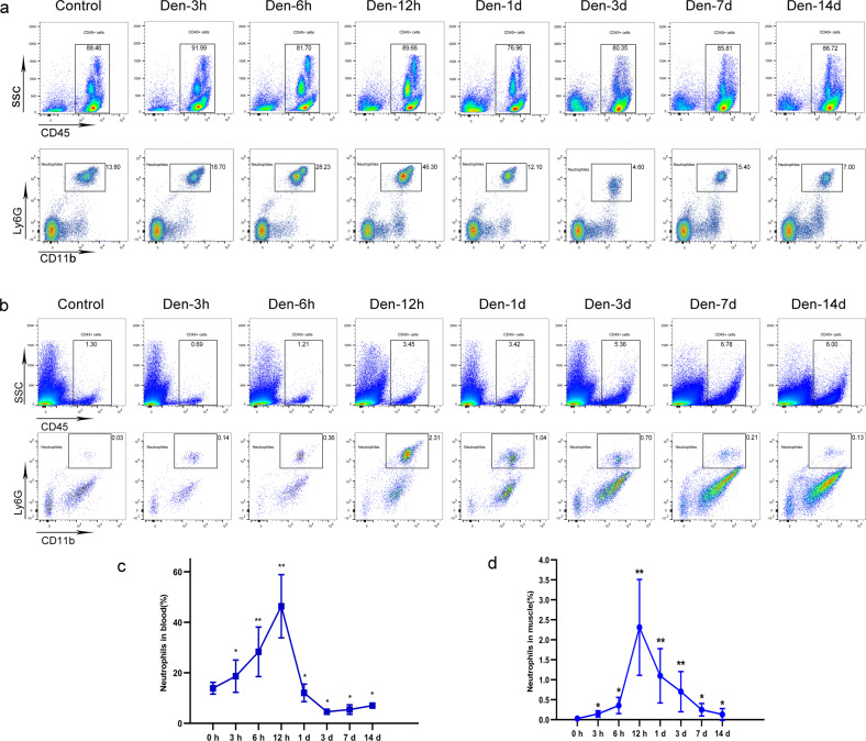
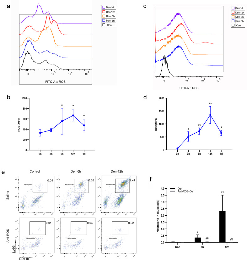
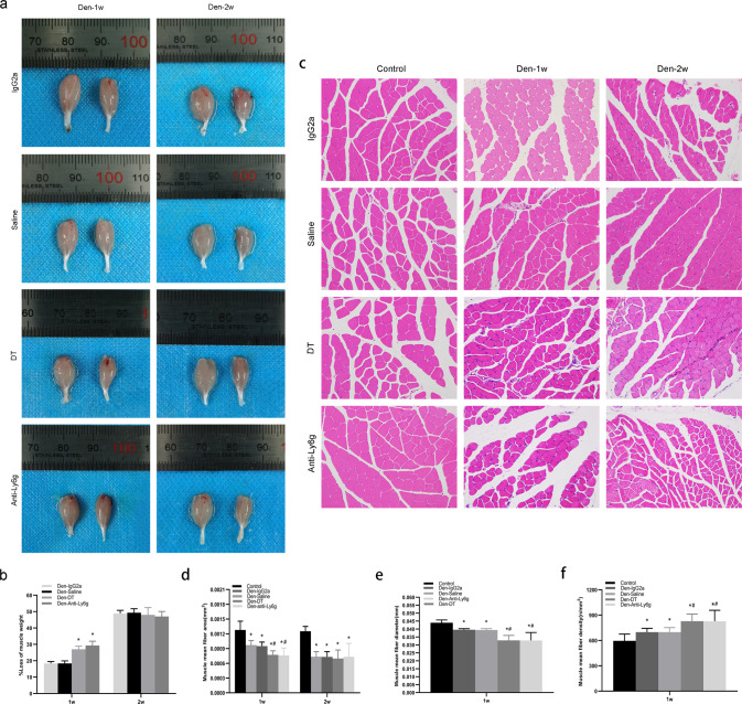
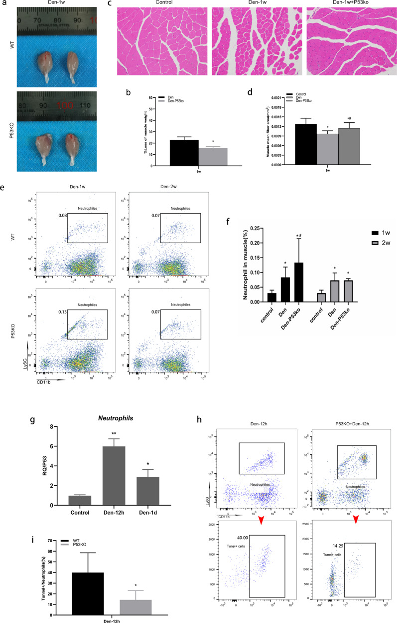
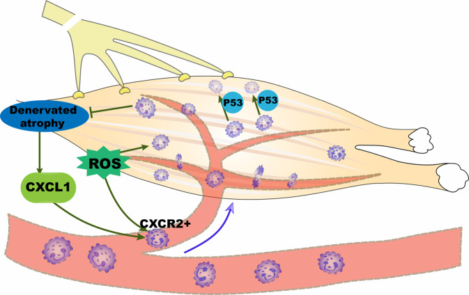
Neutrophils are the earliest master inflammatory regulator cells recruited to target tissues after direct infection or injury. Although inflammatory factors are present in muscle that has been indirectly disturbed by peripheral nerve injury, whether neutrophils are present and play a role in the associated inflammatory process remains unclear. Here, intravital imaging analysis using spinning-disk confocal intravital microscopy was employed to dynamically identify neutrophils in denervated muscle. Slice digital scanning and 3D-view reconstruction analyses demonstrated that neutrophils escape from vessels and migrate into denervated muscle tissue. Analyses using reactive oxygen species (ROS) inhibitors and flow cytometry demonstrated that enhanced ROS activate neutrophils after denervation. Transcriptome analysis revealed that the vast majority of neutrophils in denervated muscle were of the CXCR2 subtype and were recruited by CXCL1. Most of these cells gradually disappeared within 1 week via P53-mediated apoptosis. Experiments using specific blockers confirmed that neutrophils slow the process of denervated muscle atrophy. Collectively, these results indicate that activated neutrophils are recruited via chemotaxis to muscle tissue that has been indirectly damaged by denervation, where they function in delaying atrophy.


Conflict of interest statement

We declare that we do not have any commercial or associative interest that represents a conflict of interest in connection with the work submitted.

Figures

Fig. 1
Fig. 1. Dynamic changes in neutrophils in peripheral blood and denervated muscle.
a Flow cytometry analysis of neutrophils in control WT peripheral blood and blood at 3 h, 6 h, 12 h, 1 day, 3 days, 7 days, and 14 days after denervation. c Quantification of the dynamic changes in neutrophil populations as a percentage of peripheral blood cells. *P < 0.05 and **P < 0.01 versus 0 h (n = 4/group). b Flow cytometry analysis of neutrophils in control WT gastrocnemius muscle and muscle 3 h, 6 h, 12 h, 1 day, 3 days, 7 days, and 14 days after denervation. d Quantification of the dynamic changes in neutrophil populations as a percentage of cells in denervated gastrocnemius muscle. *P < 0.05 and **P < 0.01 versus 0 h (n = 4/group). WT wild type.
Fig. 2
Fig. 2. Increased ROS levels activate neutrophils after denervation.
a Flow cytometry analysis of ROS in control WT peripheral blood and blood at 3 h, 6 h, 12 h, and 1 day after denervation. b Dynamic changes in ROS in peripheral blood. *P < 0.05 versus 0 h (n = 4/group). c Flow cytometry analysis of ROS in control WT gastrocnemius muscle and muscle 3 h, 6 h, 12 h and 1 day after denervation. d Dynamic changes in ROS in denervated gastrocnemius muscle. *P < 0.05 and **P < 0.01 versus 0 h (n = 4/group). e WT mice were treated with a ROS inhibitor at the optimal dose (200 μg/mouse) or with saline before and after denervation. Flow cytometry analysis of neutrophils in control WT gastrocnemius muscle and muscle 6 and 12 h after denervation. f Quantification of neutrophils as a percentage of cells in denervated gastrocnemius muscle. *P < 0.05 and **P < 0.01 versus control; ##P < 0.01 versus Den-6h and Den-12h (n = 4/group). ROS reactive oxygen species, WT wild type.
Fig. 3
Fig. 3. Dynamic behavior of neutrophils inside blood vessels of denervated muscle.
Representative images of the accumulation of neutrophils (Ly6G: blue) and trafficking from the peripheral blood circulation to the blood vessels (PECAM-1: red) of denervated muscle in response to sciatic nerve injury in the control and 3 h, 6 h, 12 h, 1 day, 3 days, 7 days, and 14 days after denervation. Albumin (green) was injected to evaluate the patency and permeability of blood vessels, which exhibited no visible exudation. Representative images of blood vessels (large yellow arrow) and muscle fibers (small yellow arrows). Scale bars, 10 μm.
Fig. 4
Fig. 4. Neutrophils escape from blood vessels and migrate into denervated muscle.
a, b Representative 3D panoramic scanning images of recruited neutrophils (green) migrating from the blood vessels (red) to denervated muscle tissue (DAPI: blue) 12 h after sciatic nerve injury. A: Scale bars, 50 μm; B: Scale bars, 10 μm. c, d, e, f Representative 3D-view images of Imaris reconstruction showing the separated spatial distribution of blood vessels and neutrophils in denervated muscle 12 h after sciatic nerve injury. c: Scale bar, 1000 μm; d: Scale bars, 500 μm (top), 400 μm (bottom); e: Scale bars, 100 μm (top), 300 μm (bottom); f: Scale bars, 500 μm (top), 100 μm (bottom).
Fig. 5
Fig. 5. CXCL1 recruits CXCR2 neutrophils to denervated muscle.
a Flow cytometry analysis of recruited CXCR2+ and CCR2+ neutrophils in control WT gastrocnemius muscle and muscle 3 h, 6 h, 12 h 1 day, 3 days, 7 days, and 14 days after denervation. Representative gating is shown. b, c Quantification of the dynamic changes in the CXCR2+ and CCR2+ subsets as a percentage of cells in denervated gastrocnemius muscle. *P < 0.05 and **P < 0.01 versus 0 h (n = 4/group). d RNA-seq identified the top 30 GO terms associated with denervated gastrocnemius muscle. e Top 20 KEGG pathways of DEGs. f Heatmap of the top 10 upregulated DEGs in gastrocnemius muscle from control WT mice and muscle 6 h after denervation. Heatmap colors indicate directionality (red: increased; blue: decreased). g CXCL1 mRNA expression in control WT mouse gastrocnemius muscle and muscle after denervation. *P < 0.05 vs. 0 h (n = 4/group). DEG differentially expressed gene, WT wild type.
Fig. 6
Fig. 6. Neutrophils attenuate denervated muscle atrophy during the first week.
Ly6g-DTR-GFP mice were injected with DT at the optimal dose (10 μg/g body weight/mouse, 3 times/week) before and after denervation. WT mice were treated with anti-Ly6g clone 1A8 at the optimal dose (200 μg/mouse, 3 times/week) after denervation. a, b The appearance and loss of gastrocnemius muscle weight at 1 week and 2 weeks post-denervation. *P < 0.05 versus Den-Saline or Den-IgG2a (n = 6/group). c, d, e, f HE staining of muscle tissue (200×) and mean ± SEM fiber area, mean fiber diameter, and mean fiber density showing muscle atrophy that was ameliorated by neutrophils. *P < 0.05 versus Control; #P < 0.05 versus Den-Saline or Den-IgG2a (n = 6/group). WT, wild-type, Ly6g-DTR-GFP mice, heterozygous mice with ly6g gene knock-in IRES-DTRGFP; DT diphtheria toxin.
Fig. 7
Fig. 7. Neutrophils undergo apoptosis via P53 with the development of muscle atrophy.
a, b The appearance and weight loss of the gastrocnemius muscle at 1 week post-denervation. *P < 0.05 versus Den (n = 6/group). c, d HE staining of muscle tissue (200×) and the mean ± SEM fiber area; sections show that muscle atrophy was reduced following P53 knockout. *P < 0.05 versus Control; #P < 0.05 versus Den (n=6/group). e Flow cytometry analysis of neutrophils in control WT and P53 KO gastrocnemius muscle 1 week and 2 weeks after denervation. f Quantification of neutrophils as a percentage of cells of denervated gastrocnemius muscle. *P < 0.05 versus Control; #P < 0.05 versus Den (n = 4/group). g Level of P53 was measured by real-time polymerase chain reaction in neutrophils. At Control, Den-12 and Den-1d groups, the neutrophils were isolated by flow sorting, respectively. **P < 0.01 versus Control, *P < 0.05 versus Control. h Flow cytometry analysis of neutrophils and corresponding TUNEL-positive neutrophils in WT and P53 KO gastrocnemius muscle 12 h after denervation. i Quantification of TUNEL-positive cells as a percentage of neutrophils in denervated gastrocnemius muscle. *P < 0.05 versus WT (Den-12h) (n = 4/group). WT wild type, KO knockout.
Fig. 8
Fig. 8. Proposed model for the fate and function of neutrophils after denervation.
Denervation-induced atrophy immediately upregulates CXCL1 expression, which recruits ROS-activated CXCR2+ neutrophils into denervated muscle to delay skeletal muscle atrophy. Neutrophils are preprogrammed to ultimately undergo apoptosis mediated by P53.

Similar articles

Cited by

References

    1. von Grabowiecki Y, et al. Transcriptional activator TAp63 is upregulated in muscular atrophy during ALS and induces the pro-atrophic ubiquitin ligase Trim63. Elife. 2016;5:e10528. doi: 10.7554/eLife.10528. - DOI - PMC - PubMed
    1. Yang Y, et al. Development and evaluation of silk fibroin-based nerve grafts used for peripheral nerve regeneration. Biomaterials. 2007;28:5526–5535. doi: 10.1016/j.biomaterials.2007.09.001. - DOI - PubMed
    1. Gu X, Ding F, Williams DF. Neural tissue engineering options for peripheral nerve regeneration. Biomaterials. 2014;35:6143–6156. doi: 10.1016/j.biomaterials.2014.04.064. - DOI - PubMed
    1. Zhou ZB, et al. Silencing of circRNA.2837 Plays a Protective Role in Sciatic Nerve Injury by Sponging the miR-34 Family via Regulating Neuronal Autophagy. Mol. Ther. Nucleic Acids. 2018;12:718–729. doi: 10.1016/j.omtn.2018.07.011. - DOI - PMC - PubMed
    1. Avraham O, et al. Satellite glial cells promote regenerative growth in sensory neurons. Nat. Commun. 2020;11:4891. doi: 10.1038/s41467-020-18642-y. - DOI - PMC - PubMed

Publication types