. 2022 Jul 12:2022:8225499.
doi: 10.1155/2022/8225499. eCollection 2022.

The Effects of Poria cocos on Rho Signaling-Induced Regulation of Mobility and F-Actin Aggregation in MK-801-Treated B35 and C6 Cells

Affiliations

The Effects of Poria cocos on Rho Signaling-Induced Regulation of Mobility and F-Actin Aggregation in MK-801-Treated B35 and C6 Cells

Yi-Chyan Chen et al. Behav Neurol. .

Abstract

Methods: B35 neuronal cells and C6 glial cells were incubated with MK-801 for 7 days followed by MK-801, MK801 in combination with water extracts of P. cocos (PRP for P. cocos cum Radix Pini or WP for White Poria) treatment for an additional 7 days. Analysis of cell mobility, F-actin aggregation, and Rho signaling modulation was performed to clarify the roles of PRP or WP in MK-801-treated B35 and C6 cells.

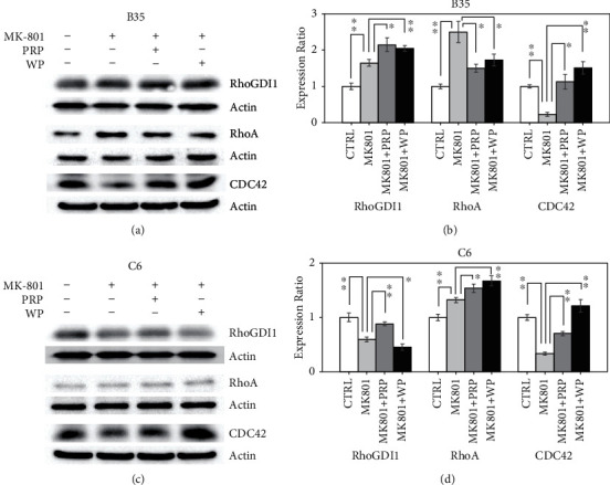
Results: MK-801 decreases B35 cell mobility, whereas the inhibited cell migration ability and F-actin aggregation in MK-801-treated B35 or C6 cells could be reversed by PRP or WP. The CDC42 expression in B35 or C6 cells would be reduced by MK-801 and restored by treating with PRP or WP. The RhoA expression was increased by MK-801 in both B35 and C6 cells but was differentially regulated by PRP or WP. In B35 cells, downregulation of PFN1, N-WASP, PAK1, and ARP2/3 induced by MK-801 can be reversely modulated by PRP or WP. PRP or WP reduced the increase in the p-MLC2 expression in B35 cells treated with MK-801. The reduction in ROCK1, PFN1, p-MLC2, and ARP2/3 expression in C6 cells induced by MK-801 was restored by PRP or WP. Reduced N-WASP and PAK1 expression was differentially regulated by PRP or WP in MK-801-treated C6 cells.


Conflict of interest statement

The authors declare that there is no conflict of interest regarding the publication of this paper.

Figures

Figure 1
Figure 1
PRP and WP differentially regulate MK-801-induced RhoGDI1, RhoA, and CDC42 regulation. Western blotting revealed the expression changes of RhoGDI1, RhoA, and CDC42 induced by PRP/WP in (a) B35 and (c) C6 cells treated with MK-801. The protein expression was quantified by using ImageJ software. Beta-actin was used as a normalization control to calculate the relative expression of the examined targets. The bar chart was constructed according to the data of three independent western blot experiments that analyzed three different batches of protein extracts of drug-treated (b) B35 and (d) C6 cells. The data was analyzed by using Student's t-test (p value<0.05; ∗∗p value<0.01) analysis.
Figure 2
Figure 2
Effects of PRP and WP on actin filament reorganization in B35 and C6 cells treated with MK-801. MK-801-treated B35 and C6 cells on coverslips were incubated with PRP or WP accordingly. ActinGreen™ 488 RreadyProbes™ reagent was used for F-actin in staining in B35 and C6 cells. (a) Red arrows show actin nucleation and blue arrows show F-actin condensation. The numbers of (b) actin nucleation and (c) F-actin condensation were obtained by counting the condensed actin spot in nuclei or condensed actin filament in extended-cytoplasm in 100 cells under different fields of view. The bar charts were made from three independent batches of experiments and analyzed by using Student's  t-test (p value <0.05; ∗∗p value <0.01) analysis.
Figure 3
Figure 3
PRP and WP regulate MK-801-induced ROCK1, PFN1, and p-MLC2 regulation. Western blotting revealed the expression changes of ROCK1, PFN1, and p-MLC2 induced by PRP/WP in (a) B35 and (c) C6 cells treated with MK-801. The protein expression was quantified by using ImageJ software. Beta-actin was used as a normalization control to calculate relative expression of examined target. The bar chart was constructed according to the data of three independent western blot experiments that analyzed three different batches of protein extracts of drug-treated (b) B35 and (d) C6 cells. The data was analyzed by using Student's t-test (p value <0.05; ∗∗p value <0.01) analysis.
Figure 4
Figure 4
Effects of MK-801, PRP, and WP on B35 and C6 cell migration. (a) Cell mobility of B35 and C6 cells was analyzed. The migrated cells were stained with propidium iodide, and then the number of cells on the membrane was counted. (b) The result in the bar chart was made from cell counts of three different drug-treated cell batches and analyzed using Student's t-test (p value <0.05; ∗∗p value <0.01).
Figure 5
Figure 5
PRP and WP regulate MK-801-induced N-WASP, PAK1, and ARP2/3 regulation. Western blotting revealed the expression changes of the indicated proteins induced by PRP/WP in (a) B35 and (c) C6 cells treated with MK-801. The protein expression was quantified by using ImageJ software. Beta-actin was used as a normalization control to calculate relative expression of examined target. The bar chart was constructed according to the data of three independent western blot experiments that analyzed three different batches of protein extracts of drug-treated (b) B35 and (d) C6 cells. The data was analyzed by using Student's t-test (p value <0.05; ∗∗p value <0.01) analysis.

Similar articles

Cited by

References

    1. Fletcher D. A., Mullins R. D. Cell mechanics and the cytoskeleton. Nature . 2010;463(7280):485–492. - PMC - PubMed
    1. Tsai F. C., Kuo G. H., Chang S. W., Tsai P. J. Ca2+ signaling in cytoskeletal reorganization, cell migration, and cancer metastasis. BioMed Research International . 2015;2015:13. doi: 10.1155/2015/409245.409245 - DOI - PMC - PubMed
    1. Tang D. D., Gerlach B. D. The roles and regulation of the actin cytoskeleton, intermediate filaments and microtubules in smooth muscle cell migration. Respiratory Research . 2017;18(1):p. 54. doi: 10.1186/s12931-017-0544-7. - DOI - PMC - PubMed
    1. Yamazaki D., Kurisu S., Takenawa T. Regulation of cancer cell motility through actin reorganization. Cancer Science . 2005;96(7):379–386. - PMC - PubMed
    1. Nakahata Y., Yasuda R. Plasticity of spine structure: local signaling, translation and cytoskeletal reorganization. Frontiers in Synaptic Neuroscience . 2018;10:p. 29. - PMC - PubMed