

Chest Wall Mass as the Dominant Presentation of Low-Grade B-Cell Non-Hodgkin's Lymphoma: A Case Report
- PMID: 35865162
- PMCID: PMC9296237
- DOI: 10.1055/s-0042-1750344
Chest Wall Mass as the Dominant Presentation of Low-Grade B-Cell Non-Hodgkin's Lymphoma: A Case Report
Abstract
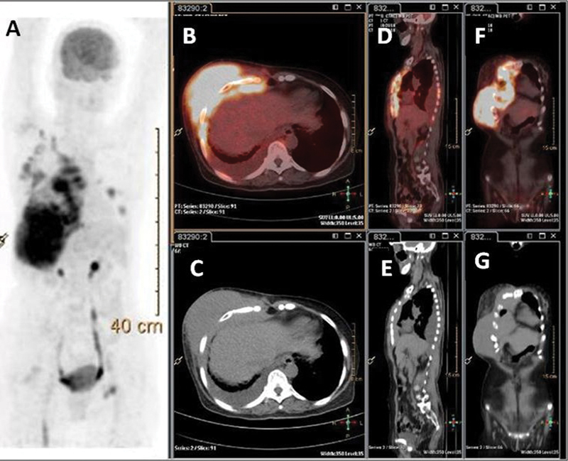
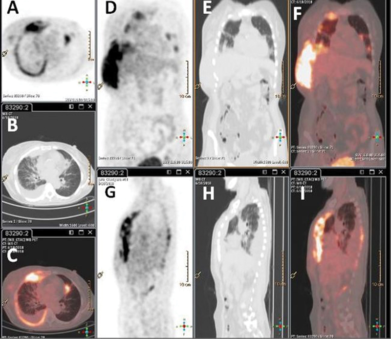
Low-grade B cell non-Hodgkin's lymphoma with dominant presentation of chest wall mass is presented in this report. The patient, a 65-year-old woman, presented with pain, rising skin temperature and redness, and swelling on the right lower chest wall. The histopathological examination revealed non-Hodgkin's lymphoma; the staging fluorodeoxyglucose-positron emission tomography/computed tomography demonstrated stage IVE disease, with hypermetabolic active disease in the right anterolateral chest wall in the form of large soft tissue mass and subcutaneous tissue with underlying bony erosion with extension into right anterior cardiophrenic space and superiorly up to right second costosternal region along the right internal mammary vessels. This was along with hypermetabolic active right axillary, right supraclavicular and left inguinal lymphadenopathy, and thickened hypermetabolic posterior right pleura with ametabolic right-sided pleural effusion. Bone marrow biopsy revealed uninvolved bone marrow. On follow-up after eight cycles of R-CHOP chemotherapy, the mass had completely resolved on contrast-enhanced computed tomography.
Keywords: B cell non-Hodgkin's lymphoma; FDG; PET/CT; primary extranodal lymphoma; staging.
World Association of Radiopharmaceutical and Molecular Therapy (WARMTH). This is an open access article published by Thieme under the terms of the Creative Commons Attribution-NonDerivative-NonCommercial License, permitting copying and reproduction so long as the original work is given appropriate credit. Contents may not be used for commercial purposes, or adapted, remixed, transformed or built upon. ( https://creativecommons.org/licenses/by-nc-nd/4.0/ ).
Conflict of interest statement
Conflict of Interest None declared.
Figures
Similar articles
-
Usefulness of (18)F-FDG positron emission tomography/computed tomography for the diagnosis of pyothorax-associated lymphoma: A report of three cases.Oncol Lett. 2010 Sep;1(5):833-836. doi: 10.3892/ol_00000146. Epub 2010 Sep 1. Oncol Lett. 2010. PMID: 22966389 Free PMC article.
-
Primary Uterine Peripheral T-cell Lymphoma: A Case Report of MRI and 18F-FDG PET/CT Findings.Medicine (Baltimore). 2016 Apr;95(17):e3532. doi: 10.1097/MD.0000000000003532. Medicine (Baltimore). 2016. PMID: 27124063 Free PMC article.
-
Fluorodeoxyglucose-positron emission tomography staging can replace bone marrow biopsy in Hodgkin's lymphoma. Results from Brazilian Hodgkin's Lymphoma Study Group.Hematol Transfus Cell Ther. 2018 Jul-Sep;40(3):245-249. doi: 10.1016/j.htct.2018.03.002. Epub 2018 Apr 24. Hematol Transfus Cell Ther. 2018. PMID: 30128433 Free PMC article.
-
Primary Hodgkin's Lymphoma of the Sternum: Report of a Case and Review of the Literature.J Clin Diagn Res. 2016 Jun;10(6):XE07-XE10. doi: 10.7860/JCDR/2016/19666.8065. Epub 2016 Jun 1. J Clin Diagn Res. 2016. PMID: 27504392 Free PMC article. Review.
-
Management of Hodgkin's lymphoma with midbrain involvement: A case report and review of literature.Hematology. 2015 Jun;20(5):272-5. doi: 10.1179/1607845414Y.0000000190. Epub 2014 Aug 18. Hematology. 2015. PMID: 25133862 Review.
References
-
- Thomas A G, Vaidhyanath R, Kirke R, Rajesh A. Extranodal lymphoma from head to toe: part 2, the trunk and extremities. AJR Am J Roentgenol. 2011;197(02):357–364. - PubMed
-
- Pairolero P C, Arnold P G. Chest wall tumors. Experience with 100 consecutive patients. J Thorac Cardiovasc Surg. 1985;90(03):367–372. - PubMed
-
- King R M, Pairolero P C, Trastek V F, Piehler J M, Payne W S, Bernatz P E. Primary chest wall tumors: factors affecting survival. Ann Thorac Surg. 1986;41(06):597–601. - PubMed
LinkOut - more resources
Full Text Sources
Research Materials