

Reduction of Interhemispheric Homotopic Connectivity in Cognitive and Visual Information Processing Pathways in Patients With Thyroid-Associated Ophthalmopathy
- PMID: 35865354
- PMCID: PMC9295451
- DOI: 10.3389/fnhum.2022.882114
Reduction of Interhemispheric Homotopic Connectivity in Cognitive and Visual Information Processing Pathways in Patients With Thyroid-Associated Ophthalmopathy
Abstract
Purpose: Thyroid-associated ophthalmopathy (TAO) is a vision threatening autoimmune and inflammatory orbital disease, and has been reported to be associated with a wide range of structural and functional abnormalities of bilateral hemispheres. However, whether the interhemisphere functional connectivity (FC) of TAO patients is altered still remain unclear. A new technique called voxel-mirrored homotopic connectivity (VMHC) combined with support vector machine (SVM) method was used in the present study to explore interhemispheric homotopic functional connectivity alterations in patients with TAO.
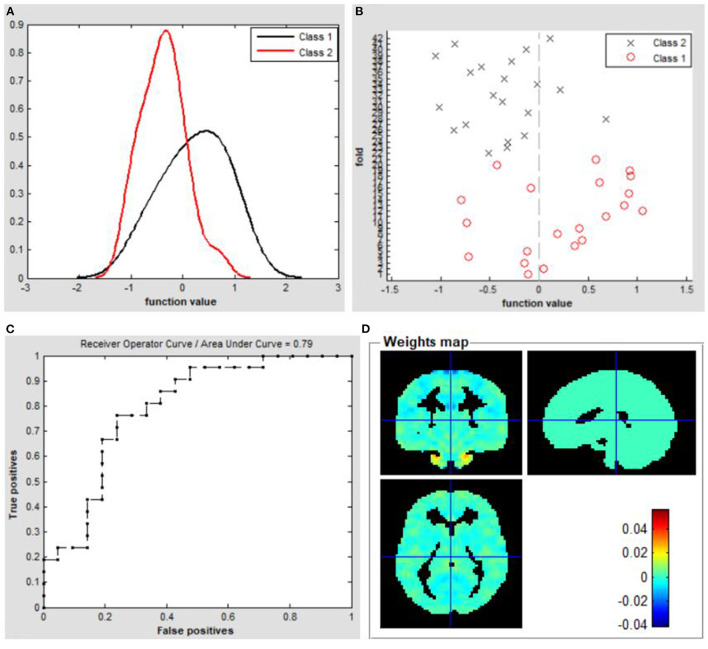
Methods: A total of 21 TAO patients (14 males and 7 females) and 21 wellmatched healthy controls (HCs, 14 males and 7 females), respectively, underwent functional magnetic resonance imaging (fMRI) scanning in the resting state. We evaluated alterations in the resting state functional connectivity between hemispheres by applying VMHC method and then selected these abnormal brain regions as seed areas for subsequent study using FC method. Furthermore, the observed changes of regions in the VMHC analysis were chosen as classification features to differentiate patients with TAO from HCs through support vector machine (SVM) method.
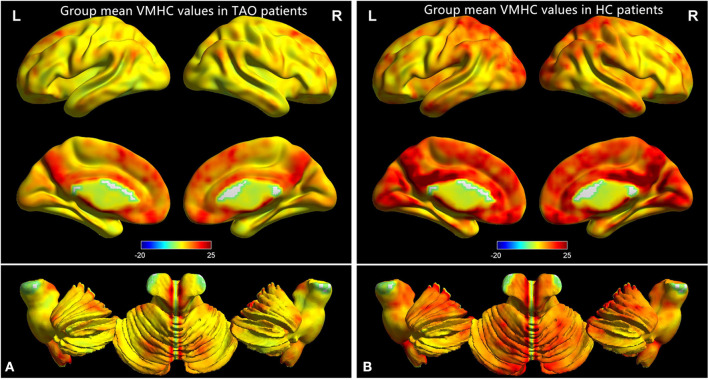
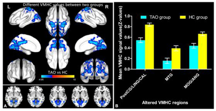
Results: The results showed that compared with HCs, TAO patients showed significantly lower VMHC values in the bilateral postcentral gyrus, lingual gyrus, calcarine, middle temporal gyrus, middle occipital gyrus and angular. Moreover, significantly decreased FC values were found between the right postcentral gyrus/lingual gyrus/calcarine and left lingual gyrus/cuneus/superior occipital gyrus, left postcentral gyrus/lingual gyrus/calcarine and right lingual gyrus/ middle temporal gyrus, right middle temporal gyrus and left cerebellum-8/lingual gyrus/middle occipital gyrus/supplementary motor area, left middle temporal gyrus and right middle occipital gyrus, right middle occipital gyrus/angular and left middle temporal pole (voxel-level p < 0.01, Gaussian random field correction, cluster-level p < 0.05). The SVM classification model achieved good performance in differentiating TAO patients from HCs (total accuracy: 73.81%; area under the curve: 0.79).
Conclusion: The present study revealed that the altered interhemisphere interaction and integration of information involved in cognitive and visual information processing pathways including the postcentral gyrus, cuneus, cerebellum, angular, widespread visual cortex and temporal cortex in patients with TAO relative to HC group. VMHC variability had potential value for accurately and specifically distinguishing patients with TAO from HCs. The new findings may provide novel insights into the neurological mechanisms underlying visual and cognitive disorders in patients with TAO.
Keywords: functional connectivity; functional magnetic resonance imaging; support vector machine; thyroid-associated ophthalmopathy; voxel-mirrored homotopic connectivity.
Copyright © 2022 Qi, Wen and Huang.
Conflict of interest statement
The authors declare that the research was conducted in the absence of any commercial or financial relationships that could be construed as a potential conflict of interest.
Figures

Similar articles
-
Local-to-Remote Brain Functional Connectivity in Patients with Thyroid-Associated Ophthalmopathy and Assessment of Its Predictive Value Using Machine Learning.Int J Gen Med. 2022 Apr 21;15:4273-4283. doi: 10.2147/IJGM.S353649. eCollection 2022. Int J Gen Med. 2022. PMID: 35480997 Free PMC article.
-
Altered Static and Dynamic Interhemispheric Resting-State Functional Connectivity in Patients With Thyroid-Associated Ophthalmopathy.Front Neurosci. 2021 Dec 6;15:799916. doi: 10.3389/fnins.2021.799916. eCollection 2021. Front Neurosci. 2021. PMID: 34938158 Free PMC article.
-
Altered Functional Connectivity of the Primary Visual Cortex in Patients With Iridocyclitis and Assessment of Its Predictive Value Using Machine Learning.Front Immunol. 2021 May 7;12:660554. doi: 10.3389/fimmu.2021.660554. eCollection 2021. Front Immunol. 2021. PMID: 34025659 Free PMC article.
-
Resting-state functional magnetic resonance imaging and support vector machines for the diagnosis of major depressive disorder in adolescents.World J Psychiatry. 2024 Nov 19;14(11):1696-1707. doi: 10.5498/wjp.v14.i11.1696. eCollection 2024 Nov 19. World J Psychiatry. 2024. PMID: 39564181 Free PMC article. Review.
-
Resting-state abnormalities in functional connectivity of the default mode network in migraine: A meta-analysis.Front Neurosci. 2023 Mar 1;17:1136790. doi: 10.3389/fnins.2023.1136790. eCollection 2023. Front Neurosci. 2023. PMID: 36937687 Free PMC article. Review.
Cited by
-
Disrupted dynamic amplitude of low-frequency fluctuations in patients with active thyroid-associated ophthalmopathy.Front Cell Dev Biol. 2023 May 12;11:1174688. doi: 10.3389/fcell.2023.1174688. eCollection 2023. Front Cell Dev Biol. 2023. PMID: 37250893 Free PMC article.
-
Functional decoding and meta-analytic connectivity modeling in thyroid-associated ophthalmopathy.Heliyon. 2023 Dec 15;10(1):e23749. doi: 10.1016/j.heliyon.2023.e23749. eCollection 2024 Jan 15. Heliyon. 2023. PMID: 38226223 Free PMC article.
-
Asthma's effect on brain connectivity and cognitive decline.Front Neurol. 2023 Feb 3;13:1065942. doi: 10.3389/fneur.2022.1065942. eCollection 2022. Front Neurol. 2023. PMID: 36818725 Free PMC article.
-
Machine learning analysis reveals aberrant dynamic changes in amplitude of low-frequency fluctuations among patients with retinal detachment.Front Neurosci. 2023 Jul 20;17:1227081. doi: 10.3389/fnins.2023.1227081. eCollection 2023. Front Neurosci. 2023. PMID: 37547140 Free PMC article.
-
Immune-related visual dysfunction in thyroid eye disease: a combined orbital and brain neuroimaging study.Eur Radiol. 2024 Jul;34(7):4516-4526. doi: 10.1007/s00330-023-10309-8. Epub 2023 Dec 19. Eur Radiol. 2024. PMID: 38112763
References
-
- Chen W., Wu Q., Chen L., Zhou J., Chen H. H., Xu X. Q., et al. . (2021a). Disrupted spontaneous neural activity in patients with thyroid-associated ophthalmopathy: a resting-state fMRI study using amplitude of low-frequency fluctuation. Front. Hum. Neurosci. 15, 676967. 10.3389/fnhum.2021.676967 - DOI - PMC - PubMed
-
- Chen Y., Meng Z., Zhang Z., Zhu Y., Gao R., Cao X., et al. . (2019a). The right thalamic glutamate level correlates with functional connectivity with right dorsal anterior cingulate cortex/middle occipital gyrus in unmedicated obsessive-compulsive disorder: a combined fMRI and 1H-MRS study. Aust. n Z J. Psychiatry. 53, 207–218. 10.1177/0004867418806370 - DOI - PubMed
LinkOut - more resources
Full Text Sources