Case Reports
. 2022 Jul 14;17(9):3349-3354.
doi: 10.1016/j.radcr.2022.06.048. eCollection 2022 Sep.

Primary cardiac epithelioid angiosarcoma: A case report

Affiliations
Case Reports

Primary cardiac epithelioid angiosarcoma: A case report

Thanh Hoa Do et al. Radiol Case Rep. .

Abstract

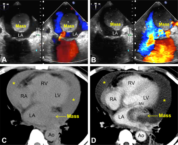
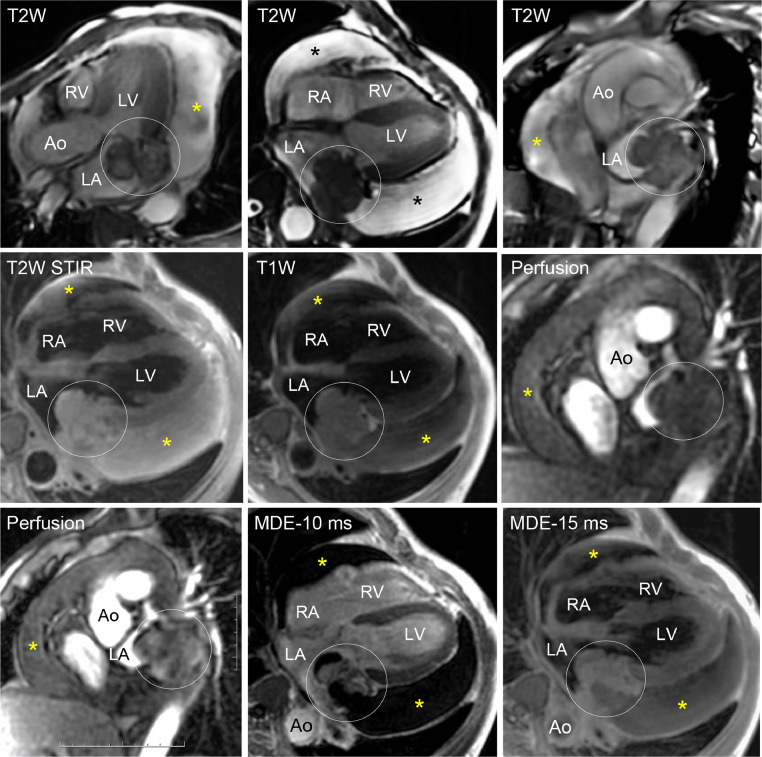
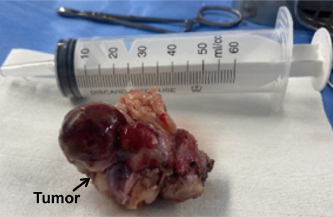
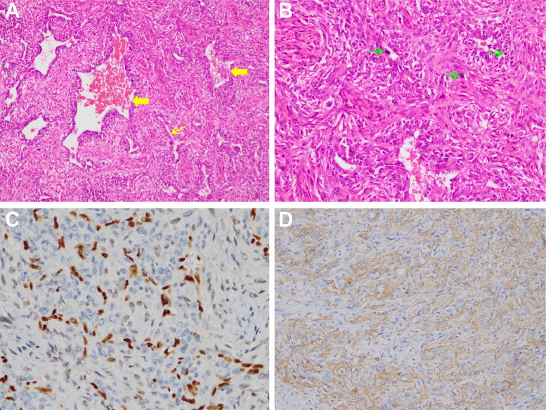
Primary cardiac angiosarcoma is an extremely rare, high-grade malignancy. Here, we describe the case of a 44-year-old male patient with a heart tumor in the left atrium wall, which caused a large amount of pericardial effusion that invaded the surrounding area and is visible on transthoracic echocardiography, computed tomography, and magnetic resonance imaging. The postoperative histopathological results confirmed this case as a primary cardiac epithelioid angiosarcoma.

Keywords: Computed tomography (CT); Heart tumor; Magnetic resonance imaging (MRI); Primary cardiac epithelioid angiosarcoma; Transthoracic echocardiography (TTE).


Figures

Fig 1
Fig. 1
Transthoracic echocardiography (A, B): Two chamber views showed a hyperechoic mass originating from the left atrium and mild mitral valve regurgitation. Chest CT axial images at the precontrast (C) and arterial phases (D) showed a left atrial chamber mass (yellow arrow) that heterogeneously enhances and infiltrates the posterior left atrial wall and involves the mitral valve with an extension into the left atrium and pericardial effusion (asterisks). MV: mitral valve, RA: right atrium, LA: left atrium, RV: right ventricle, LV: left ventricle, Ao: descending aorta.
Fig 2
Fig. 2
Cardiac MRI showed a cardiac tumor (circle), dimensions: 5.3 × 4.9 × 5.0 cm in the left atrium with pericardial effusion (asterisks). The tumor origin rose from the posterior left atrial wall, adhered to the posterior mitral valve, and extended into the lung hilum (T2W and T1W images). The lesion demonstrated late and heterogeneous enhancement (Perfusion and MDE images). Ao: aorta, RA: right atrium, LA: left atrium, RV: right ventricle, LV: left ventricle, MDE: myocardial delayed enhancement, STIR: short tau inversion recovery.
Fig 3
Fig. 3
Postoperative macroscopic image of the tumor, with an irregular border and size of 5 cm × 7 cm.
Fig 4
Fig. 4
Histological cardiac angiosarcoma image. (A) Hematoxylin-eosin stain showed a high density of the spindle cells, interspersed uneven blood vessels (yellow bold arrows), and staghorn branched blood vessels (yellow thin arrows). (B) Higher magnification (400x) expressed large spindle cells with atypical nucleus and mitosis (green thin arrows), arranging haphazard clusters. (C) ERG-positive (in brown) in the tumor area with various densities. (D) CD34 immunostain of heart angiosarcoma showing positive vascular maker expression (200x).

Similar articles

Cited by

References

    1. Blackmon S.H., Reardon M.J. Surgical treatment of primary cardiac sarcomas. Tex Heart Inst J. 2009;36(5):451–452. - PMC - PubMed
    1. Patel S.D., Peterson A., Bartczak A., Lee S., Chojnowski S., Gajewski P., et al. Primary cardiac angiosarcoma - a review. Med Sci Monit. 2014;20:103–109. - PMC - PubMed
    1. Sachdeva S., Patel N., Gupta S., Isufi M., Saeed T., Dalal S., et al., Abstract 12847: left atrial cardiac angiosarcoma: a systematic review of case reports.2021. 144(Suppl_1): p. A12847-A12847.
    1. Best A.K., Dobson R.L., Ahmad A.R. Best cases from the AFIP: cardiac angiosarcoma. Radiographics, 2003. 2022:S141–S145. 23 Spec No. - PubMed
    1. Janigan D.T., Husain A., Robinson N.A. Cardiac angiosarcomas. A review and a case report. Cancer. 1986;57(4):852–859. - PubMed

Publication types

LinkOut - more resources