

Identification of Leishmania tropica from Pediatric Visceral Leishmaniasis in Southern Mediterranean Region of Turkey
- PMID: 35865400
- PMCID: PMC9266607
- DOI: 10.4084/MJHID.2022.053
Identification of Leishmania tropica from Pediatric Visceral Leishmaniasis in Southern Mediterranean Region of Turkey
Abstract
Background and objective: Protozoa of the genus Leishmania are obligate intracellular parasites, and Leishmania species cause a spectrum of species-specific clinical symptoms known as cutaneous, mucocutaneous, and visceral leishmaniasis. For example, Leishmania major and Leishmania tropica cause cutaneous leishmaniasis, while Leishmania infantum and Leishmania donovani cause visceral leishmaniasis (VL). However, molecular studies in recent years have shown that Leishmania species cause different clinical symptoms.
Objectives: Our aim was to evaluate the relationship between the clinical and molecular characterization of leishmania isolates in children with VL defined in Turkey, an intercontinental transitional region.
Methods: The clinical diagnosis of VL was confirmed by detecting amastigotes in the bone marrow aspirate and/or the rK39 test and/or molecular methods (genus-specific PCR, Real-Time PCR, ITS1 PCR-RFLP, DNA sequencing).
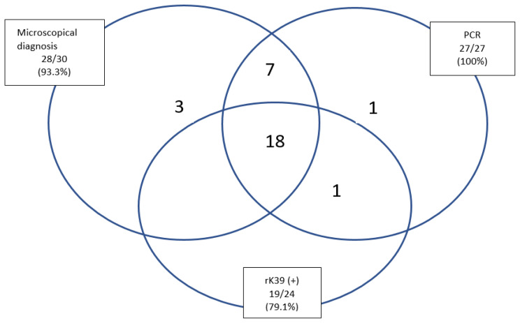
Results: Most of the VL patients were referred from the districts of Adana (53.3%) and others from neighboring provinces; Hatay (16.6%), Osmaniye (3%), Gaziantep (3%), Adıyaman (3%), and 20% case were Syrian immigrants A clinical diagnosis of VL was confirmed in 30 patients with different diagnostic methods. 93% was found positive with microscopic examination, 79.1% with rK39 dipstick test, and 60% with genus-specific PCR assay in clinical samples. The Leishmania isolates were identified as L. infantum (40%), L. donovani (26.7%), and L. tropica (23.3%) using Real-Time PCR, ITS1 PCR-RFLP, and DNA sequencing. There was no cutaneous finding in any case in clinical examination.The most common clinical findings were fever (93.3%) and splenomegaly (90%), followed by hepatomegaly (76.6%). The most common laboratory finding was thrombocytopenia (86.6%), followed by anemia (70%). In addition, hemophagocytic lymphohistiocytosis was detected in bone marrow aspiration in two of our patients. Since pentavalent antimony salts treatment initially failed in four patients, it was necessary to switch to Liposomal-Amphotericin B with treatment success.
Conclusions: The presence of L. tropica in VL patients, despite the absence of cutaneous findings in any of the cases, shows that this strain can cause VL, contrary to conventional knowledge. In the Adana province, where this study was carried out, L. infantum from CL cases in previous studies should be taken into account, and visceral spread in CL cases and accompanying cutaneous lesions in VL cases should be investigated in detail.
Keywords: DNA Sequencing; Leishmania donovani; Leishmania infantum; Leishmania tropica; Visceral leishmaniasis.
Conflict of interest statement
Competing interests: The authors declare no conflict of Interest.
Figures
Similar articles
-
Leishmaniasis in Turkey: Visceral and cutaneous leishmaniasis caused by Leishmania donovani in Turkey.Acta Trop. 2017 Sep;173:90-96. doi: 10.1016/j.actatropica.2017.05.032. Epub 2017 Jun 3. Acta Trop. 2017. PMID: 28587839
-
Clinical manifestations and genetic variation of Leishmania infantum and Leishmania tropica in Southern Turkey.Exp Parasitol. 2015 Jul;154:67-74. doi: 10.1016/j.exppara.2015.04.014. Epub 2015 Apr 22. Exp Parasitol. 2015. PMID: 25913665
-
A real-time ITS1-PCR based method in the diagnosis and species identification of Leishmania parasite from human and dog clinical samples in Turkey.PLoS Negl Trop Dis. 2013 May 9;7(5):e2205. doi: 10.1371/journal.pntd.0002205. Print 2013. PLoS Negl Trop Dis. 2013. PMID: 23675543 Free PMC article.
-
The Geographical Distribution of Human Cutaneous and Visceral Leishmania Species Identified by Molecular Methods in Iran: A Systematic Review With Meta-Analysis.Front Public Health. 2021 Jun 25;9:661674. doi: 10.3389/fpubh.2021.661674. eCollection 2021. Front Public Health. 2021. PMID: 34249836 Free PMC article.
-
Leishmaniasis in Turkey.Acta Trop. 2002 Oct;84(1):43-8. doi: 10.1016/s0001-706x(02)00134-1. Acta Trop. 2002. PMID: 12387909 Review.
Cited by
-
Refugees/Immigrants and leishmaniasis in the world's largest hosting country, Türkiye: A systematic review.PLoS Negl Trop Dis. 2025 Apr 7;19(4):e0012947. doi: 10.1371/journal.pntd.0012947. eCollection 2025 Apr. PLoS Negl Trop Dis. 2025. PMID: 40193403 Free PMC article.
-
Role of Molecular Diagnosis in Imported Cutaneous Leishmaniasis and Its Public Health Significance in India.Pathogens. 2025 Apr 30;14(5):436. doi: 10.3390/pathogens14050436. Pathogens. 2025. PMID: 40430757 Free PMC article.
References
-
- Leishmaniasis. Jan 8, 2022. https://www.who.int/news-room/fact-sheets/detail/leishmaniasis .